


Figure 7.
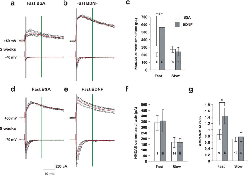
Differential regulation of NMDAR-EPSCs and AMPAR/NMDAR ratio by fast and slow BDNF application.
Whole-cell, voltage-clamp recordings were performed on CA1 pyramidal neurons of hippocampal slices. BSA or BDNF was perfused at slow (25 ml/hr) or fast (240 ml/hr) rate. (a, b, d and e) Representative traces of NMDAR (upper) and AMPAR (lower) currents evoked by extracellular stimulating presynaptic neurons at 50 μA in 2- and 8-week old animals. Black: individual current traces; Red: average waveform. Shaded grey and green rectangular areas indicate where measurements were taken to determine AMPA (at −70 mV) and NMDA (at +50 mV) current amplitudes, respectively. (c) NMDAR currents in 2-week old slices. Note that the average amplitude was significantly increased (p=0.009) after fast but not slow BDNF perfusion. (f) No differences in NMDAR currents were observed between fast and slow BDNF application in 8-week old CA1 neurons. (g) AMPA/NMDA current ratio in 8-week old CA1 neurons. The ratio was significantly increased (p<0.05) after fast but not slow BDNF perfusion.