


Figure 5.
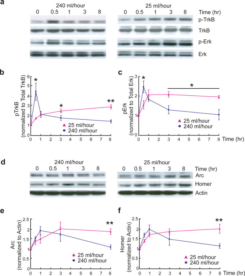
Differential signaling of fast and slow BDNF stimulation in hippocampal slices.
(a–c) Differential kinetics of TrkB and Erk activation induced by fast and slow rates of BDNF stimulation. The CA1 areas from adult hippocampal slices were quickly dissected at the indicated time points after slow (25 ml/hour) or fast (240 ml/hour) BDNF (8 nM, 200 ng/ml) perfusion. TrkB and Erks were used as loading controls. Representative Western blots (a) and quantitative plots (b–c) are presented. n = 3–5 slices/time point. N = 3 independent experiments.
(d–f) Differential expression of Homer1a and Arc in adult hippocampal slices induced by slow and fast BDNF perfusion. The experiments were performed identically as in (a). Representative Western blots (d) and quantification of data (e–f) (N = 3 repetitions of pooled slices) are shown. The same experiment was repeated using entirely independent samples, and the same results were obtained.