


Figure 4. SOD1+/A4V Motor Neurons Exhibit Mitochondrial Defects That are Rescued by Genetic Correction of the SOD1A4V Mutation.
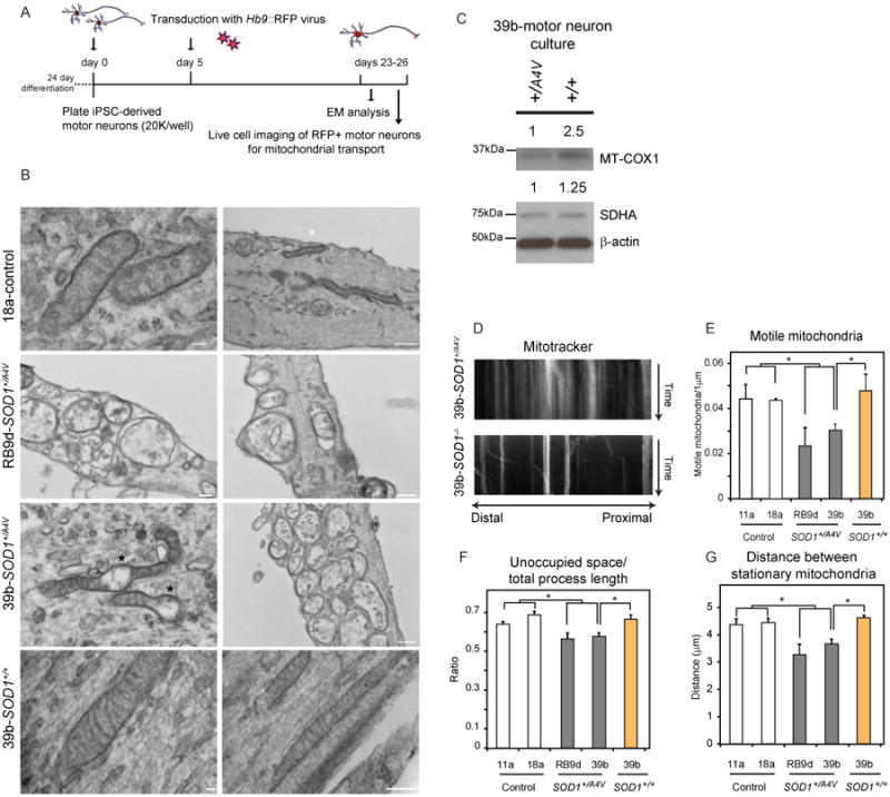
(A) Experimental outline: SOD1+/A4V and isogenic control MN cultures were plated at low densities, transduced with an Hb9∷RFP lentivirus on day 5 and analyzed on days 23-26. (B) Electron microscopy analysis demonstrating that in contrast to control neurons (top panel), SOD1+/A4V neurons (middle panels) often exhibited swelling, morphological disorganization and clustering in neurites. Mitochondria in genetically corrected neurons (bottom panel) exhibited normal morphological characteristics. Representative images out of 3 independent experiments are shown. (C) WB analysis of mitochondrial proteins. (D) Representative kymographs of MitoTracker-stained mitochondria in MNs after live cell imaging. Distal and proximal orientation relative to cell body is indicated. SOD1+/A4V MNs exhibit mitochondria transportation deficiencies relative to controls with (E) fewer motile mitochondria, (F) less space unoccupied by mitochondria relative to axon length and (G) shorter distances between stationary mitochondria (n=4, +/-SEM, P<0.05).