


Figure 4.
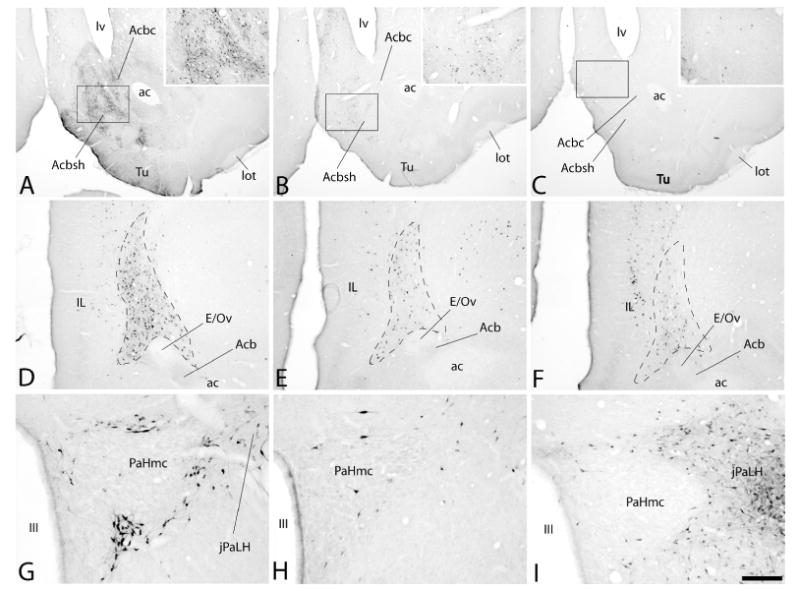
Photomicrographs illustrating retrogradely labeled neurons in the accumbens (Acb in A-C), a heavily labeled structure lodged beneath the infralimbic cortex (outlined area to the right of IL in D-F) and the magnocelluar division of the hypothalamic paraventricular nucleus (PaHmc in G-I) following injections of cholera toxin β subunit (Ctβ) into the ventral tegmental area (VTA, panels A, D and G), rostromedial tegmental nucleus (RMTg, panels B, E and H) and lateral habenula (LHb, panels C, F and I). Note the density of retrogradely labeled neurons in the Acb (A), beneath the infralimbic cortex (D) and surrounding the PaHmc (G) is greatest after injection of tracer into the VTA (A, D and G), whereas it is densest in the juxta-paraventricular lateral hypothalamus (jPaLH in I) following tracer injections into the LHb (C, F and I). Insets in A, B and C are enlargements of the respective boxed areas in the photomicrographs. See list for additional abbreviations. Scale bar: 1 mm in A-C, 200 μm in D-F and 100 μm in G-I.