



Abstract
Background
Epithelial barrier dysfunction is thought to play a role in many mucosal diseases including asthma, chronic rhinosinusitis (CRS), and eosinophilic esophagitis (EoE).
Objective
The objective of this study was to investigate the role of OSM in epithelial barrier dysfunction in human mucosal disease.
Methods
OSM expression was measured in tissue extracts, nasal secretions, and BAL. Effects of OSM stimulation on barrier function of normal human bronchial epithelial (NHBE) cells and nasal epithelial cells (NEC) cultured at air liquid interface (ALI) were assessed using transepithelial electrical resistance (TEER) and FITC dextran flux. Dual color immunofluorescence was used to evaluate integrity of tight junction structures in cultured epithelial cells.
Results
Analysis of CRS samples showed that OSM mRNA and protein were highly increased in nasal polyps compared to control uncinate tissue (p<0.05). OSM was also elevated in BAL of allergic asthmatics following segmental allergen challenge and in esophageal biopsies from EoE patients. OSM stimulation of ALI cultures resulted in reduced barrier function measured by decreased TEER and increased FITC dextran flux (p<0.05). Alterations in barrier function by OSM were reversible, and the viability of epithelial cells was unaffected. OSM levels in lysates of nasal polyps and UT positively correlated with α2-macroglobulin, a marker of epithelial leak, in localized nasal secretions (r=0.4855, p<0.05).
Conclusions
These results suggest that OSM may play a role in epithelial barrier dysfunction in CRS and other mucosal diseases.
Keywords: Oncostatin M, Epithelial barrier, Chronic rhinosinusitis, Eosinophilic esophagitis, Atopic asthma, Transepithelial electrical resistance, tight junctions
Introduction
Mucosal epithelium maintains tissue defense and homeostasis under normal circumstances through many functions, including mucociliary action, the production of mucus and antimicrobial peptides, maintenance of air-surface liquid and ion transport1. The epithelial barrier also serves as a first line of defense by creating a physical barrier to pathogens, allergens and other environmental factors. The integrity of the epithelial barrier is largely determined by the structure of intercellular epithelial junctions, including tight junctions, adherens junctions, and desmosomes2. Upon breach of the epithelial barrier, pathogens, allergens and environmental factors can synergize with alarmins and danger associated molecular patterns (DAMP) that are released in response to cellular damage, thereby activating innate and adaptive immune responses3. The ability of the immune system to neutralize the pathogenic stimulus and repair the tissue damage is critical for the restoration of the tissue to a normal homeostatic state.
Dysfunction of epithelial maintenance and repair of barrier has been implicated in the pathogenesis of many diseases, including asthma, atopic dermatitis, eosinophilic esophagitis (EoE), and chronic rhinosinusitis (CRS). The airway epithelium of allergic asthmatic patients has been shown to have a number of defects, including decreased expression of intercellular junction proteins, increased susceptibility to injury, dysregulated repair, and increased proliferation4, 5, 6, 7. Dysfunctional epithelial barrier function, shown through abnormal electrical and ion permeability, has also been demonstrated in CRS8, 9, 10, 11. Furthermore, sinus epithelium in nasal polyps of patients with CRS was shown to have morphological changes consistent with epithelial dysfunction, including basal cell proliferation and goblet cell hyperplasia, in addition to increased epithelial damage, and decreased tissue resistance compared to control8, 12, 13, 14.
Oncostatin M (OSM) is a member of the IL-6 family of cytokines, and has been shown to signal through 2 receptors; the type 1 receptor is a heterodimer of LIFR and gp130 and the type 2 receptor is a heterodimer of OSM receptor β chain (OSMR) and gp130. OSM has been shown to be expressed by multiple cell types of hematopoietic lineage, but it is not known to be expressed by epithelium, fibroblasts, or smooth muscle, the cells that express the different forms of the OSM receptor15. Previous studies have reported elevated levels of OSM in sinus tissue from allergic rhinitis patients, psoriatic skin and sputum of asthmatic patients with irreversible airflow obstruction16, 17, 18. Moreover, intratracheal administration of adenoviral OSM to mice was shown to be sufficient to induce a type 2 immune response in the lung that was characterized by goblet cell hyperplasia, formation of bronchial associated lymphoid tissue (BALT), eosinophil infiltration, production of IL-4, IL-5, IL-13 and eotaxins, and airway hyperresponsiveness in a mouse model19, 20. Studies using reconstituted epidermis have shown that OSM treatment decreased expression of filaggrin, a molecule that is important for barrier function in keratinocytes, suggesting that OSM could contribute to barrier dysfunction in the skin21. OSM has also been shown to decrease barrier function in endothelial cells22. Given that OSM is sufficient to induce a type 2 immune response in the lung19, and has been shown to potentially contribute to barrier dysfunction in the endothelium and the skin, we tested whether OSM was elevated in CRS and contributed to epithelial barrier dysfunction in this disease.
Methods
Patients and tissue sample collection
Control subjects and patients with CRS were recruited from the Allergy-Immunology and Otolaryngology clinics of the Northwestern Medical Faculty Foundation and the Northwestern Sinus Center at Northwestern Medicine. Uncinate tissue (UT), nasal polyps, epithelial scrapings from inferior turbinate (IT), nasal lavage, and sponge based nasal secretions were obtained during routine functional endoscopic sinus surgery from patients with CRS, as defined by the American Academy of Otolaryngology—Head and Neck Surgery Chronic Rhinosinusitis Task Force23. Patients with established immunodeficiency, pregnancy, coagulation disorder, classic allergic fungal sinusitis, or cystic fibrosis were excluded from this study. Control subjects did not have a history of sinonasal inflammation, and tissue was collected during endoscopic skull-base tumor excisions, intranasal procedures for obstructive sleep apnea, and facial fracture repairs. Characteristics of patients donating sinus samples are shown in Table E1 in this article's Online Repository at www.jacionline.org. Nasal secretions from the middle meatus were collected by an endoscopically placed 0.375 inch polyvinyl alcohol (PVA) sponge (Medtronic, Minneapolis, MN) that was inserted between the middle turbinate and the adjacent uncinate process for ten minutes before removal. The polyvinyl alcohol sponges were then centrifuged at 14,000 RPM for 10 minutes, and extracted with a further 100 μl of PBS and 1% protease inhibitor cocktail (PIC) to collect the nasal secretions. Segmental allergen challenge and bronchoalveolar lavage (BAL) was performed as previously described24, 25. The patient cohort used for segmental allergen challenge was previously described24. Biopsies from control and EoE patients were obtained by endoscopic collection on the initial visit, as previously described, and the characteristics of these patients is shown in Table E2 in this article's Online Repository at www.jacionline.org26. Diagnosis of EoE was based on an eosinophil count of 15 or more per high-powered field in an esophageal biopsy section.
Quantitative RT-PCR and microarray analysis
RNA isolated from tissue samples was prepared, as previously described27. Single-strand cDNA was synthesized from 0.5μg of total RNA with Superscript II reverse transcriptase and random primers (Invitrogen, Carlsbad, Calif). Semiquantitative realtime RT-PCR was performed using an Applied Biosystems 7500 Sequence Detection System (Applied Biosystems, Foster City, Calif) in 20μL reactions (10μL of 2× TaqMan Master mix [Applied Biosystems], 400nmol/L of each primer and 200nmol/L of TaqMan probe plus 10μg of cDNA). Primer and probe sets for OSM (Hs00968300_g1), OSMR (Hs00384276.m1), and β-glucuronidase (4326320E) were purchased from Applied Biosystems. Several housekeeping genes were screened for expression in UT and nasal polyps; β-glucuronidase was found to have the most consistent expression between these two tissue types, and was used as a housekeeping gene when analyzing gene expression in both UT and nasal polyps. Primers (Forward: CTGGCCGGGACCTGACT, Reverse: GCAGCCGTGGCCATCTC) and probe (6-VIC-CACCACCACGGCCGA-MGB) for β-actin were synthesized by IDT technologies (IDT Technologies, Coralville, IA) and used to detect the housekeeping gene in EoE studies. Microarray analysis was performed, as previously described 28.
Protein detection
OSM protein was detected in tissue and nasal secretions using a bead array assay for OSM, according to the manufacturer's instructions (EMD Millipore, Billerica, Mass). Sinus tissue extracts were prepared, as previously described29. OSM levels were analyzed on a Luminex 200 instrument (Life Technologies, Grand Island, NY). Alpha 2-macroglobulin protein was detected in nasal secretions using an ELISA assay, according to the manufacturer's instructions (Alpco, Salem, NH). Albumin and OSM protein were detected in BAL following segmental antigen challenge, as previously described24.
Cell culture and barrier assessment
Normal human bronchial epithelial (NHBE) (Lonza, Walkersville, MD) and nasal epithelial cells (NEC) from our clinical repository were seeded onto transwells and grown to confluence in bronchial epithelial growth medium (BEGM) (Lonza). Once confluent, the media from the upper chamber of the transwell was removed, constituting the transition to air liquid interface (ALI) culture conditions. NHBE and NEC were fully differentiated at day 21 of ALI culture. Barrier function was measured using two methods; transepithelial electrical resistance (TEER) and FITC dextran flux. Fully differentiated NHBE and NEC were stimulated with recombinant human OSM (R&D systems, Minneapolis, MN) at 100 ng/mL for 48 hours, unless otherwise noted. TEER was measured using a voltohmmeter (World Precision Instruments, Sarasota, FL). Dextran flux was measured by placing 10kD FITC-conjugated dextran (Sigma, St Louis, MO) in the upper chamber of the transwell for 30 minutes to assess barrier, after which supernatants were harvested from the lower chamber and a Molecular Devices Spectramax Gemini EM fluorimeter (Molecular Devices, Sunnyvale, Calif) was used to measure the amount of FITC-dextran that crossed through the cell layer. Cytotoxicity of OSM treatment was measured using a Cytotoxicity Detection Kit Plus (Roche Applied Science, Indianapolis, IN). Four day ex vivo culture of UT and nasal polyps was performed as previously described28.
Immunofluorescence staining
NHBE and NEC cell monolayers grown on transwell inserts were fixed in 10% formalin before washing with PBS. The cell layers were blocked with PBS containing 5% goat serum (Vector Laboratories, Burlingame, Calif), 1% BSA (Sigma), and 1.6% Triton X-100 (Sigma) for 1 hour at room temperature (RT), then incubated with rabbit anti-occludin for 1 hour at RT (1:200, 71-1500, Life Technologies). The membranes were then incubated with secondary antibody for 1 hour at RT with Alexa Fluor 488 goat anti-rabbit antibody (Life Technologies). Slides were mounted with slowfade gold antifade reagent with DAPI counter stain (Life Technologies). Imaging was performed using a Nikon A1R confocal microscope using the 40× objective. Images were processed using ImageJ software.
Statistical analysis
All data were analyzed using GraphPad Prism 6 software (GraphPad Software, La Jolla, Calif.). All data were reported as mean ± SEM. Differences between groups were analyzed using a non-parametric Kruskal-Wallis ANOVA or Mann-Whitney U test. Correlations were assessed using the Pearson correlation. Significance: *p<0.05, **p<0.01, ***p<0.001, ****p<.0001.
Results
OSM was elevated in CRS
We first wanted to determine whether OSM expression was elevated in polyps from CRSwNP patients. Using data from a previously published microarray study (GEO dataset GSE36830)30, we found that OSM expression was elevated in nasal polyps of CRS patients compared to UT from control patients (figure E1A in this article's Online Repository at www.jacionline.org). We then went on to verify this finding at the mRNA and protein level, using a separate set of samples. Consistent with our array results, we found that OSM mRNA in nasal polyps was elevated compared to UT from control patients (figure 1A). In addition, OSM protein was elevated in nasal polyps compared to control UT and in nasal lavage fluids from CRSwNP patients compared to CRSsNP patients (figures 1B and C). Analysis of supernatants from an ex vivo 4 day culture of sinonasal tissues also showed elevated levels of OSM in the supernatants of cultures of nasal polyp tissues compared to UT from CRSsNP and CRSwNP (figure 1D)28. To test whether the tissue may contain OSM-responsive cells, we evaluated expression of the OSM receptor (OSMR) and found that OSMR mRNA was also elevated in nasal polyps compared to control UT (Figure E1B in this article's Online Repository at www.jacionline.org).
Figure 1. OSM was elevated in CRs.

(A) OSM mRNA was increased in nasal polyps compared to UT from control, CRSsNP and CRSwNP patients, (1.76 ± .85 vs. .06 ± .013 respectively, p< .01, n=11-13, Kruskal-Wallis test). (B) OSM was increased in nasal polyp tissue compared to control UT as measured by Luminex, (3.38 pg/mg ± 1.95 vs. 47.49 pg/mg ± 17.98 respectively, p<.01, n=11-19, Kruskal-Wallis test). (C) OSM protein was increased in nasal lavage of CRSwNP patients compared to CRSsNP, (78.50pg ± 44.16 vs. 327.1pg ± 78.83 respectively, n=10-40, Kruskal-Wallis test). (D) OSM protein was increased in culture supernatants of nasal polyp tissue compared to UT from CRSsNP and CRSwNP, (19.1ng ± 2.6ng vs. 4.7ng ± 1.1 and 4.9ng ± 0.89 respectively, n=4-23, Kruskal-Wallis test).
OSM decreased barrier function in cultured airway epithelium
To study the effect of OSM stimulation on airway epithelium, we utilized an air-liquid interface culture system, which allowed us to study differentiated epithelium in vitro. Fully differentiated normal human bronchial epithelial (NHBE) cells were left unstimulated, or stimulated with various concentrations of OSM, 1-100ng/mL for 48h. Barrier function of the NHBE cultures was assessed using TEER, which measures the resistance of the cell layer, and paracellular dextran flux. In an intact barrier, TEER will be high, while dextran flux will be low. OSM stimulation of NHBE cells induced a concentration-dependent decrease in TEER (figure 2A) and an increase in dextran flux (figure 2B), which are both characteristic of significant loss of barrier integrity. ALI monolayer cultures of nasal epithelial cells (NEC) also showed decreased TEER (figure E2A in this article's Online Repository at www.jacionline.org), and increased dextran flux (figure E2B in this article's Online Repository at www.jacionline.org), when stimulated with 100 ng/mL OSM. Importantly, OSM stimulation of NHBE did not decrease viability of the cells, as assessed by trypan blue staining (data not shown). In addition, there was no difference in lactate dehydrogenase (LDH) levels in the supernatants of OSM-treated cells when compared to controls (figure E2D in this article's Online Repository at www.jacionline.org). In order to determine whether the effects of OSM treatment on epithelial barrier function were reversible, NEC cultures were stimulated with a high concentration of OSM for 2 days (100 ng/ml), and then washed and allowed to recover for 7 days in ALI culture media. We found no difference between the TEER measurements following the 7 day recovery period and the TEER measured prior to OSM stimulation (baseline), indicating that the effect of OSM on barrier function was reversible (Figure E2C in this article's Online Repository at www.jacionline.org).
Figure 2. OSM decreased barrier function in airway epithelium.

ALI cultures of NHBE cells were grown until fully differentiated at day 21 of culture, and then the cells were left unstimulated, or stimulated with OSM at the specified concentration. (A) OSM stimulation resulted in a concentration dependent decrease of TEER, data is presented as the change in resistance over the 48 hour stimulation normalized to resistance at 0h to show the percentage of resistance lost during stimulation (((0h TEER- 48h TEER)/ 0h TEER)*100) (p< .0001, n=7-11, Kruskal-Wallis test). (B) Dextran flux across the cell layer was increased with OSM stimulation, suggesting decreased barrier function following OSM stimulation (p< .001, n=5, Kruskal-Wallis test). Data represent mean ± SEM.
OSM stimulation altered organization of intercellular junctions
Intercellular junctions, particularly tight junctions, are largely responsible for the barrier function of mucosal epithelium. In order to determine whether OSM stimulation altered expression of key junctional proteins, we analyzed mRNA expression of occludin, claudin 1, E-cadherin, and ZO-1. We found that OSM stimulation of NHBEs had no effect on the level of gene expression of these molecules (data not shown). Additionally, OSM stimulation did not change occludin protein expression as measured using western blot (data not shown). We next determined whether OSM disrupted the localization of junctional proteins. Using immunofluorescence analysis of occludin, we found the characteristic lattice-like structure of occludin+ tight junctions between epithelial cells of both unstimulated NHBE and NEC cultures (Figure 3). However, following OSM stimulation at 100 ng/mL in both NHBE and NEC, it was clear that the organization of the tight junction had been lost (figure 3), indicating that OSM could disrupt tight junction localization, even though it did not affect expression of tight junction proteins. Additionally, when NHBE were stimulated with various concentrations of OSM, tight junction structure was also lost at 10ng/mL OSM while it was intact at 1ng/mL (figure 3).
Figure 3. OSM disrupted organization of tight junctions.

NHBE were left unstimulated, or stimulated with indicated concentrations of OSM for 48hrs. NEC were either left unstimulated, or stimulated with 100ng/mL OSM. The tight junction protein occludin is stained in green, cell nuclei are in blue. Representative of NHBE, n=3, and NEC, n=3.
OSM was associated with impaired barrier function in CRS
In order to determine whether there was an epithelial cell-intrinsic defect in CRS, we next assessed whether NEC from CRS patients had decreased barrier function compared to controls. NEC collected in vivo from the IT from CRSsNP, CRSwNP, and control patients were cultured ex vivo at ALI, and TEER was measured weekly. NEC from each group developed robust barrier function over the course of the assay, and no differences were seen among the groups (Figure 4A). This suggests that the cause of barrier dysfunction in CRS is probably extrinsic to the epithelium. Since OSM was elevated in CRS, and significantly decreased epithelial barrier function in vitro, we next wanted to determine whether OSM might contribute to epithelial barrier dysfunction in vivo. We collected nasal secretions from specific locations within the middle meatus using a sponge in the sinus cavity placed adjacent to the polyp tissue, and measured α2-macroglobulin, which has been used as a macromolecular marker of epithelial leak, in the nasal secretions collected from the sponges31, 32. Levels of OSM in tissue lysates from CRSsNP and CRSwNP patients were significantly positively correlated with levels of α2-macroglobulin in adjacent nasal secretions (p<.05; r=0.4855; Figure 4B), suggesting that there was a relationship between OSM levels and epithelial barrier dysfunction in vivo.
Figure 4. OSM expression correlated with a marker of epithelial leak in vivo.

(A) ALI cultures of NEC from IT from control, CRSsNP, and CRSwNP patients show no difference in TEER (n=7-11). Data represent mean ± SEM. (B) OSM levels in nasal polyps and UT from CRSsNP patients correlated with epithelial leak as determined by assessment of α2-macroglobulin levels in adjacent nasal secretions (Pearson r=.4855, n=11-13, p<.05). Grey dots signify nasal polyp tissue and black dots signify UT of CRSsNP patients.
OSM was elevated in EoE and atopic asthma
We next wanted to determine whether OSM was elevated in other mucosal disorders with dysregulated type 2 immune responses and barrier dysfunction. First, we analyzed mRNA from endoscopic esophageal biopsies from control and EoE patients. We found that OSM expression was elevated in EoE biopsies compared to controls (Figure 5A). We next analyzed levels of OSM protein in the BAL of allergic asthmatic patients following segmental antigen challenge24, 25. While OSM was not present in the BAL of the allergic asthmatic patients when challenged with saline, OSM levels were significantly elevated by 18-20 hours following allergen challenge (Figure 5B). Additionally, we measured levels of human serum albumin, another macromolecular marker associated with epithelial leak, in the BAL of these patients, and we found that HSA levels correlated positively with OSM in the BAL (p<.001; r=0.8062; figure 5C). Collectively, these data indicate that OSM was associated with barrier dysfunction in other mucosal diseases mediated by type 2 inflammation.
Figure 5. OSM levels were increased in EoE and upon allergen challenge in allergic asthmatics.

(A) OSM mRNA was increased in esophageal biopsies of EoE patients compared to control, (6.4e-005 ± 1.8e-005 vs. .00024 ± 4.9e-005 respectively, p<.01, n=8-13, Mann-Whitney U test). (B) OSM protein was increased in BAL of allergic asthmatics following segmental allergen challenge compared to BAL challenged with saline, (.56 ± .56 vs. 22.8 ± 6.3 respectively, p<.0001, n=16, Mann-Whitney U test. (C) OSM levels in BAL following allergen challenge in allergic asthmatics correlate with levels of HSA, (Pearson r=.8062, p<.001).
Discussion
We have found that OSM was elevated in nasal polyp tissues derived from CRSwNP patients (Figure 1). Furthermore, studies using cultured bronchial and nasal epithelium demonstrated that stimulation with OSM at concentrations similar to those seen in vivo were sufficient to cause significant loss of barrier function and disorganization of tight junction structure (Figures 2 and 3). Additionally, OSM expression detected in nasal polyp tissue lysates correlated with nasal lavage levels of α2-macroglobulin, a marker of epithelial leak in localized nasal secretions (Figure 4). Together, these data suggest that OSM may be mediating epithelial barrier dysfunction in CRS, potentially leading to a chronic inflammatory state through increased exposure of the tissue to environmental factors including pathogens, allergens, and pollutants (figure 6). In support of this hypothesis, we found increased OSM mRNA in esophageal biopsies of EoE patients compared to controls (Figure 5), and increased OSM protein following allergen challenge in allergic asthmatics that correlated with epithelial leak in the BAL (Figure 5). Although these findings suggest that OSM may play a role in other diseases of mucosal epithelium that are also associated with type 2 inflammation, further studies are needed to verify these findings in other diseases.
Figure 6. Proposed model for the role of OSM in epithelial barrier dysfunction in CRS.
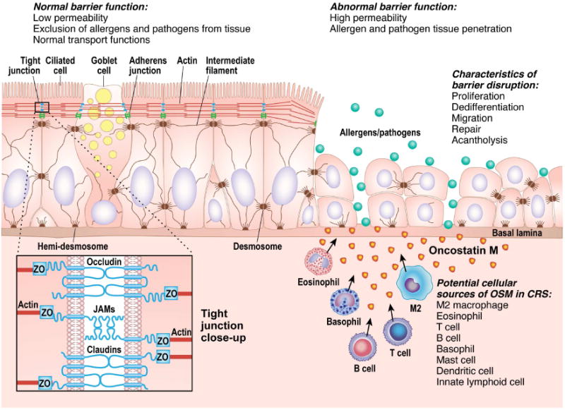
Airway epithelium develops barrier function through a complex network of intercellular junctions that adhere the cells to each other and the cytoskeleton, illustrated on the left side of the figure. Barrier dysfunction, as illustrated on the right side of the figure, may occur following exposure to OSM and lead to a chronic inflammatory state due to the presence of environmental factors that would not otherwise have access to the tissue.
The repair of epithelial damage occurs in two basic steps. First, basal cells proliferate and migrate into the wound to cover the damaged area, and second, the progenitor cells stop proliferating and differentiate into ciliated or goblet cells, and intercellular junctions are then formed to reestablish barrier function in the repaired tissue33. While the mechanism of OSM mediated barrier dysfunction in mucosal epithelium in vitro is currently unknown, elevated OSM may be one signal that initiates the first step of repair following cellular damage. However, in vivo in a diseased state, high levels of OSM, or the absence of a signal to initiate the second step of the repair process, could potentially prevent the induction of the second step, leading to a chronically proliferative state in which differentiation and establishment of barrier function does not occur. OSM might achieve its barrier disrupting effects in vitro via a number of signaling pathways, including STAT1, STAT3, STAT5, phosphatidylinositol-3-kinase (PI3K) and the MAPK pathway34, 35, 36, 37, 38. Future studies will be aimed at determining which of these pathways is responsible for the in vitro effects of OSM. Whether OSM disrupts barrier in vivo, and the molecular pathway by which it might do so, will be challenging to assess. The STAT3 pathway is of particular interest since it mediates both host defense responses as well as barrier maintenance and repair. In vivo studies of STAT3 phosphorylation in fresh primary epithelial cells from either healthy subjects or patients with CRS have proven to be technically difficult. Although we have previously shown a decrease in phosphorylation of STAT3 in whole nasal polyp tissue, it is quite possible that the level of STAT3 activation in epithelial cells differs from that of the whole polyp tissue39, 40. The results of in vitro studies of possible signaling pathways for the OSM effects will hopefully point the way toward the pathways that should be the focus of future in vivo studies.
A recent study by Soyka et al investigated whether ex vivo sinus tissue from CRS patients had decreased resistance compared to control tissue. Large sinus tissue biopsies were obtained and resistance of the tissue was measured in an Ussing chamber. They found that tissue from CRSwNP patients had profoundly decreased resistance compared to control tissue. This is an important finding because it showed that the in vivo barrier function in CRSwNP sinus tissue was indeed impaired. The Soyka, et al study also showed decreased TEER in ex vivo cultured epithelia grown at air liquid interface from CRSwNP patients compared to epithelia from control patients13.
While the in vivo findings of Soyka et al. are in agreement with the present findings, the in vitro studies using epithelial cells are discordant with our results. As shown in figure 4A, we detected no differences in TEER among cells derived from CRS groups and control subjects, suggesting that barrier dysfunction in CRS is a cell extrinsic event. Many factors may explain the discrepancy between these findings, including differences in culture technique, nasal scraping technique, the anatomical location of the scrapings, timing of the measurements, or differences in levels of OSM in vivo, among others.
It is not yet clear which cells are responsible for the elevated production of OSM found in diseased mucosal tissues, but ongoing studies in our lab are aimed at identifying these cells in CRS. Many types of hematopoietic cells that are known to be present in CRS tissue have been shown to make OSM in vitro, including activated T cells, dendritic cells, mast cells, eosinophils, neutrophils, and macrophages41, 42, 43, 44. It would be particularly interesting if M2 macrophages produce OSM in CRS. Our laboratory and others have previously shown that M2 macrophages are increased in nasal polyps compared to control UT, and these cells produced factor XIII-A and contributed to excessive fibrin deposition in nasal polyps45, 46, 47, 48. Interestingly, OSM also has been shown to upregulate the production of fibrinogen, which is cleaved into fibrin by thrombin to provide substrate for fibrin deposition49. Thus, future studies are needed to determine if OSM also plays a role in the fibrin deposition and tissue remodeling that occurs in nasal polyps.
Of the mucosal diseases analyzed in this manuscript, the pathogenesis of asthma has been most studied. Asthma is classically thought to involve a predisposition to the development of allergic or type 2 immune responses, leading to persistent inflammation of the airways and subsequent tissue remodeling. However, this allergic/type 2 immunity model fails to fully explain many clinical features of asthma, including the heterogeneity of asthma phenotypes and the lack of association between airway eosinophilia and airway hyper-responsiveness50, 51, 52, 53, 54. An alternative model of asthma pathogenesis has been proposed, in which the generation of allergy and type 2 immunity is the result of epithelial barrier dysfunction55. This barrier hypothesis is perhaps best supported by the finding that single nucleotide polymorphisms in the skin barrier gene, filaggrin, are associated with the development of atopic dermatitis and asthma, suggesting that barrier dysfunction precedes development of atopy, or at least sensitization56. Epithelial dysfunction has been characterized in asthma patients compared to control patients. Asthma patients have decreased expression of intercellular junction proteins including occludin, ZO-1, and E-cadherin and have decreased barrier function assessed by TEER and dextran permeability7, 57, 58. Additionally, reduced caveolin-1 expression in airway epithelium from asthma patients was shown to associate with decreased E-cadherin expression, and promoted epithelial expression of TSLP and IL-33, suggesting that barrier dysfunction may initiate type 2 immune responses57. Thus, therapeutic intervention focused on either preventing barrier dysfunction, or restoring barrier once it is lost, may be effective in the treatment of mucosal diseases including asthma, CRS, and EoE.
Importantly, we have shown that the OSM-mediated decrease in barrier function in cultured NEC from CRS patients was reversible (Figure E2C), which suggests that therapeutic intervention targeting OSM has the potential to be beneficial in the treatment of CRS through the restoration of epithelial barrier function. OSM has been therapeutically targeted in rheumatoid arthritis (RA) through the neutralization of OSM, and the inhibition of the JAK/STAT pathway59, 60. JAK inhibitors CP-690,550 and INCB028050, which inhibit JAK-1, JAK-2 and JAK-3, were shown to effectively block the activation of STAT1, STAT3 and STAT5 in OSM-treated fibroblasts in vitro60. In contrast, a phase II trial in rheumatoid arthritis (RA) using an anti-OSM monoclonal antibody, GSK315234, did not show any benefit of blocking OSM, potentially due to low affinity OSM binding. However, the drug was well tolerated in patients and the study investigators concluded that a high-affinity anti-OSM antibody could be beneficial in the treatment of RA59. Other potential strategies for the therapeutic neutralization of OSM include using soluble gp130 (sgp130), or receptor fusion proteins. Sgp130 is a natural inhibitor of IL-6 family cytokines, and treatment with this protein could potentially be effective in preventing the downstream effects of OSM. However, sgp130 is not specific to OSM and would also prevent downstream signaling of other IL-6 cytokine family members including IL-6, LIF, IL-11 and CNTF61. A receptor fusion protein (RFP) for the inhibition of OSM has been recently described in a murine model. The RFP consists of OSM ligand binding subunits of OSMR and gp130 connected by a flexible linker domain, and it was shown to be a highly potent and specific inhibitor of OSM62.
In summary, we have shown that OSM was elevated in the nasal polyps of patients with CRS, profoundly decreased barrier function of airway epithelium in vitro, and correlated with epithelial leak in nasal secretions in vivo. In addition, our data suggest that OSM may be increased in EoE and allergic asthma. Thus, OSM may play a role in barrier dysfunction in mucosal disease, and inhibition of OSM and/or its downstream effects may be beneficial in the treatment of diseases associated with type 2 inflammation and loss of barrier function by potentially restoring the barrier function of mucosal epithelial cells.
Supplementary Material
Clinical Implications.
Elevated OSM was seen in mucosal diseases including CRS, asthma and EoE, and OSM mediated barrier dysfunction of airway epithelium in vitro, suggesting that therapeutic inhibition of OSM and its downstream signaling may be advantageous in the treatment of mucosal diseases associated with type 2 inflammation.
Acknowledgments
The authors would like to thank Rebecca A. Krier-Burris, Jingfei Li, Christopher J. Ocampo, and Joshua B. Wechsler for their technical assistance, manuscript review, lively discussion, and helpful suggestions.
This study was supported in part by Grants R37HL068546, R01HL078860, U19AI106683, and T32AI007476-16 from the NIH, and by the Ernest S. Bazley Foundation
Imaging work was performed at the Northwestern University Center for Advanced Microscopy generously supported by NCI CCSG P30 CA060553 awarded to the Robert H Lurie Comprehensive Cancer Center.
Abbreviations
- ALI
Air-Liquid Interface
- BAL
Bronchoalveolar lavage
- CRS
Chronic rhinosinusitis
- CRSsNP
Non-polypoid CRS
- CRSwNP
Polypoid CRS
- EoE
Eosinophilic esophagitis
- NEC
Nasal epithelial cells
- NHBE
Normal human bronchial epithelial cells
- OSM
Oncostatin M
- STAT
Signal transducer and activator of transcription
- TEER
Transepithelial electrical resistance
- UT
Uncinate tissue
Footnotes
Publisher's Disclaimer: This is a PDF file of an unedited manuscript that has been accepted for publication. As a service to our customers we are providing this early version of the manuscript. The manuscript will undergo copyediting, typesetting, and review of the resulting proof before it is published in its final citable form. Please note that during the production process errors may be discovered which could affect the content, and all legal disclaimers that apply to the journal pertain.
References
- 1.Podolsky DK. Mucosal immunity and inflammation. V. Innate mechanisms of mucosal defense and repair: the best offense is a good defense. The American journal of physiology. 1999;277(3 Pt 1):G495–499. doi: 10.1152/ajpgi.1999.277.3.G495. [DOI] [PubMed] [Google Scholar]
- 2.Fanning AS, Jameson BJ, Jesaitis LA, Anderson JM. The tight junction protein ZO-1 establishes a link between the transmembrane protein occludin and the actin cytoskeleton. The Journal of biological chemistry. 1998;273(45):29745–29753. doi: 10.1074/jbc.273.45.29745. [DOI] [PubMed] [Google Scholar]
- 3.Oppenheim JJ, Tewary P, de la Rosa G, Yang D. Alarmins initiate host defense. Advances in experimental medicine and biology. 2007;601:185–194. doi: 10.1007/978-0-387-72005-0_19. [DOI] [PubMed] [Google Scholar]
- 4.Trautmann A, Kruger K, Akdis M, Muller-Wening D, Akkaya A, Brocker EB, et al. Apoptosis and loss of adhesion of bronchial epithelial cells in asthma. International archives of allergy and immunology. 2005;138(2):142–150. doi: 10.1159/000088436. [DOI] [PubMed] [Google Scholar]
- 5.Holgate ST, Lackie P, Wilson S, Roche W, Davies D. Bronchial epithelium as a key regulator of airway allergen sensitization and remodeling in asthma. American journal of respiratory and critical care medicine. 2000;162(3 Pt 2):S113–117. doi: 10.1164/ajrccm.162.supplement_2.ras-12. [DOI] [PubMed] [Google Scholar]
- 6.Vignola AM, Chiappara G, Siena L, Bruno A, Gagliardo R, Merendino AM, et al. Proliferation and activation of bronchial epithelial cells in corticosteroid-dependent asthma. The Journal of allergy and clinical immunology. 2001;108(5):738–746. doi: 10.1067/mai.2001.119160. [DOI] [PubMed] [Google Scholar]
- 7.Xiao C, Puddicombe SM, Field S, Haywood J, Broughton-Head V, Puxeddu I, et al. Defective epithelial barrier function in asthma. The Journal of allergy and clinical immunology. 2011;128(3):549–556. e541–512. doi: 10.1016/j.jaci.2011.05.038. [DOI] [PubMed] [Google Scholar]
- 8.Dejima K, Randell SH, Stutts MJ, Senior BA, Boucher RC. Potential role of abnormal ion transport in the pathogenesis of chronic sinusitis. Archives of otolaryngology--head & neck surgery. 2006;132(12):1352–1362. doi: 10.1001/archotol.132.12.1352. [DOI] [PubMed] [Google Scholar]
- 9.Bernstein JM, Gorfien J, Noble B, Yankaskas JR. Nasal polyposis: immunohistochemistry and bioelectrical findings (a hypothesis for the development of nasal polyps) The Journal of allergy and clinical immunology. 1997;99(2):165–175. doi: 10.1016/s0091-6749(97)70091-5. [DOI] [PubMed] [Google Scholar]
- 10.Bernstein JM, Yankaskas JR. Increased ion transport in cultured nasal polyp epithelial cells. Archives of otolaryngology--head & neck surgery. 1994;120(9):993–996. doi: 10.1001/archotol.1994.01880330071013. [DOI] [PubMed] [Google Scholar]
- 11.Wise SK, Laury AM, Katz EH, Den Beste KA, Parkos CA, Nusrat A. Interleukin-4 and interleukin-13 compromise the sinonasal epithelial barrier and perturb intercellular junction protein expression. International forum of allergy & rhinology. 2014;4(5):361–370. doi: 10.1002/alr.21298. [DOI] [PMC free article] [PubMed] [Google Scholar]
- 12.Saitoh T, Kusunoki T, Yao T, Kawano K, Kojima Y, Miyahara K, et al. Relationship between epithelial damage or basement membrane thickness and eosinophilic infiltration in nasal polyps with chronic rhinosinusitis. Rhinology. 2009;47(3):275–279. doi: 10.4193/Rhin08.109. [DOI] [PubMed] [Google Scholar]
- 13.Soyka MB, Wawrzyniak P, Eiwegger T, Holzmann D, Treis A, Wanke K, et al. Defective epithelial barrier in chronic rhinosinusitis: the regulation of tight junctions by IFN-gamma and IL-4. The Journal of allergy and clinical immunology. 2012;130(5):1087–1096. e1010. doi: 10.1016/j.jaci.2012.05.052. [DOI] [PubMed] [Google Scholar]
- 14.Kern RC, Conley DB, Walsh W, Chandra R, Kato A, Tripathi-Peters A, et al. Perspectives on the etiology of chronic rhinosinusitis: an immune barrier hypothesis. American journal of rhinology. 2008;22(6):549–559. doi: 10.2500/ajr.2008.22.3228. [DOI] [PMC free article] [PubMed] [Google Scholar]
- 15.Scaffidi AK, Mutsaers SE, Moodley YP, McAnulty RJ, Laurent GJ, Thompson PJ, et al. Oncostatin M stimulates proliferation, induces collagen production and inhibits apoptosis of human lung fibroblasts. British journal of pharmacology. 2002;136(5):793–801. doi: 10.1038/sj.bjp.0704769. [DOI] [PMC free article] [PubMed] [Google Scholar]
- 16.Simpson JL, Baines KJ, Boyle MJ, Scott RJ, Gibson PG. Oncostatin M (OSM) is increased in asthma with incompletely reversible airflow obstruction. Experimental lung research. 2009;35(9):781–794. doi: 10.3109/01902140902906412. [DOI] [PubMed] [Google Scholar]
- 17.Kang HJ, Kang JS, Lee SH, Hwang SJ, Chae SW, Woo JS, et al. Upregulation of oncostatin m in allergic rhinitis. The Laryngoscope. 2005;115(12):2213–2216. doi: 10.1097/01.mlg.0000187819.89889.4a. [DOI] [PubMed] [Google Scholar]
- 18.Boniface K, Diveu C, Morel F, Pedretti N, Froger J, Ravon E, et al. Oncostatin M secreted by skin infiltrating T lymphocytes is a potent keratinocyte activator involved in skin inflammation. Journal of immunology. 2007;178(7):4615–4622. doi: 10.4049/jimmunol.178.7.4615. [DOI] [PubMed] [Google Scholar]
- 19.Fritz DK, Kerr C, Fattouh R, Llop-Guevara A, Khan WI, Jordana M, et al. A mouse model of airway disease: oncostatin M-induced pulmonary eosinophilia, goblet cell hyperplasia, and airway hyperresponsiveness are STAT6 dependent, and interstitial pulmonary fibrosis is STAT6 independent. Journal of immunology. 2011;186(2):1107–1118. doi: 10.4049/jimmunol.0903476. [DOI] [PubMed] [Google Scholar]
- 20.Botelho FM, Rangel-Moreno J, Fritz D, Randall TD, Xing Z, Richards CD. Pulmonary expression of oncostatin M (OSM) promotes inducible BALT formation independently of IL-6, despite a role for IL-6 in OSM-driven pulmonary inflammation. Journal of immunology. 2013;191(3):1453–1464. doi: 10.4049/jimmunol.1203318. [DOI] [PMC free article] [PubMed] [Google Scholar]
- 21.Gazel A, Rosdy M, Bertino B, Tornier C, Sahuc F, Blumenberg M. A characteristic subset of psoriasis-associated genes is induced by oncostatin-M in reconstituted epidermis. The Journal of investigative dermatology. 2006;126(12):2647–2657. doi: 10.1038/sj.jid.5700461. [DOI] [PubMed] [Google Scholar]
- 22.Takata F, Sumi N, Nishioku T, Harada E, Wakigawa T, Shuto H, et al. Oncostatin M induces functional and structural impairment of blood-brain barriers comprised of rat brain capillary endothelial cells. Neuroscience letters. 2008;441(2):163–166. doi: 10.1016/j.neulet.2008.06.030. [DOI] [PubMed] [Google Scholar]
- 23.Meltzer EO, Hamilos DL, Hadley JA, Lanza DC, Marple BF, Nicklas RA, et al. Rhinosinusitis: establishing definitions for clinical research and patient care. The Journal of allergy and clinical immunology. 2004;114(6 Suppl):155–212. doi: 10.1016/j.jaci.2004.09.029. [DOI] [PMC free article] [PubMed] [Google Scholar]
- 24.Kato A, Xiao H, Chustz RT, Liu MC, Schleimer RP. Local release of B cell-activating factor of the TNF family after segmental allergen challenge of allergic subjects. The Journal of allergy and clinical immunology. 2009;123(2):369–375. doi: 10.1016/j.jaci.2008.11.022. [DOI] [PMC free article] [PubMed] [Google Scholar]
- 25.Liu MC, Bleecker ER, Lichtenstein LM, Kagey-Sobotka A, Niv Y, McLemore TL, et al. Evidence for elevated levels of histamine, prostaglandin D2, and other bronchoconstricting prostaglandins in the airways of subjects with mild asthma. The American review of respiratory disease. 1990;142(1):126–132. doi: 10.1164/ajrccm/142.1.126. [DOI] [PubMed] [Google Scholar]
- 26.Nicodeme F, Hirano I, Chen J, Robinson K, Lin Z, Xiao Y, et al. Esophageal distensibility as a measure of disease severity in patients with eosinophilic esophagitis. Clinical gastroenterology and hepatology : the official clinical practice journal of the American Gastroenterological Association. 2013;11(9):1101–1107. e1101. doi: 10.1016/j.cgh.2013.03.020. [DOI] [PMC free article] [PubMed] [Google Scholar]
- 27.Kato A, Peters A, Suh L, Carter R, Harris KE, Chandra R, et al. Evidence of a role for B cell-activating factor of the TNF family in the pathogenesis of chronic rhinosinusitis with nasal polyps. The Journal of allergy and clinical immunology. 2008;121(6):1385–1392. 1392 e1381–1382. doi: 10.1016/j.jaci.2008.03.002. [DOI] [PMC free article] [PubMed] [Google Scholar]
- 28.Hulse KE, Norton JE, Suh L, Zhong Q, Mahdavinia M, Simon P, et al. Chronic rhinosinusitis with nasal polyps is characterized by B-cell inflammation and EBV-induced protein 2 expression. The Journal of allergy and clinical immunology. 2013;131(4):1075–1083. 1083 e1071–1077. doi: 10.1016/j.jaci.2013.01.043. [DOI] [PMC free article] [PubMed] [Google Scholar]
- 29.Tieu DD, Peters AT, Carter RG, Suh L, Conley DB, Chandra R, et al. Evidence for diminished levels of epithelial psoriasin and calprotectin in chronic rhinosinusitis. The Journal of allergy and clinical immunology. 2010;125(3):667–675. doi: 10.1016/j.jaci.2009.11.045. [DOI] [PMC free article] [PubMed] [Google Scholar]
- 30.Seshadri S, Lin DC, Rosati M, Carter RG, Norton JE, Suh L, et al. Reduced expression of antimicrobial PLUNC proteins in nasal polyp tissues of patients with chronic rhinosinusitis. Allergy. 2012;67(7):920–928. doi: 10.1111/j.1398-9995.2012.02848.x. [DOI] [PMC free article] [PubMed] [Google Scholar]
- 31.Holter JF, Weiland JE, Pacht ER, Gadek JE, Davis WB. Protein permeability in the adult respiratory distress syndrome. Loss of size selectivity of the alveolar epithelium. The Journal of clinical investigation. 1986;78(6):1513–1522. doi: 10.1172/JCI112743. [DOI] [PMC free article] [PubMed] [Google Scholar]
- 32.Meyer P, Persson CG, Andersson M, Wollmer P, Linden M, Svensson C, et al. Alpha2-macroglobulin and eosinophil cationic protein in the allergic airway mucosa in seasonal allergic rhinitis. The European respiratory journal. 1999;13(3):633–637. doi: 10.1183/09031936.99.13363399. [DOI] [PubMed] [Google Scholar]
- 33.Yamaguchi Y, Yoshikawa K. Cutaneous wound healing: an update. The Journal of dermatology. 2001;28(10):521–534. doi: 10.1111/j.1346-8138.2001.tb00025.x. [DOI] [PubMed] [Google Scholar]
- 34.Lutticken C, Wegenka UM, Yuan J, Buschmann J, Schindler C, Ziemiecki A, et al. Association of transcription factor APRF and protein kinase Jak1 with the interleukin-6 signal transducer gp130. Science. 1994;263(5143):89–92. doi: 10.1126/science.8272872. [DOI] [PubMed] [Google Scholar]
- 35.Ernst M, Oates A, Dunn AR. Gp130-mediated signal transduction in embryonic stem cells involves activation of Jak and Ras/mitogen-activated protein kinase pathways. The Journal of biological chemistry. 1996;271(47):30136–30143. doi: 10.1074/jbc.271.47.30136. [DOI] [PubMed] [Google Scholar]
- 36.Lamb P, Seidel HM, Haslam J, Milocco L, Kessler LV, Stein RB, et al. STAT protein complexes activated by interferon-gamma and gp130 signaling molecules differ in their sequence preferences and transcriptional induction properties. Nucleic acids research. 1995;23(16):3283–3289. doi: 10.1093/nar/23.16.3283. [DOI] [PMC free article] [PubMed] [Google Scholar]
- 37.Kuropatwinski KK, De Imus C, Gearing D, Baumann H, Mosley B. Influence of subunit combinations on signaling by receptors for oncostatin M, leukemia inhibitory factor, and interleukin-6. The Journal of biological chemistry. 1997;272(24):15135–15144. doi: 10.1074/jbc.272.24.15135. [DOI] [PubMed] [Google Scholar]
- 38.Rose TM, Bruce AG. Oncostatin M is a member of a cytokine family that includes leukemia-inhibitory factor, granulocyte colony-stimulating factor, and interleukin 6. Proceedings of the National Academy of Sciences of the United States of America. 1991;88(19):8641–8645. doi: 10.1073/pnas.88.19.8641. [DOI] [PMC free article] [PubMed] [Google Scholar]
- 39.Hulse KE, Chaung K, Seshadri S, Suh L, Norton JE, Carter RG, et al. Suppressor of cytokine signaling 3 expression is diminished in sinonasal tissues from patients with chronic rhinosinusitis with nasal polyps. The Journal of allergy and clinical immunology. 2014;133(1):275–277. e271. doi: 10.1016/j.jaci.2013.08.015. [DOI] [PMC free article] [PubMed] [Google Scholar]
- 40.Peters AT, Kato A, Zhang N, Conley DB, Suh L, Tancowny B, et al. Evidence for altered activity of the IL-6 pathway in chronic rhinosinusitis with nasal polyps. The Journal of allergy and clinical immunology. 2010;125(2):397–403. e310. doi: 10.1016/j.jaci.2009.10.072. [DOI] [PMC free article] [PubMed] [Google Scholar]
- 41.Salamon P, Shoham NG, Puxeddu I, Paitan Y, Levi-Schaffer F, Mekori YA. Human mast cells release oncostatin M on contact with activated T cells: possible biologic relevance. The Journal of allergy and clinical immunology. 2008;121(2):448–455. e445. doi: 10.1016/j.jaci.2007.08.054. [DOI] [PubMed] [Google Scholar]
- 42.Suda T, Chida K, Todate A, Ide K, Asada K, Nakamura Y, et al. Oncostatin M production by human dendritic cells in response to bacterial products. Cytokine. 2002;17(6):335–340. doi: 10.1006/cyto.2002.1023. [DOI] [PubMed] [Google Scholar]
- 43.Brown TJ, Lioubin MN, Marquardt H. Purification and characterization of cytostatic lymphokines produced by activated human T lymphocytes. Synergistic antiproliferative activity of transforming growth factor beta 1, interferon-gamma, and oncostatin M for human melanoma cells. Journal of immunology. 1987;139(9):2977–2983. [PubMed] [Google Scholar]
- 44.Tamura S, Morikawa Y, Miyajima A, Senba E. Expression of oncostatin M in hematopoietic organs. Developmental dynamics : an official publication of the American Association of Anatomists. 2002;225(3):327–331. doi: 10.1002/dvdy.10156. [DOI] [PubMed] [Google Scholar]
- 45.Peterson S, Poposki JA, Nagarkar DR, Chustz RT, Peters AT, Suh LA, et al. Increased expression of CC chemokine ligand 18 in patients with chronic rhinosinusitis with nasal polyps. The Journal of allergy and clinical immunology. 2012;129(1):119–127. e111–119. doi: 10.1016/j.jaci.2011.08.021. [DOI] [PMC free article] [PubMed] [Google Scholar]
- 46.Krysko O, Holtappels G, Zhang N, Kubica M, Deswarte K, Derycke L, et al. Alternatively activated macrophages and impaired phagocytosis of S. aureus in chronic rhinosinusitis. Allergy. 2011;66(3):396–403. doi: 10.1111/j.1398-9995.2010.02498.x. [DOI] [PubMed] [Google Scholar]
- 47.Takabayashi T, Kato A, Peters AT, Hulse KE, Suh LA, Carter R, et al. Increased expression of factor XIII-A in patients with chronic rhinosinusitis with nasal polyps. The Journal of allergy and clinical immunology. 2013;132(3):584–592. e584. doi: 10.1016/j.jaci.2013.02.003. [DOI] [PMC free article] [PubMed] [Google Scholar]
- 48.Takabayashi T, Kato A, Peters AT, Hulse KE, Suh LA, Carter R, et al. Excessive fibrin deposition in nasal polyps caused by fibrinolytic impairment through reduction of tissue plasminogen activator expression. American journal of respiratory and critical care medicine. 2013;187(1):49–57. doi: 10.1164/rccm.201207-1292OC. [DOI] [PMC free article] [PubMed] [Google Scholar]
- 49.Vasse M, Paysant J, Soria J, Collet JP, Vannier JP, Soria C. Regulation of fibrinogen biosynthesis by cytokines, consequences on the vascular risk. Haemostasis. 1996;26 Suppl 4:331–339. doi: 10.1159/000217313. [DOI] [PubMed] [Google Scholar]
- 50.Brightling CE, Bradding P, Symon FA, Holgate ST, Wardlaw AJ, Pavord ID. Mast-cell infiltration of airway smooth muscle in asthma. The New England journal of medicine. 2002;346(22):1699–1705. doi: 10.1056/NEJMoa012705. [DOI] [PubMed] [Google Scholar]
- 51.Wardlaw AJ, Brightling CE, Green R, Woltmann G, Bradding P, Pavord ID. New insights into the relationship between airway inflammation and asthma. Clinical science. 2002;103(2):201–211. doi: 10.1042/cs1030201. [DOI] [PubMed] [Google Scholar]
- 52.Leckie MJ, ten Brinke A, Khan J, Diamant Z, O'Connor BJ, Walls CM, et al. Effects of an interleukin-5 blocking monoclonal antibody on eosinophils, airway hyper-responsiveness, and the late asthmatic response. Lancet. 2000;356(9248):2144–2148. doi: 10.1016/s0140-6736(00)03496-6. [DOI] [PubMed] [Google Scholar]
- 53.Wenzel S, Wilbraham D, Fuller R, Getz EB, Longphre M. Effect of an interleukin-4 variant on late phase asthmatic response to allergen challenge in asthmatic patients: results of two phase 2a studies. Lancet. 2007;370(9596):1422–1431. doi: 10.1016/S0140-6736(07)61600-6. [DOI] [PubMed] [Google Scholar]
- 54.Wenzel SE. Asthma phenotypes: the evolution from clinical to molecular approaches. Nature medicine. 2012;18(5):716–725. doi: 10.1038/nm.2678. [DOI] [PubMed] [Google Scholar]
- 55.Roth HM, Wadsworth SJ, Kahn M, Knight DA. The airway epithelium in asthma: developmental issues that scar the airways for life? Pulmonary pharmacology & therapeutics. 2012;25(6):420–426. doi: 10.1016/j.pupt.2012.09.004. [DOI] [PubMed] [Google Scholar]
- 56.Palmer CN, Irvine AD, Terron-Kwiatkowski A, Zhao Y, Liao H, Lee SP, et al. Common loss-of-function variants of the epidermal barrier protein filaggrin are a major predisposing factor for atopic dermatitis. Nature genetics. 2006;38(4):441–446. doi: 10.1038/ng1767. [DOI] [PubMed] [Google Scholar]
- 57.Hackett TL, de Bruin HG, Shaheen F, van den Berge M, van Oosterhout AJ, Postma DS, et al. Caveolin-1 controls airway epithelial barrier function. Implications for asthma. American journal of respiratory cell and molecular biology. 2013;49(4):662–671. doi: 10.1165/rcmb.2013-0124OC. [DOI] [PubMed] [Google Scholar]
- 58.Hackett TL, Singhera GK, Shaheen F, Hayden P, Jackson GR, Hegele RG, et al. Intrinsic phenotypic differences of asthmatic epithelium and its inflammatory responses to respiratory syncytial virus and air pollution. American journal of respiratory cell and molecular biology. 2011;45(5):1090–1100. doi: 10.1165/rcmb.2011-0031OC. [DOI] [PubMed] [Google Scholar]
- 59.Choy EH, Bendit M, McAleer D, Liu F, Feeney M, Brett S, et al. Safety, tolerability, pharmacokinetics and pharmacodynamics of an anti- oncostatin M monoclonal antibody in rheumatoid arthritis: results from phase II randomized, placebo-controlled trials. Arthritis research & therapy. 2013;15(5):R132. doi: 10.1186/ar4312. [DOI] [PMC free article] [PubMed] [Google Scholar]
- 60.Migita K, Izumi Y, Torigoshi T, Satomura K, Izumi M, Nishino Y, et al. Inhibition of Janus kinase/signal transducer and activator of transcription (JAK/STAT) signalling pathway in rheumatoid synovial fibroblasts using small molecule compounds. Clinical and experimental immunology. 2013;174(3):356–363. doi: 10.1111/cei.12190. [DOI] [PMC free article] [PubMed] [Google Scholar]
- 61.Jostock T, Mullberg J, Ozbek S, Atreya R, Blinn G, Voltz N, et al. Soluble gp130 is the natural inhibitor of soluble interleukin-6 receptor transsignaling responses. European journal of biochemistry / FEBS. 2001;268(1):160–167. doi: 10.1046/j.1432-1327.2001.01867.x. [DOI] [PubMed] [Google Scholar]
- 62.Brolund L, Kuster A, Korr S, Vogt M, Muller-Newen G. A receptor fusion protein for the inhibition of murine oncostatin M. BMC biotechnology. 2011;11:3. doi: 10.1186/1472-6750-11-3. [DOI] [PMC free article] [PubMed] [Google Scholar]
Associated Data
This section collects any data citations, data availability statements, or supplementary materials included in this article.