


Figure 4.
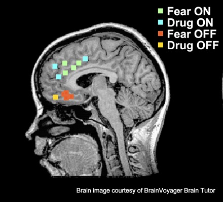
Human homologs of rodent prefrontal areas that modulate fear and addiction. Green dots represent regions of human dACC correlated with fear expression, as assessed by fMRI (Phelps et al. 2004; Milad et al. 2007a). Blue dots represent regions in human addicts correlated with cocaine craving after exposure to cocaine-related cues, as assessed by fMRI (Garavan et al. 2000) or PET mapping of cerebral blood flow using 15O-labeled water (Childress et al. 1999). Red dots depict approximate regions of vmPFC that are correlated with fear extinction recall, as assessed by fMRI (Phelps et al. 2004; Kalisch et al. 2006; Milad et al. 2007b). Yellow dot represents the vmPFC equivalent in addicted subjects. This region is deactivated, as assessed by PET metabolic mapping with 2-deoxyglucose, during states of cocaine craving, suggesting a failure to engage extinction (Bonson et al. 2002). Collectively, these studies suggest that this vmPFC is homologous to rodent IL, whereas the dorsal regions of dACC are homologous to rodent PL. (MRI brain image duplicated with permission from BrainVoyager Brain Tutor software, by Brain Innovation BV, Maastricht, Netherlands.)