


Figure 1. TH17 cells lose IL-17A and acquire IL-10 in vivo.
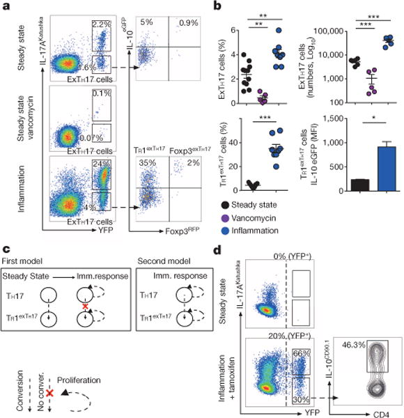
a, Flow cytometric analysis of small intestinal CD4+ T cells. Steady state, vancomycin-treated, or treated with anti-CD3 monoclonal antibody (inflammation) depicted. b, Number and frequencies of exTH17 cells (gated on CD4+ T cells) and TR1exTH17 cells (gated on exTH17 cells) are cumulative of two and three independent experiments respectively. IL-10 mean fluorescence intensity (MFI) data (n = 3 biological replicates) of one representative experiment out of three are shown. Mean ± s.e.m.; *P ≤ 0.05, **P ≤ 0.005, ***P ≤ 0.0005 by ANOVA (Bonferroni’s multiple comparison test) or by t-test for percentage (MannWhitney U-test, two tailed) and MFI (paired t-test, two-tailed) of TR1exTH17 cells. c, Hypotheses: first model, expansion of pre-existing TR1exTH17; second model, conversion of TH17 cells expanded/induced over the course of immune response. d, Flow cytometric analysis of small intestinal CD4+ T cells isolated from iFate mice in steady state, and upon anti-CD3 monoclonal antibody and tamoxifen (Inflammation + tamoxifen) treatment. Frequencies of YFP+ cells, TH17 and exTH17 (gated on YFP+ cells), and of IL-10+ cells (gated on exTH17) are representative of two experiments (n = 6 biological replicates).