


Figure 4.
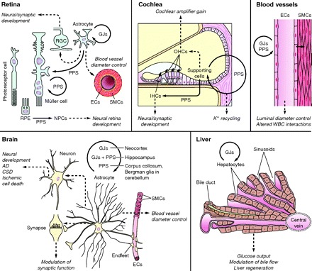
Overview of mechanisms and functions of intercellular Ca2+ waves (ICWs), based on ex vivo and in vivo evidence from the organs indicated. ICWs between cells of the same type are indicated by circular arrows; noncircular arrows indicate ICWs between different cell types. Gap junctions (GJs) and paracrine purinergic signaling (PPS) are involved in most cases, but only the most prevalent mechanism is depicted. Dashed arrows indicate proposed functions (in italics). Retina: ICWs that propagate in astrocytes and Müller cells may provoke electrical activity in retinal ganglion cells (RGCs) and influence the blood vessel caliber. Retinal pigment epithelium cells (RPE) show ICWs which may influence proliferation and differentiation of neural progenitor cells (NPCs). Cochlea: ICWs in supporting cells may be involved in K+ recycling and may influence outer hair cells (OHCs) via ATP release to alter the cochlear amplifier gain and inner hair cells (IHCs) to modify electrical activity and synaptic connectivity during development. Blood vessels: ICWs propagate along the vessel wall via endothelial cells (ECs) and modulate white blood cell (WBC) interactions with ECs as well as smooth muscle cell (SMC) contractility via direct (myoendothelial gap junctions) or indirect mechanism to control vessel diameter. Brain: mechanisms of ICW propagation differ with brain region; GJs, GJs+PPS, or PPS mediate ICWs in the neocortex, hippocampus, and corpus callosum and Bergman glia, respectively. ICWs of astrocytes propagate via astrocyte endfeet to small-diameter blood vessels to influence blood vessel diameter. ICWs of astrocytes may also modulate synaptic signaling as well as contribute to neural development or disease processes (AD, Alzheimer disease; CSD, cortical spreading depression). Liver: ICWs propagating between hepatocytes may influence hepatic glucose output, bile production, and tissue regeneration.