


Figure 4. GDC-0941 activated Wnt signaling in MDA-MB-231 cells.
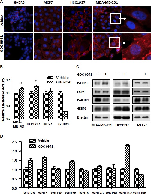
(A) Cells were seeded on coverslips the day before the experiment and treated with 0.5 μM GDC-0941 for 18 hr. The immunofluorescence (IF) of beta-catenin is indicated as red fluorescence, and double staining with DAPI is blue. Magnification: 400 X for the left three panels. The right panel shows enlarged images from the white squares. (B) Cells were transfected with 400 ng of the TOPflash plasmid and 25 ng of the Renilla plasmid for dual luciferase assays in 24-well plates in various breast cancer cell lines. Two days after transfection, the cells were treated with 0.5 μM GDC-0941 for 3 hr. The cells were then lysed for luminescence measurements. TOPflash activity was normalized against Renilla (n=3). * p<0.05. (C) MDA-MB-231, HCC1937 and MCF7 cells were treated with 0.5 μM GDC-0941 for 1 hr, and cell lysates were used for immunoblot analyses. (D) MDA-MB-231 cells were treated with 0.5 μM GDC-0941 for 24 hr. The relative mRNA expression of WNT ligands was analyzed using RT-qPCR. Black bars represent the SEM (n=3).