


Figure 5. Structures of the juxtamembrane latch and the transmembrane helices of EGFR.
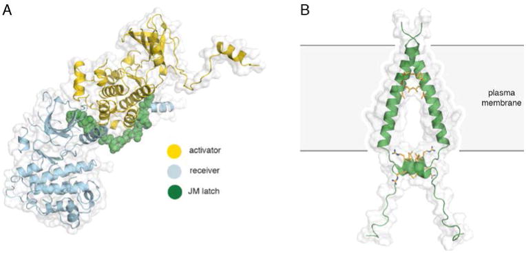
A) The asymmetric dimer of EGFR with the juxtamembrane latch highlighted in green (PDB ID 3GOP). B) An NMR structure of the transmembrane and JM-A helices of EGFR in lipid bicelles (PDB ID 2M20). Interactions between the N-terminal GxxxG-like motifs on the transmembrane segments and the LRRLL motifs of the juxtamembrane segments are highlighted in yellow.