


Figure 4. Kinase domain structures of the EGFR family members.
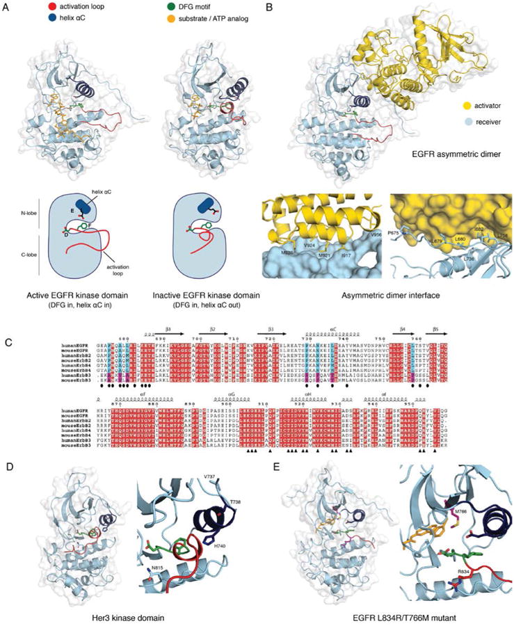
A) The active (PDB ID 2GS6) and the inactive (PDB ID 2GS7) conformation of the EGFR kinase domain. Helix αC is colored blue, the DFG motif green, and the activation loop red. The active structure has an ATP analog - peptide conjugate bound, and the inactive structure has AMP-PNP bound (colored yellow). B) The asymmetric dimer of the EGFR kinase domain (PDB ID 2GS6). The activator kinase is colored yellow, and the receiver (enzymatically active) kinase is colored blue. Residue contacts important on the activator and the receiver are highlighted. C) A sequence alignment of the EGFR family members from human and mouse. Two regions containing the residues involved in the N- and C-lobe faces of the dimer interface are shown in the upper and lower panels, respectively. Identical residues are colored in red. Residues in the N- and C-lobe faces of the dimer interface are denoted by ovals and triangles, respectively. Blue and magenta highlight residues in the dimer interface that are conserved among EGFR, Her2, and Her4 but not in Her3. D) Structure of the Her3 kinase domain (PDB ID 3KEX) with a zoom-in of the active site, and residues resulting in a catalytically impaired kinase are highlighted. The catalytic Asp 813 in EGFR is replaced by Asn 815 in Her3, the critical Glu 738 in helix αC of EGFR is replaced with His 740 in Her3, and Val 737 and Thr 738 in Her3 stabilize the inactive conformation of helix αC. E) A structure of the L834R/T766M double mutant EGFR kinase domain (PDB ID 4LL0) with a zoom-in of the active site. The bound inhibitor, PD168393, is colored yellow, and the mutations are highlighted.