


Fig 2. Purkinje-tree percepts.
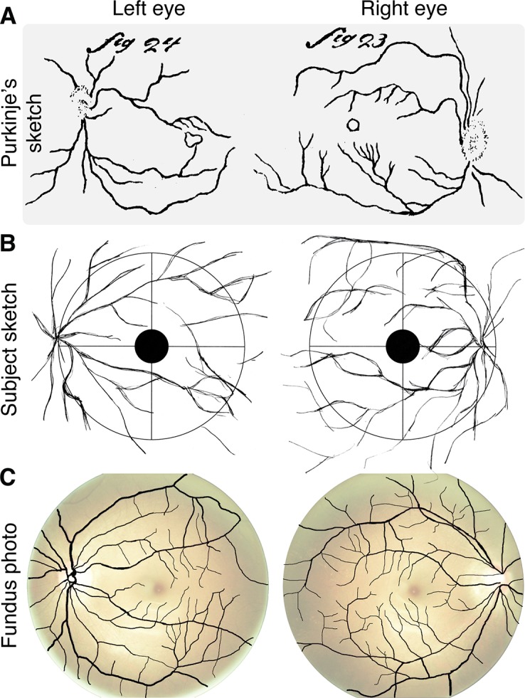
A: Sketch of entoptic visualization of retinal blood vessels from Purkinje ([7]; Figs 23 and 24). B: Sketch produced by a naïve observer in our study while viewing penumbral cone directed flicker at 16 Hz. C: Fundus photographs with overlaid extracted retinal blood vessels (see detailed methods in Appendix). The contrast and brightness of the fundus photographs were adjusted, and then made transparent for visualization purposes (see Fig 5 for raw fundus photographs). The fundus photos were obtained after the observer produced her sketches.