


Figure 4. Crystal structure of Lys33-linked triUb.
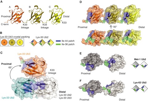
(A) The crystal structure of Lys33-linked triUb. (B) Representative diagram of how a Ub moiety in an ASU is arranged into triUb (left) and how Ile44 and Ile36 patches are located in Lys33-linked triUb (right) (C) Superposition of the distal Ub moieties (white) of Lys33-linked diUb and Lys33-linked triUb, shown in cartoon and surface representations. Hydrophobic patches are coloured as in Figure 3D. Only two Ub moieties are shown for Lys33-linked triUb. In comparison with the proximal Ub of Lys33 diUb (teal), the proximal Ub of Lys33 triUb (orange) moves approximately 65° from to the more open conformation. (D–F) A semi-transparent surface of Lys33 triUb (D), Met1 diUb (E) and Lys63 diUb (F) coloured as in Figure 3D (PDB 2W9N, 2JF5 [13]).