


Figure 8. MiR-21 regulates IL-10 expression via PDCD4.
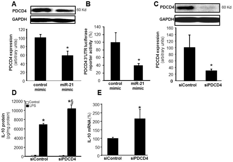
A, PDCD4 expression in MDM transfected with miRIDIAN hsa-miR-21 mimic or control-mimic. PDCD4 levels were determined using Western blot. Quantification of PDCD4 level was performed using densitometry and the data were normalized to β-actin. Data are mean ± SD (n=3). *, p<0.05 compared to control mimic transfected cells. B, To demonstrate that PDCD4 is a direct target for miR-21, HEK 293 cells were infected with lenti Luc-PDCD4–3′UTR plasmid along with either miR-21 mimic or control mimic. Data represent mean ± SD (n=4). *, p<0.05 compared to control transfected cells. C, PDCD4 protein expression was measured using Western Blot in MDM transfected with SiPDCD4 (100nM) or siControl. Quantification of PDCD4 level was performed using densitometry. Data are mean ± SD (n=3). *, p<0.05 compared to siControl transfected cells. D–E, MDM were transfected with siPDCD4 or siControl. Cells were activated with LPS (1ug/ml) for 24h after knockdown of PDCD4 in MDM. D, IL-10 protein and E, IL-10 mRNA were determined using ELISA and quantitative PCR respectively. Data are mean ±SD (n = 4); *, p<0.05 compared to macrophage transfected with siControl non-LPS. §, p<0.05 compared to siControl LPS treated group.