


Figure 9.
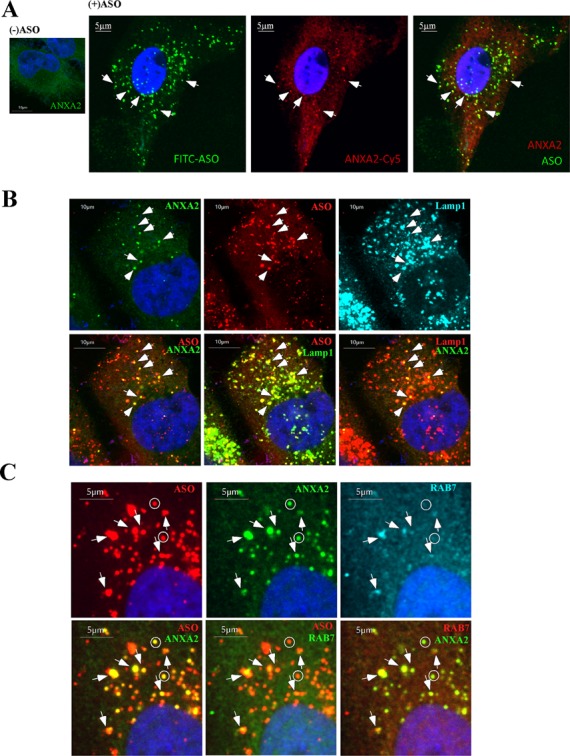
ANXA2 can co-localize with ASOs in late endosomes/lysosomes upon free uptake. (A) ANXA2 staining in control cells (left panel) or cells incubated with 2-μM FITC-labeled ASOs for 24 h (right panels). ANXA2-stained structures that co-localize with ASOs are exemplified by arrows. Scale bars: 5 μm. (B) Co-staining of ANXA2 and Lamp1 in cells incubated with 2-μM Cy3-labeled ASOs for 24 h. Arrows indicate the co-localization of ASO, ANXA2 and Lamp1. Scale bars: 10 μm. (C) Co-staining of ANXA2 and Rab7 in cells incubated with 2-μM Cy3-labeled ASOs for 24 h. Co-localization of ASO/ANXA2/Rab7 is exemplified by arrows; whereas some ANXA2/ASO containing structures that do not co-localize with Rab7 are indicated by circles. Scale bars: 5 μm.