



Abstract
Rationale: Chronic obstructive pulmonary disease (COPD) occurs in a minority of smokers and is characterized by intermittent exacerbations and clinical subphenotypes such as emphysema and chronic bronchitis. Although sphingolipids as a class are implicated in the pathogenesis of COPD, the particular sphingolipid species associated with COPD subphenotypes remain unknown.
Objectives: To use mass spectrometry to determine which plasma sphingolipids are associated with subphenotypes of COPD.
Methods: One hundred twenty-nine current and former smokers from the COPDGene cohort had 69 distinct sphingolipid species detected in plasma by targeted mass spectrometry. Of these, 23 were also measured in 131 plasma samples (117 independent subjects) using an untargeted platform in an independent laboratory. Regression analysis with adjustment for clinical covariates, correction for false discovery rate, and metaanalysis were used to test associations between COPD subphenotypes and sphingolipids. Peripheral blood mononuclear cells were used to test associations between sphingolipid gene expression and plasma sphingolipids.
Measurements and Main Results: Of the measured plasma sphingolipids, five sphingomyelins were associated with emphysema; four trihexosylceramides and three dihexosylceramides were associated with COPD exacerbations. Three sphingolipids were strongly associated with sphingolipid gene expression, and 15 sphingolipid gene/metabolite pairs were differentially regulated between COPD cases and control subjects.
Conclusions: There is evidence of systemic dysregulation of sphingolipid metabolism in patients with COPD. Subphenotyping suggests that sphingomyelins are strongly associated with emphysema and glycosphingolipids are associated with COPD exacerbations.
Keywords: ceramides, sphingomyelins, emphysema, metabolomics, genomics
At a Glance Commentary
Scientific Knowledge on the Subject
In preclinical studies, ceramides, the basic unit of sphingolipids, has been linked to emphysema development. However, the complexity of the sphingolipid metabolism generates multiple sphingolipid species with distinct biological functions, and chronic obstructive pulmonary disease (COPD) is a common disease with multiple phenotypes, including emphysema. Recent data suggest an association between circulating and sputum levels of sphingolipids and COPD, but only two classes have been studied (ceramides and sphingomyelin), and these findings have been neither validated in larger cohorts nor studied in relationship with distinct COPD phenotypes.
What This Study Adds to the Field
This study investigates the associations of plasma sphingolipids with COPD subtypes such as airflow obstruction, emphysema, chronic bronchitis, and frequent exacerbations in a well-characterized cohort of current or former smokers. Our findings add to previous studies reporting associations of sphingomyelin and ceramides with airflow obstruction and emphysema. In addition, we identified novel sphingolipids associated with COPD phenotypes such as frequent COPD exacerbations. We showed that COPD subphenotypes are associated with dysregulated gene/metabolite pathways for sphingolipids, adding further evidence to their role in the pathogenesis of COPD.
Chronic obstructive pulmonary disease (COPD) is the third leading cause of death in the United States (1). Although smoking is the major risk factor for COPD, most smokers do not develop COPD, and those who do have variable clinical phenotypes such as airflow obstruction, emphysema, chronic bronchitis, and frequent COPD exacerbations (2). There is little known about what predisposes certain smokers to develop a particular COPD phenotype. Recently, there have been reports suggesting that alteration in sphingolipid metabolism may be linked to disease susceptibility (3–6), although large-scale human studies of specific sphingolipids have been lacking.
Sphingolipids contain a backbone of sphingoid bases and, depending on the O-linked R group, are subclassified into sphingosines, sphingomyelins, ceramides, or glycosphingolipids. In mammals, there are more than 100 species of sphingolipids and a multitude of enzymes that regulate sphingolipid metabolism. Sphingolipids are found primarily in cell membranes, but also in biofluids such as plasma. Having pleiotropic effects, specific sphingolipids have been implicated in diverse biologic processes (7) that include: cell death, proliferation, differentiation, autophagy, senescence, migration, and efferocytosis.
Sphingolipids have been implicated in several lung diseases, including COPD (3, 8, 9). The majority of sphingolipid lung research has focused on ceramides, the central intermediate metabolites in the sphingolipid metabolism and key proapoptotic signaling molecules, and on sphingosine-1-phosphate (S1P), a downstream metabolite of ceramide and key prosurvival and immune regulator. Smokers (10) and patients with COPD (11) have increased ceramides in their lungs, and ceramides have been implicated in tobacco smoke–induced pulmonary vascular cell apoptosis (12) and clearance of apoptotic cells by alveolar macrophages (13). S1P receptor 5 expression is reduced the lungs of patients with COPD (14), and S1P analogs inhibit emphysema development in animal models (15). Recently, in a small cohort, sphingolipids were found elevated in the sputum in COPD (6).
These findings suggest that sphingolipid metabolism is dysregulated by smoking and COPD and that these derangements may be pathogenic and could serve as biomarkers (10). However, previous studies have been limited by small sample sizes, limited clinical phenotyping, and limited focus on a particular subclass of sphingolipids. To overcome these limitations and further investigate sphingolipids for their association with clinical COPD phenotypes, such as emphysema, chronic bronchitis, and frequent exacerbations, we used two mass spectrometry–based approaches to quantitate 69 plasma sphingolipids in more than 250 smokers with and without COPD from the COPDGene study. Furthermore, we integrated plasma metabolomics analyses with recently published microarray profiling of this cohort (4) to discover relationships that might elucidate mechanisms of sphingolipid dysregulation in COPD.
Methods
Study Population
The institutional review boards of participating institutions approved this study. All subjects were from the COPDGene cohort, which is a National Institutes of Health–sponsored multicentered study of the genetic epidemiology of COPD (16). From this cohort we selected a subset of 129 subjects (Table 1) for targeted sphingolipid analysis to obtain a wide range of COPD phenotypes (defined below). Additionally, 131 subjects who participated in a microarray study of gene expression in peripheral blood mononuclear cells (GEO accession number GSE 42057) (4) were selected for untargeted sphingolipid analysis. Additional details are provided in the online supplement.
Table 1.
Characteristics of Subjects
| Targeted Cohort (N = 129) | Untargeted Cohort (N = 131) | P Value | |
|---|---|---|---|
| Age, yr | 63 (58–71) | 64 (57–70) | 0.96 |
| Sex, % men | 57 | 56 | 0.79 |
| Current smokers, % | 23 | 23 | 0.95 |
| Pack-years | 46 (34–58) | 42 (30–64) | 0.71 |
| Body mass index | 27 (24–31) | 27 (24–32) | 0.58 |
| FEV1 % | 64 (40–92) | 66 (42–89) | 0.8 |
| FEV1/FVC | 0.56 (0.39–0.72) | 0.61 (0.44–0.74) | 0.15 |
| Emphysema, % | 11.5 ± 11.4 | 9.18 ± 10.4 | 0.09 |
| Chronic bronchitis, % | 19 | 18 | 0.95 |
| Exacerbations/yr, moderate | 0.7 ± 1.3 | 0.6 ± 1 | 0.3 |
| Exacerbations/yr, severe | 0.2 ± 0.7 | 0.1 ± 0.3 | 0.17 |
Data are presented as median (interquartile range) or mean ± SD. Emphysema was defined as percent of lung attenuation voxels below −950 Hounsfield units; chronic bronchitis was defined by daily productive cough for at least 3 months in the previous 2 consecutive years; exacerbations were defined as moderate (treated with either antibiotics or corticosteroids) or severe (leading to hospitalization) in the previous year.
Clinical Data Collection and Definitions of COPD Phenotypes
Each subject was classified by four different clinical phenotypes: airflow obstruction (COPD), chronic bronchitis, emphysema, and exacerbations (see Table E1 in the online supplement). COPD was defined using Global Initiative for Chronic Obstructive Lung Disease criteria (17). Emphysema was measured using quantitative high-resolution computed tomography (HRCT) as described (18). Exacerbations were defined by acutely worse cough, sputum, and dyspnea in those with and without COPD. Only moderate exacerbations (treated by corticosteroids and/or antibiotics) or severe exacerbations (causing hospitalization) were counted. Chronic bronchitis was defined as cough that produces sputum daily for 3 consecutive months for at least 2 consecutive years.
Sphingolipid Measurements
Sphingolipid measurements were performed independently in two separate laboratories using two separate protocols (see online supplement for more details). A targeted, quantitative, mass spectrometry panel (Washington University) included 69 sphingolipids (Table E2). Sphingomyelins, dihydrosphingomyelins, ceramides, and dihydroceramides were extracted using a modified Bligh-Dyer extraction method, in the presence of internal standards. Sphingoid bases, ceramide-1-phosphate, monohexosylsphingosine, monohexosylceramides, dihexosylceramides, trihexosylceramides, monohydroxylated monohexosylceramides, monohydroxylated dihexosylceramides, sulfatides, and gangliosides were extracted after protein precipitation with methanol, followed by supernatant collection, drying, and reconstituting with 1:1 methanol/water, in the presence of internal standards. A second untargeted protocol (National Jewish Health) was performed as detailed elsewhere (19).
Statistical Analysis
Differences in demographic characteristics of study subjects were analyzed using a t test for continuous variables and a Chi-square test for categorical variables. Regression modeling and covariates are described further in the online supplement. Because the sphingolipid levels were highly correlated within class (Figure E1), we also computed the first principal component of each sphingolipid class (Tables E3 and E4) using prcomp function in R.
Replication between the targeted and untargeted platforms was determined using the Stouffer-Liptak Z-score method, which converts the P values from the two studies to normal quantiles and averages them to obtain a combined P value (20, 21). Each of the 23 sphingolipids that overlapped between the two studies was tested, and consistency in the direction of the effect on the phenotype was taken into account.
Results
Study Subjects and Baseline Characteristics
Demographics, physiology, quantitative HRCT measurements, and patient-reported outcomes for each group are listed in Table 1 and Table E1. Except for slightly more subjects with emphysema in the untargeted cohort, there were no statistically significant differences in the baseline characteristics between the targeted and untargeted cohorts.
Targeted Identification of Plasma Sphingolipids
Our previous results suggested that sphingolipids were candidate biomarkers for COPD (4); we therefore performed targeted measurement of multiple sphingolipid classes. These included: sphingomyelins (SM d18:1), dihydrosphingomyelins (SM d18:0), ceramides (Cer d18:1), dihydroceramides (Cer d18:0), sphingoid bases, ceramide-1-phosphate, monohexosylsphingosine, monohexosylceramides, dihexosylceramides, trihexosylceramides, monohydroxylated monohexosylceramides, monohydroxylated dihexosylceramides, sulfatides, and gangliosides. After filtering out species that exhibited no or very low peaks, overlapped with other peaks, exhibited multiple peaks with retention times in close proximity, or had large coefficients of variance, 69 sphingolipid species were used for quantitative comparisons (Table E2). Multiple sphingolipids were associated with clinical covariates such as age, sex, body mass index (BMI), and current smoking (Table E3). Three sphingolipid species showed a negative correlation with age (correlation test P value < 0.01), seven species showed a negative correlation with BMI (correlation P value < 0.01), and 32 species showed higher levels in female subjects (t test P value < 0.01). Although not significant, select species showed trends for alterations depending on current smoking status. Because of these associations, we adjusted for covariates in all our models.
Because strong correlations were noted within classes of sphingolipids (Figure E1), sphingolipid species were further grouped into 10 classes (Table E4), which further revealed robust interclass collinearities. For instance, plasma sphingomyelins were strongly correlated with ceramides (P < 0.001), and both sphingomyelins and ceramides were inversely correlated with S1P (P < 0.001). Because of the high intraclass correlations for plasma sphingolipids, each class of sphingolipids was also represented in analyses by the first principal component from the intraclass principal component analysis.
Plasma Sphingolipids Association with Different Phenotypes of COPD
Patients with COPD exhibit heterogeneity in clinical presentation, with different morbidities and prognoses for each phenotype; therefore, we investigated if specific sphingolipids or sphingolipid classes are significantly associated with individual phenotypes. The most statistically significant negative (inverse) association between specific COPD phenotypes and sphingolipid classes was for emphysema and sphingomyelins; the most statistically significant positive (direct) association was between COPD exacerbations and trihexosylceramides (Figure 1).
Figure 1.

Heat map showing sphingolipids associated with specific chronic obstructive pulmonary disease (COPD) phenotypes. Each class was represented by the first principle component. Emphysema was defined as percent of lung attenuation voxels below −950 Hounsfield units; chronic bronchitis was defined by daily productive cough for at least 3 months in the previous 2 consecutive years; exacerbations (severe) were the number of hospital admissions for a COPD admission in the previous year. Each phenotype was modeled using regression with covariates (age, sex, smoking status, body mass index, and airflow obstruction [FEV1] where appropriate), detailed in the online methods. The color legend shows shading based on the signed −10 log of the P value with negative (−) associations shown in blue and green and positive (+) associations shown in orange and red for each sphingolipid class. Non–statistically significant associations are shown in yellow and light orange.
Individual sphingolipids associated with emphysema
We identified 26 sphingolipids that were significantly associated with emphysema after adjustment for covariates, with 8 remaining significant after adjustment for false discovery rate (FDR) (Table E5). The most highly represented group was that of sphingomyelins (10 species associated with emphysema, including 5 after adjustment for false discovery), all of which had negative associations with emphysema. The three plasma sphingolipids most significantly associated with emphysema were ceramide d18.1.N16.0 (P = 0.0006), ganglioside GM3 d18.1.N16.0 (P = 0.0004), and sphingomyelin d18.1.N16.0 (P = 0.0009), all being inversely associated with emphysema (Figure 2), followed by monohexosylceramide d18.1.N16.0 (P = 0.002; Table E5). When stratified by subjects with less severe airflow obstruction (i.e., milder COPD), the receiver operating characteristic curves demonstrated that these top sphingolipids improved the ability to diagnose moderate to severe emphysema beyond just clinical and physiologic covariates (Table E6).
Figure 2.
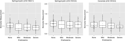
Plasma ceramides are inversely associated with emphysema severity. Ceramide (d18:1/N16:0), sphingomyelin (d18:1/N24:1), and sphingomyelin (d18:1/N16:0) are representative examples (false discovery rate < 0.02 in Table E5). Shown are box plots of median, quartiles, and outliers. A total of 33, 91, 97, and 100% of subjects with no, mild, moderate, or severe emphysema, respectively, had airflow obstruction (post-bronchodilator FEV1/FVC < 0.7).
Individual sphingolipids associated with COPD exacerbations
After adjustment for covariates and correction for FDR, we identified 11 sphingolipids that were associated with severe COPD exacerbations, including four trihexosylceramides, three dihexosylceramides, sulfatide d18.1.N16.0, ganglioside GD1.d18.1.N16.0, sphingomyelin SM.d18.1.N14.1, and S1P (Table E7). All associations were positive except for S1P and SM.d18.1.N14.1, and the strongest signal was the positive association of severe COPD exacerbations with plasma trihexosylceramides d18.1.N18.0 (adjusted P = 1.6 × 10⁻⁶). Most of these associations were also identified with COPD exacerbations of lesser severity (Table E8). Receiver operating characteristic analysis demonstrates that these sphingolipids improved the ability to diagnose severe exacerbations beyond just clinical and physiologic covariates (Table E9). Interestingly, for less severe exacerbations, only in subjects with less airflow obstruction, Cer.d18.1.N18.0 showed enhanced association with exacerbations beyond just clinical and physiologic covariates (Figure 3 and Table E10).
Figure 3.

Receiver operating characteristics (ROC) curve analysis and area under the curve (AUC) for sphingolipids significantly associated with moderate chronic obstructive pulmonary disease exacerbations. The first model uses only the demographic and clinical covariates (Covariates only), including covariates age, sex, smoking status, body mass index, and airflow obstruction (FEV1). The second model includes the same covariates and adds plasma levels of Cer.d18.1.N18.0, which was the sphingolipid that most improved ROC.
Sphingolipids associated with other COPD phenotypes (airflow obstruction and chronic bronchitis)
There were no plasma sphingolipid species significantly associated with chronic bronchitis (Table E11), FEV1 (data not shown), FEV1/FVC (Table E11), or bronchodilator responsiveness (data not shown) after correcting for FDR; however, several metabolomic trends noted in these subgroups were used in combination with genomic data for phenotype-metabolomic interpretations.
Replication of Associations between COPD Phenotypes and Sphingolipids
For replication, we used an independently generated metabolomics dataset (detected via untargeted mass spectrometry using orthogonal methods in an independent laboratory) from plasma of 131 subjects in COPDGene. Between the two datasets, we identified 23 overlapping sphingolipids (8 sphingomyelins, 6 ceramides, 3 gangliosides, 2 sulfogalactosylceramides, dihydroceramide, trihexosylceramide, S1P, and sphingosine). For overlapping sphingolipids, we performed metaanalysis of the two datasets accounting for the direction of association. Seven of 23 sphingolipids showed negative associations with emphysema, of which five sphingomyelins (Table 2) exhibited the strongest signal for replication between the two platforms (FDR < 0.10). Four sphingolipids demonstrated associations with moderate and severe exacerbations (Table 3). Except for S1P, all were positively associated with exacerbations (FDR < 0.10).
Table 2.
Metaanalysis of Sphingolipids Associated with Emphysema
| Sphingolipid | Targeted Cohort |
Untargeted Cohort |
Metaanalysis P Value | ||
|---|---|---|---|---|---|
| Slope | P Value | Slope | P Value | ||
| Ganglioside GM3 (d18:1/16:0) | −0.7654 | 0.0004 | −0.1277 | 0.3768 | 0.0018* |
| Sphingomyelin(d18:0/24:1(15Z)) | −0.4534 | 0.0057 | −0.0623 | 0.5349 | 0.0167* |
| Sphingomyelin(d18:1/14:0) | −0.4335 | 0.0040 | −0.1070 | 0.3650 | 0.0075* |
| Sphingomyelin(d18:1/16:0) | −0.8964 | 0.0010 | −0.1867 | 0.4258 | 0.0037* |
| Sphingomyelin(d18:1/16:1) | −0.5198 | 0.0054 | −0.1459 | 0.3621 | 0.0090* |
| Sphingomyelin(d18:1/24:1(15Z)) | −0.6062 | 0.0009 | −0.2182 | 0.0829 | 0.0004* |
| Sphingomyelin(d18:2/14:0) | −0.3520 | 0.0237 | −0.0673 | 0.5444 | 0.0425 |
False discovery rate < 0.10.
Table 3.
Metaanalysis of Sphingolipids Associated with Exacerbations
| Sphingolipid | Targeted Cohort |
Untargeted Cohort |
Metaanalysis P Value | ||
|---|---|---|---|---|---|
| Slope | P Value | Slope | P Value | ||
| Sphingosine 1-phosphate | −0.7051 | 0.1838 | −0.6928 | 0.0004 | 0.0006* |
| Trihexosylceramide (d18:1/16:0) | 0.8804 | 0.0693 | 0.9839 | 0.0059 | 0.0012* |
| 3-O-Sulfogalactosylceramide (d18:1/16:0) | 0.8262 | 0.0507 | 0.5191 | 0.0792 | 0.0087* |
| Galabiosylceramide (d18:1/24:1(15Z)) | 0.6077 | 0.1741 | 0.4177 | 0.0641 | 0.0232 |
False discovery rate < 0.10.
Relationship between Plasma Sphingolipids and Gene Expression in Smokers with and without COPD
All subjects in the untargeted metabolomics dataset had microarray data generated from peripheral blood mononuclear cells collected from the same plasma analyzed for metabolomics. Thus, we were able to explore relationships between plasma sphingolipid metabolites and potentially related sphingolipid pathway genes (Table E13). The most significant correlations were between N-(tetradecanoyl)-sphing-4-enine-1-(2-aminoethylphosphonate) and GM2 ganglioside activator (ρ = −0.39; P = 0.00001; FDR = 0.04), between SM(d18:1/12:0) and lysosomal sialidase (ρ = −0.36; P = 0.00007; FDR = 0.12) (Figure E2), and between sphingosine and Ceroid-Lipofuscinosis, Neuronal 8 (ρ = −0.34; P = 0.0002; FDR = 0.03).
After interrogation of correlations between sphingolipid metabolite and gene expression and clinical phenotype, subjects who reported moderate or severe COPD exacerbations were most likely to have discordant correlations for gene–metabolite pairs (Table 4). For example, the gene–metabolite pair C9orf47 and NeuGcα2-8NeuGcα2-3Galβ1-4GlcNAcβ1-3Galβ1-4Glcβ-Cer(d18:1/24:1[15Z]) had correlations −0.19 and 0.64 in subjects without or with severe COPD exacerbations, respectively (P = 0.00001; FDR = 0.01) (Figure 4).
Table 4.
Differential Metabolite–Gene Expression Correlations for Sphingolipids by Chronic Obstructive Pulmonary Disease Phenotype
| Sphingolipid | Gene | COPD Subphenotype | ρ without Phenotype | ρ with Phenotype | P Value | FDR |
|---|---|---|---|---|---|---|
| N-(tetradecanoyl)-sphing-4-enine-1-(2-aminoethylphosphonate)* | KDSR | Moderate or severe exacerbation | −0.17 | 0.50 | 0.00005 | 0.06 |
| SM(d18:1/16:1) | GM2A | −0.15 | 0.54 | 0.00001 | 0.04 | |
| CerP(d18:1/24:1(15Z))* | ST8SIA2 | Severe exacerbation | −0.05 | 0.63 | 0.00006 | 0.05 |
| NeuGcα2-3Galβ1-4GlcNAcβ1-3Galβ1-4Glcβ-Cer(d18:1/24:1(15Z))* | C9orf47 | −0.19 | 0.64 | 0.00001 | 0.01 | |
| Cer d18:1 N16:0 | ACER3 | 0.11 | 0.73 | 0.00001 | 0.01 | |
| *1-3Galα1-3Galα1-3Galα1-4Galβ1-4Glcβ-Cer(d18:1/24:1(15Z))* | C9orf47 | −0.23 | 0.56 | 0.00009 | 0.06 | |
| Cer d18:1 N16:0 | VAPA | −0.03 | 0.62 | 0.00014 | 0.08 | |
| Cer(d18:1/24:1(15Z))* | COL4A3BP | −0.07 | 0.60 | 0.00019 | 0.08 | |
| Cer(d16:1/23:0)* | COL4A3BP | −0.12 | 0.56 | 0.00040 | 0.15 | |
| Cer d18:1 N16:0 | KDSR | −0.10 | 0.55 | 0.00055 | 0.15 | |
| Cer d18:1 N16:0 | ORMDL1 | −0.14 | 0.54 | 0.00051 | 0.15 | |
| SM(d18:0/24:1(15Z))* | CYR61 | −0.18 | 0.53 | 0.00048 | 0.15 | |
| First PC ceramides | COL4A3BP | 0.09 | 0.54 | 0.00242 | 0.16 | |
| First PC sphingosines | ST8SIA3 | Emphysema (upper lobe) | −0.47 | 0.19 | 0.00039 | 0.06 |
| First PC sphingosines | CYR61 | Airflow obstruction | 0.28 | −0.39 | 0.00011 | 0.02 |
Definition of abbreviations: COPD = chronic obstructive pulmonary disease; FDR = false discovery rate; PC = principal component.
Discovered in the untargeted profiling.
Figure 4.

Disruption of sphingolipid metabolite–gene correlations by chronic obstructive pulmonary disease (COPD) phenotype. Scatterplot of NeuGcα2-3Galβ1-4GlcNAcβ1-3Galβ1-4Glcβ-Cer(d18:1/24:1(15Z)) and C9orf47 gene expression. Subjects without severe COPD exacerbations had a correlation of −0.19, whereas subjects with a history of severe COPD exacerbations had a correlation of 0.64 (P = 0.00001; false discovery rate = 0.01).
Discussion
This study of more than 250 subjects from the COPDGene cohort is the largest and the only combined targeted and nontargeted metabolomic study of plasma sphingolipid species in smokers with and without COPD; it is the first to link specific sphingolipids with distinct COPD phenotypes. We identified novel sphingolipid subclasses such as gangliosides and sphingomyelins as potential biomarkers inversely associated with severe emphysema and trihexosylceramide as a potential biomarker positively associated with frequent COPD exacerbations. All these associations were noted even after adjusting for covariates of age, smoking status, airflow obstruction, sex, and BMI; most were replicated in an independent cohort using an orthogonal mass spectrometry approach in a different laboratory.
Sphingomyelins are integral components of the plasma membrane, typically enriched in lipid microdomains of caveolae and clathrin-coated pits (22). Sphingomyelins are produced from ceramides via sphingomyelin synthase, or they can generate ceramides through hydrolysis by sphingomyelinases (Figure 5). In addition to rapidly generating ceramides as second messengers, sphingomyelins may themselves play a role in cell migration, apoptosis, autophagy, and cell survival/proliferation (22). We identified 10 specific sphingomyelins (e.g., SM.d18.1.N16.0) that were inversely associated with emphysema, whereas others (e.g., SM.d18.1.N18.0) were not. These species-specific effects could only be studied with mass spectrometry; however, we also noted strong correlations among the whole subclass of sphingomyelins. A recent report (5) found no statistically significant association between HRCT-measured emphysema severity at baseline and sphingomyelin, measured with less accurate spectrophotometric assays (23). However, low baseline plasma sphingomyelin was associated with worse COPD (lower FEV1), whereas high levels were associated with more rapid progression of emphysema. Our data support the finding that worse COPD is associated with lower plasma sphingomyelins (5), but the apparent paradoxical observation of higher plasma sphingomyelins with progression of emphysema cannot be validated by our experimental design. Such observation might be explained if low sphingomyelin levels reflect low lung mass and progression of emphysema occurs in lungs that are not yet severely emphysematous. This could be explored by careful kinetics of specific sphingomyelins accompanied by concomitant HRCT measurements of the lung.
Figure 5.
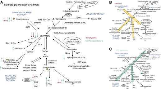
Sphingolipids metabolo-genomic pathway patterns associated with chronic obstructive pulmonary disease (COPD) subphenotypes. (A) Detailed sphingolipid metabolic pathway with the major ceramide synthetic pathways noted as sphingomyelinase, de novo, and recycling pathways (abbreviations list in the text). Individual or classes of metabolites measured in the study are identified as precursors and/or products relative to synthetic or catabolic reactions with directionality marked by black arrows. Catalytic enzymes are noted next to the middle of the reactions arrow. Circled are enzymes whose genes were significantly related with COPD phenotypes in genomic analyses. In color are classes (arrowheads) or species (arrows) of metabolites that were associated with respective (color-coded) COPD phenotypes. Solid arrows indicate: 16–18 carbon chain sphingolipids; dotted arrows 24–26 carbon chain sphingolipids; single dash arrows: 14 carbon chain sphingolipids. The direction of the arrowhead or arrow marks the direction of association (i.e., up: increased; down: decreased). Closed-headed arrows represent changes with significance levels P < 0.05. (B, C) Superimposed on the sphingolipid metabolic pathways are deducted directions of sphingolipid metabolic flux, based on the results of the metabolo-genomic study, by disease phenotype, as noted.
Plasma ceramides may be generated by activated plasma sphingomyelinases (24) (Figure 5) and could signal outside-in (25) and/or be further metabolized to sphingosine (by plasma ceramidases). To the extent that plasma levels reflect cellular metabolism, the decrease in both sphingomyelins and ceramides with subsequent increases in ceramide metabolites suggests that low sphingomyelin levels are not due to a blockage in sphingomyelin synthase (which would result in ceramide accumulation) but due to increased catabolism coupled with decreased availability of its building block precursor, ceramide (Figure 5). In such conditions, supplementation of sphingomyelin may fuel ceramide production and catabolism, by a “revved up” system. Plasma sphingomyelins might also reflect the burden or quality of circulating (shed) cellular plasma membranes, such as apoptotic microparticles or exosomes. Clearly, these unbiased metabolomic results raise important mechanistic questions that can be explored in models of COPD.
Ceramides (especially Cer.d18.1.N16.0), gangliosides (especially GM3.d18.1.N16.0), and monohexosylceramides (especially monohexcer.d18.1.N16.0) also showed an inverse correlation with emphysema. Although ceramides have been known to play a role in the pathogenesis of emphysema for several years (10, 26), the involvement of gangliosides and monohexosylceramides has not been described. An up-regulation of ceramide production has been typically associated with models of emphysema, by causing endothelial and epithelial cell apoptosis (3) or by inhibiting alveolar macrophage clearance of apoptotic cells (13). In this cross-sectional sampling, the inverse correlation of plasma ceramides with emphysema appears surprising. However, if our global metabolomics assessment of plasma sphingolipids reflects pulmonary cellular events, worse emphysema may be linked to increased activity of both sphingomyelinase and recycling (from gangliosides) pathways, followed by ceramide consumption/degradation, rather than accumulation (Figure 5), possibly via plasma ceramidases. Because plasma S1P was not increased and tended to be negatively correlated with emphysema and COPD exacerbations, unlike sphingosine, it is possible that the sphingolipid metabolism is accelerated toward ceramide production and degradation but inhibited at the level of sphingosine kinase activity (Figure 5). The reduced production of S1P may be associated with decreased cellular survival and proliferation and increased apoptosis of alveolar cells (15). Much less is known about the significance of decreased gangliosides (or glycoceramides) in the lung, reports showing association with diabetes or hearing loss (27). Although our earlier genomic analysis hinted at this involvement (4), this is the first report of decreased monosialodihexosylganglioside GM3.d18.1.N16.0 as putative biomarker of emphysema. Similarly, the role of monohexosylceramides in the lung is unknown, but they were shown to bind to surfactant protein D (28) and to decrease on activation of acid sphingomyelinase and acid β-glucosidase1, as a sign of ceramide production via recycling from gangliosides during inflammation (29). These results may embolden future investigations of lung sphingolipid metabolomic signatures and the functional role of the gangliosides and monohexosylceramides in COPD.
The strongest positive association with COPD exacerbations was for trihexosylceramides (trihexcer.d18.1.N22.0 and trihexcer.d18.1.N24.0), a lipid class with unknown lung effects; one case report of Fabry disease (characterized by overabundance of trihexosylceramides) showed association with COPD (30). Other sphingolipids such as ceramide (d18.1.N18.0) and S1P were inversely associated with COPD exacerbations. This, along with negative correlations of plasma sphingomyelins and ceramides with S1P, suggests a rapid flux of sphingosine-to-ceramide-to glycosylated ceramides metabolism, with potential disruption of ceramide-to-S1P metabolism (Figure 5) in those with COPD exacerbations. Alternatively, whether tissues or cells may differentially contribute to the abundance of plasma metabolites in these individuals could be explored by comparing plasma with lung tissue metabolomics signatures. Because these samples were collected at a time remote from an actual exacerbation, metabolomic signatures are unlikely to reflect corticosteroid or antibiotic treatment or acute infections. Rather, they may indicate specific changes associated with developing COPD exacerbations. For example, shunting ceramide metabolism toward trihexosylceramides may render the lungs more susceptible to inflammation, due to defects in lymphocyte function, as learned from a mouse model of Fabry disease (31).
To further translate biomarker findings to disease pathogenesis, we explored alterations of gene–metabolite associations with disease phenotypes. We identified a negative association of sphingosine levels with the expression of CYR61 only in subjects with severe COPD. Although the significance of this finding remains to be tested, the extracellular matrix protein CCN1/Cyr61 is activated by S1P via receptor 2 (32), which also activates rho kinase to cause lung damage during cigarette smoke exposure (33, 34). Other disrupted gene–metabolite associations of interest in those with severe COPD exacerbations were between ceramide and alkaline ceramidase, which degrades unsaturated long chain ceramides (35); ORMDL1, which regulates de novo ceramide biosynthesis, recently involved in asthma (36); and 3-ketodihydrosphingosine reductase, which reduces 3-ketodihydrosphingosine to dihydrosphingosine (37). Another intriguing association was that between circulating ceramide and COL4A3BP, also known as CERTL, a gene encoding for ceramide transporter protein Goodpasture antigen-binding protein, which mediates the nonvesicular intracellular transport of ceramides (38). Superimposing potential sphingolipid biomarkers with genetic signatures helped map the directionality of sphingolipid metabolism in individuals with COPD exacerbations (Figure 5). Using similar approaches may strengthen the associations of sphingolipid metabolism and chronic bronchitis or obstructive ventilatory defects in COPD, which were weak at the plasma metabolite levels, but in conjunction with genomic findings suggested a potential metabolic flux toward higher S1P and de novo pathway activation, respectively (Figure E3).
In summary, this study demonstrates that specific sphingolipids, such as ceramides, sphingomyelins, and gangliosides, may be biomarker candidates of COPD phenotypes, such as emphysema and frequent COPD exacerbations. Our study’s strengths include the large number of highly phenotyped subjects, the multiple specific sphingolipid measurements, and independent replication. Limitations of our study include lack of healthy nonsmoking subjects, of longitudinal studies, and of lung tissue, bronchoalveolar lavage fluid, or sputum analyses. The latter would have allowed us to probe if plasma findings parallel those in the lung and to validate a small study that suggested sputum sphingolipids are associated with COPD (6). Our results in a much larger cohort of patients with COPD corroborate those reported in the sputum, in particular that there are abnormalities in sphingolipid metabolism and glycosphingolipid changes in COPD. The complementary nature of these studies conducted in different compartments may help interpret our results in plasma, which indicate an overall accelerated metabolism/catabolism of ceramide at the systemic level, in contrast to the increased ceramides and dihydroceramides recovered in the sputum in COPD. This may be due to active transfer and use of plasma sphingolipids in the lung with release of ceramides in sputum; alternatively, it may be due to different dynamics of sphingolipid metabolism in circulating cells and endothelium compared with airway macrophages or epithelial cells that may contribute to sputum sphingolipid content.
Our results strengthen the evidence implicating ceramide in emphysema and suggest a previously unsuspected role for ceramide metabolism in COPD exacerbations. Furthermore, our data suggest that additional subclasses of sphingolipids such as sphingomyelins, gangliosides, monohexosylceramides, and trihexosylceramides should be investigated, to validate their role as biomarkers and as potential contributors to COPD pathogenesis.
Acknowledgments
Acknowledgment
The authors thank Christina Schnell, Taylor Thorn, and Cori Fratelli for assistance with recruiting subjects and preparing samples and Kristina Watson for administrative support.
Footnotes
Funded by National Heart, Lung, and Blood Institute grants U01 HL089856 and U01 HL089897 (Edwin Silverman and James Crapo); National Center for Research Resources/National Institutes of Health grants UL1 RR025780 and S10 RR023703; National Institutes of Health/National Center for Advancing Translational Sciences Colorado Clinical and Translational Science Awards UL1 TR000154 (Ron Sokol), P20 HL-113444 (J.E.S.), and P20 HL-113445 (R.P.B., I.P., and N.R.).
Author Contributions: R.P.B., N.R., and K.K. designed the study; C.C., D.S.O., J.E.S., and N.R. generated data; S.J., C.C., G.J.H., C.S., and K.K. analyzed data; R.P.B., I.P., N.R., and K.K. interpreted data and wrote the manuscript.
Originally Published in Press as DOI: 10.1164/rccm.201410-1771OC on December 10, 2014
This article has an online supplement, which is accessible from this issue’s table of contents at www.atsjournals.org
Author disclosures are available with the text of this article at www.atsjournals.org.
References
- 1.Murphy SL, Xu J, Kochanek KD. Deaths: final data for 2010. Natl Vital Stat Rep. 2013;61:1–117. [PubMed] [Google Scholar]
- 2.Friedlander AL, Lynch D, Dyar LA, Bowler RP. Phenotypes of chronic obstructive pulmonary disease. COPD. 2007;4:355–384. doi: 10.1080/15412550701629663. [DOI] [PubMed] [Google Scholar]
- 3.Petrache I, Petrusca DN, Bowler RP, Kamocki K. Involvement of ceramide in cell death responses in the pulmonary circulation. Proc Am Thorac Soc. 2011;8:492–496. doi: 10.1513/pats.201104-034MW. [DOI] [PMC free article] [PubMed] [Google Scholar]
- 4.Bahr TM, Hughes GJ, Armstrong M, Reisdorph R, Coldren CD, Edwards MG, Schnell C, Kedl R, LaFlamme DJ, Reisdorph N, et al. Peripheral blood mononuclear cell gene expression in chronic obstructive pulmonary disease. Am J Respir Cell Mol Biol. 2013;49:316–323. doi: 10.1165/rcmb.2012-0230OC. [DOI] [PMC free article] [PubMed] [Google Scholar]
- 5.Ahmed FS, Jiang XC, Schwartz JE, Hoffman EA, Yeboah J, Shea S, Burkart KM, Barr RG. Plasma sphingomyelin and longitudinal change in percent emphysema on CT. The MESA lung study. Biomarkers. 2014;19:207–213. doi: 10.3109/1354750X.2014.896414. [DOI] [PMC free article] [PubMed] [Google Scholar]
- 6.Telenga ED, Hoffmann RF, Ruben t’Kindt, Hoonhorst SJ, Willemse BW, van Oosterhout AJ, Heijink IH, van den Berge M, Jorge L, Sandra P, et al. Untargeted lipidomic analysis in chronic obstructive pulmonary disease. Uncovering sphingolipids. Am J Respir Crit Care Med. 2014;190:155–164. doi: 10.1164/rccm.201312-2210OC. [DOI] [PubMed] [Google Scholar]
- 7.Hannun YA, Obeid LM. Principles of bioactive lipid signalling: lessons from sphingolipids. Nat Rev Mol Cell Biol. 2008;9:139–150. doi: 10.1038/nrm2329. [DOI] [PubMed] [Google Scholar]
- 8.Uhlig S, Gulbins E. Sphingolipids in the lungs. Am J Respir Crit Care Med. 2008;178:1100–1114. doi: 10.1164/rccm.200804-595SO. [DOI] [PubMed] [Google Scholar]
- 9.Yang Y, Uhlig S. The role of sphingolipids in respiratory disease. Ther Adv Respir Dis. 2011;5:325–344. doi: 10.1177/1753465811406772. [DOI] [PubMed] [Google Scholar]
- 10.Petrache I, Natarajan V, Zhen L, Medler TR, Richter AT, Cho C, Hubbard WC, Berdyshev EV, Tuder RM. Ceramide upregulation causes pulmonary cell apoptosis and emphysema-like disease in mice. Nat Med. 2005;11:491–498. doi: 10.1038/nm1238. [DOI] [PMC free article] [PubMed] [Google Scholar]
- 11.Scarpa MC, Baraldo S, Marian E, Turato G, Calabrese F, Saetta M, Maestrelli P. Ceramide expression and cell homeostasis in chronic obstructive pulmonary disease. Respiration. 2013;85:342–349. doi: 10.1159/000341185. [DOI] [PubMed] [Google Scholar]
- 12.Petrusca DN, Van Demark M, Gu Y, Justice MJ, Rogozea A, Hubbard WC, Petrache I. Smoking exposure induces human lung endothelial cell adaptation to apoptotic stress. Am J Respir Cell Mol Biol. 2014;50:513–525. doi: 10.1165/rcmb.2013-0023OC. [DOI] [PMC free article] [PubMed] [Google Scholar]
- 13.Petrusca DN, Gu Y, Adamowicz JJ, Rush NI, Hubbard WC, Smith PA, Berdyshev EV, Birukov KG, Lee CH, Tuder RM, et al. Sphingolipid-mediated inhibition of apoptotic cell clearance by alveolar macrophages. J Biol Chem. 2010;285:40322–40332. doi: 10.1074/jbc.M110.137604. [DOI] [PMC free article] [PubMed] [Google Scholar]
- 14.Cordts F, Pitson S, Tabeling C, Gibbins I, Moffat DF, Jersmann H, Hodge S, Haberberger RV. Expression profile of the sphingosine kinase signalling system in the lung of patients with chronic obstructive pulmonary disease. Life Sci. 2011;89:806–811. doi: 10.1016/j.lfs.2011.08.018. [DOI] [PubMed] [Google Scholar]
- 15.Diab KJ, Adamowicz JJ, Kamocki K, Rush NI, Garrison J, Gu Y, Schweitzer KS, Skobeleva A, Rajashekhar G, Hubbard WC, et al. Stimulation of sphingosine 1-phosphate signaling as an alveolar cell survival strategy in emphysema. Am J Respir Crit Care Med. 2010;181:344–352. doi: 10.1164/rccm.200906-0826OC. [DOI] [PMC free article] [PubMed] [Google Scholar]
- 16.Regan EA, Hokanson JE, Murphy JR, Make B, Lynch DA, Beaty TH, Curran-Everett D, Silverman EK, Crapo JD. Genetic epidemiology of COPD (COPDGene) study design. COPD. 2010;7:32–43. doi: 10.3109/15412550903499522. [DOI] [PMC free article] [PubMed] [Google Scholar]
- 17.Pauwels RA, Buist AS, Ma P, Jenkins CR, Hurd SS GOLD Scientific Committee. Global strategy for the diagnosis, management, and prevention of chronic obstructive pulmonary disease: National Heart, Lung, and Blood Institute and World Health Organization Global Initiative for Chronic Obstructive Lung Disease (GOLD): executive summary. Respir Care. 2001;46:798–825. [PubMed] [Google Scholar]
- 18.Carolan BJ, Kim YI, Williams AA, Kechris K, Lutz S, Reisdorph N, Bowler RP. The association of adiponectin with computed tomography phenotypes in chronic obstructive pulmonary disease. Am J Respir Crit Care Med. 2013;188:561–566. doi: 10.1164/rccm.201212-2299OC. [DOI] [PMC free article] [PubMed] [Google Scholar]
- 19.Yang Y, Cruickshank C, Armstrong M, Mahaffey S, Reisdorph R, Reisdorph N. New sample preparation approach for mass spectrometry-based profiling of plasma results in improved coverage of metabolome. J Chromatogr A. 2013;1300:217–226. doi: 10.1016/j.chroma.2013.04.030. [DOI] [PMC free article] [PubMed] [Google Scholar]
- 20.Stouffer SA. Princeton: Princeton University Press; 1949. The American soldier. [Google Scholar]
- 21.Liptak T. On the combination of independent tests. Magyar Tudomanyos Akademia Matematikai Kutato Intezetenek Kozlemenyei. 1958;3:171–179. [Google Scholar]
- 22.Taniguchi M, Okazaki T. The role of sphingomyelin and sphingomyelin synthases in cell death, proliferation and migration-from cell and animal models to human disorders. Biochim Biophys Acta. 2014;1841:692–703. doi: 10.1016/j.bbalip.2013.12.003. [DOI] [PubMed] [Google Scholar]
- 23.Hojjati MR, Jiang XC. Rapid, specific, and sensitive measurements of plasma sphingomyelin and phosphatidylcholine. J Lipid Res. 2006;47:673–676. doi: 10.1194/jlr.D500040-JLR200. [DOI] [PubMed] [Google Scholar]
- 24.Levy M, Khan E, Careaga M, Goldkorn T. Neutral sphingomyelinase 2 is activated by cigarette smoke to augment ceramide-induced apoptosis in lung cell death. Am J Physiol Lung Cell Mol Physiol. 2009;297:L125–L133. doi: 10.1152/ajplung.00031.2009. [DOI] [PMC free article] [PubMed] [Google Scholar]
- 25.Medler TR, Petrusca DN, Lee PJ, Hubbard WC, Berdyshev EV, Skirball J, Kamocki K, Schuchman E, Tuder RM, Petrache I. Apoptotic sphingolipid signaling by ceramides in lung endothelial cells. Am J Respir Cell Mol Biol. 2008;38:639–646. doi: 10.1165/rcmb.2007-0274OC. [DOI] [PMC free article] [PubMed] [Google Scholar]
- 26.Petrache I, Petrusca DN.The involvement of sphingolipids in chronic obstructive pulmonary diseasesIn: Gulbins E, Petrache I.editorsSphingolipids in disease. Handbook of experimental pharmacologyVol. 216Heidelberg, Germany: Springer-Verlag GmbH; 2013. pp. 247–264 [DOI] [PMC free article] [PubMed] [Google Scholar]
- 27.Inokuchi J. Physiopathological function of hematoside (GM3 ganglioside) Proc Jpn Acad, Ser B, Phys Biol Sci. 2011;87:179–198. doi: 10.2183/pjab.87.179. [DOI] [PMC free article] [PubMed] [Google Scholar]
- 28.Kuroki Y, Gasa S, Ogasawara Y, Shiratori M, Makita A, Akino T. Binding specificity of lung surfactant protein SP-D for glucosylceramide. Biochem Biophys Res Commun. 1992;187:963–969. doi: 10.1016/0006-291x(92)91291-w. [DOI] [PubMed] [Google Scholar]
- 29.Kitatani K, Sheldon K, Rajagopalan V, Anelli V, Jenkins RW, Sun Y, Grabowski GA, Obeid LM, Hannun YA. Involvement of acid beta-glucosidase 1 in the salvage pathway of ceramide formation. J Biol Chem. 2009;284:12972–12978. doi: 10.1074/jbc.M802790200. [DOI] [PMC free article] [PubMed] [Google Scholar]
- 30.Wang RY, Abe JT, Cohen AH, Wilcox WR. Enzyme replacement therapy stabilizes obstructive pulmonary Fabry disease associated with respiratory globotriaosylceramide storage. J Inherit Metab Dis. 2008;31:S369–S374. doi: 10.1007/s10545-008-0930-x. [DOI] [PubMed] [Google Scholar]
- 31.Pereira CS, Azevedo O, Maia ML, Dias AF, Sa-Miranda C, Macedo MF. Invariant natural killer T cells are phenotypically and functionally altered in Fabry disease. Mol Genet Metab. 2013;108:241–248. doi: 10.1016/j.ymgme.2013.01.018. [DOI] [PubMed] [Google Scholar]
- 32.Young N, Van Brocklyn JR. Roles of sphingosine-1-phosphate (S1P) receptors in malignant behavior of glioma cells. Differential effects of S1P2 on cell migration and invasiveness. Exp Cell Res. 2007;313:1615–1627. doi: 10.1016/j.yexcr.2007.02.009. [DOI] [PMC free article] [PubMed] [Google Scholar]
- 33.Richens TR, Linderman DJ, Horstmann SA, Lambert C, Xiao YQ, Keith RL, Boé DM, Morimoto K, Bowler RP, Day BJ, et al. Cigarette smoke impairs clearance of apoptotic cells through oxidant-dependent activation of RhoA. Am J Respir Crit Care Med. 2009;179:1011–1021. doi: 10.1164/rccm.200807-1148OC. [DOI] [PMC free article] [PubMed] [Google Scholar]
- 34.Schweitzer KS, Hatoum H, Brown MB, Gupta M, Justice MJ, Beteck B, Van Demark M, Gu Y, Presson RG, Jr, Hubbard WC, et al. Mechanisms of lung endothelial barrier disruption induced by cigarette smoke: role of oxidative stress and ceramides. Am J Physiol Lung Cell Mol Physiol. 2011;301:L836–L846. doi: 10.1152/ajplung.00385.2010. [DOI] [PMC free article] [PubMed] [Google Scholar]
- 35.Hu W, Xu R, Sun W, Szulc ZM, Bielawski J, Obeid LM, Mao C. Alkaline ceramidase 3 (ACER3) hydrolyzes unsaturated long-chain ceramides, and its down-regulation inhibits both cell proliferation and apoptosis. J Biol Chem. 2010;285:7964–7976. doi: 10.1074/jbc.M109.063586. [DOI] [PMC free article] [PubMed] [Google Scholar]
- 36.Michel S, Busato F, Genuneit J, Pekkanen J, Dalphin JC, Riedler J, Mazaleyrat N, Weber J, Karvonen AM, Hirvonen MR, et al. PASTURE study group. Farm exposure and time trends in early childhood may influence DNA methylation in genes related to asthma and allergy. Allergy. 2013;68:355–364. doi: 10.1111/all.12097. [DOI] [PubMed] [Google Scholar]
- 37.Kihara A, Igarashi Y. FVT-1 is a mammalian 3-ketodihydrosphingosine reductase with an active site that faces the cytosolic side of the endoplasmic reticulum membrane. J Biol Chem. 2004;279:49243–49250. doi: 10.1074/jbc.M405915200. [DOI] [PubMed] [Google Scholar]
- 38.Kudo N, Kumagai K, Tomishige N, Yamaji T, Wakatsuki S, Nishijima M, Hanada K, Kato R. Structural basis for specific lipid recognition by CERT responsible for nonvesicular trafficking of ceramide. Proc Natl Acad Sci USA. 2008;105:488–493. doi: 10.1073/pnas.0709191105. [DOI] [PMC free article] [PubMed] [Google Scholar]