


Abstract
Coronary artery aneurysm is defined as any coronary artery dilation exceeding the diameter of normal adjacent segments or the diameter of the largest coronary artery by 1.5 times. Giant coronary aneurysm refers to an aneurysm with a diameter larger than 20 mm. Giant coronary artery aneurysms have an incidence of 0.02%–2%. Here we describe a patient who had giant coronary aneurysms involving all three coronary arteries. He was managed conservatively with no cardiac events on a follow up of 1 year.
Keywords: Coronary artery aneurysm, Atherosclerosis, Non ST elevation myocardial infarction
Coronary artery aneurysm is defined as any coronary artery dilation exceeding the diameter of normal adjacent segments or the diameter of the largest coronary artery by 1.5 times.1 Giant coronary aneurysm refers to an aneurysm with a diameter of 20 mm or more.2 Giant coronary artery aneurysms have an incidence of 0.02%–2%.1 Here we describe a patient who had giant coronary aneurysms involving all three coronary arteries. He was managed conservatively with no cardiac events on a follow up of 1 year.
A 63 year old male presented to us with history of left sided shoulder pain of one day duration. The pain was persistent, radiating to the back and left hand, associated with sweating and mild dyspnea. The pain started after exertion in the form of lifting a heavy weight. There was a past history of similar episodes of shoulder pain after carrying heavy weight in the past 10yrs, disabling the patient for half an hour each time. He was an ex-smoker with no diabetes, hypertension, dyslipidemia or family history.
Examination revealed a pulse rate of 90 beats per minute, blood pressure of 100/60 mm of Hg, there was no frozen shoulder on the left side and the rest of the systemic examination was normal. His electrocardiogram revealed dynamic T inversions in V4 to V6 and troponin I was elevated (Trop I – 1.4 ng/dl, the upper limit being 0.05 ng/dl).
2D Trans thoracic Echocardiography showed normal valves with mild dilatation of aortic root (4.1 cm), no regional wall motion abnormalities and normal biventricular function with left ventricular ejection fraction of 65%. The right coronary artery (RCA) was aneurysmal at origin. The left anterior descending artery (LAD) and the left circumflex artery (LCX) were seen to be dilated in the parasternal short axis view at the level of aortic valve (Fig. 1).The left main coronary artery appeared normal.
Fig. 1.
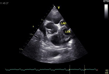
Echocardiographic parasternal short axis view at the level of the aortic valve. The LAD and LCX are aneurysmal and seen in relation to the left coronary sinus.
In view of non ST elevation myocardial infarction, the patient was taken for a coronary angiogram which was done via the right radial route. There were aneurysms involving all three coronary arteries mainly involving their proximal segments. The LAD and the LCX were aneurysmal in their proximal segments but RCA was aneurysmal in its entire length (Figs. 2 and 3, Videos 1 and 2). There was a 95% stenosis of distal LCX distal to the stenosis. An aortogram was also performed which showed a normal ascending aorta, arch and descending aorta until its bifurcation into the iliac arteries. The aortic arch vessels were also normal.
Fig. 2.

RAO caudal view during coronary angiogram showing the aneurysms of both LAD and LCX with a 95% stenosis of distal LCX distal to the stenosis.
Fig. 3.

LAO view of coronary angiogram showing the RCA aneurysm involving its entire length.
Supplementary video related to this article can be found at http://dx.doi.org/10.1016/j.ihj.2014.11.003
The following are the supplementary video related to this article:
RAO caudal view showing the aneurysmal LAD and LCX coronary arteries. LCX has 90% stenosis distal to the aneurysm.
LAO view of RCA showing aneurysm in its entire length with significant slow flow.
A coronary CT angiography was done in order to delineate the actual size of the aneurysm and also to look for the presence of thrombus. The CT revealed aneurysmal dilatation of LAD up to 4.3 cm in length and 1.2 cm in its widest dimension (Fig. 4). There was a fusiform aneurysm of LCX of 4.9 cm in length and 4.5 cm in its maximum dimension with mural thrombus (Fig. 4). There was a long 8.1 cm fusiform aneurysm of RCA with 1 cm width in its maximum dimension (Fig. 5).
Fig. 4.

CT angiogram showing the aneurysms in the proximal LCX and Proximal LAD.
Fig. 5.
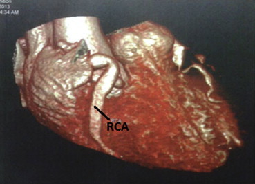
CT angiogram showing the aneurysm of the RCA involving its entire length.
The patient's CRP was elevated (CRP – 7.9 mg/dl) but ESR was normal (ESR – 16 mm), ANA and other autoimmune markers were negative ruling out active connective tissue disease. The rare possibility of an undetected Kawasaki disease during childhood with coronary aneurysms was entertained. The patient was advised to undergo a coronary artery bypass surgery as the distal vessels were relatively uninvolved. But as the patient opted for medical management, he was managed conservatively with dual antiplatelets, high dose statins, betablockers and ACE inhibitors. Patient had no further angina and has been on regular follow up for the past 1 year.
1. Discussion
Coronary artery aneurysm was first described by Morgagni in 1761. The first case series of 21 cases of aneurysm was reported in 1929.3 Coronary artery aneurysms have a prevalence of 4.9% according to the Coronary Artery Surgery Study (CASS) registry.4 An Indian study reported an incidence of 10–12%, the highest incidence reported so far.5 The right coronary artery was the most commonly affected (40–87% of aneurysms), followed by the left circumflex or left anterior descending artery.6 Three-vessel involvement is rare, as seen in this patient.
The majority of cases of coronary artery aneurysms in adults are acquired usually as a result of atherosclerosis, however other causes include Kawasaki disease, autoimmune disease (systemic lupus erythematosus, Behcet's disease, and rheumatoid arthritis), infection, syphilis, congenital, dissection or iatrogenic.1 The patients with atherosclerotic cause are divided into coronary aneurysms with and without atherosclerotic coronary artery disease (ACAD). This is important as the actuarial 5-year survival is significantly lower in patients with aneurysm and ACAD.4 Most studies have shown no significant difference in events or survival,4 except for a retrospective study which showed more 5-year mortality in patients with aneurysms irrespective of ACAD.7 Patients with aneurysms and ACAD were compared in a retrospective study from Greece with a group of patients with ACAD alone, matched for all risk factors. Both the groups had a similar incidence of angina, but patients with aneurysms alone had a significantly increased exercise tolerance time.7
It has also been postulated that patients with aneurysms and angina can experience a paradoxical worsening of ischemia after using nitroglycerin via a mechanism termed dilated coronaropathy.8 Hence our patient was not started on nitrates. Two interventional techniques have been described: obliteration with PTFE-covered stents11,12 or surgical therapy (coronary artery bypass grafting with or without resection or ligation of the aneurysm). From the largest experience in adults, on surgical management, which typically includes bypass grafting is based on the CASS sub-study. No difference in survival after CABG was noted between patients with ACAD and aneurysms (over 500 patients) and ACAD alone.9,13
Percutaneous treatment is a new option, with a markedly smaller data set, and includes stenting and coiling. It is recommended for patients with aneurysm less than 10 mm in diameter.10,14
2. Conclusion
We present a rare case of coronary artery aneurysm involving all three coronary arteries who presented with a non ST elevation myocardial infarction. He showed a pure coronary involvement with a normal aorta. As the natural history of coronary artery aneurysms are generally benign according to most studies and the patient's choice, he was managed conservatively with no further events in the 1 year of follow-up. Such a case joins one of the few reports of aneurysms involving all three coronary arteries.15
Conflicts of interest
All authors have none to declare.
References
- 1.Syed M., Lesch M. Coronary artery aneurysm: a review. Prog Cardiovasc Dis. 1997;40:77–84. doi: 10.1016/s0033-0620(97)80024-2. [DOI] [PubMed] [Google Scholar]
- 2.Li D., Wu Q., Sun L. Surgical treatment of giant coronary artery aneurysm. J Thorac Cardiovasc Surg. 2005;130:817–821. doi: 10.1016/j.jtcvs.2005.04.004. [DOI] [PubMed] [Google Scholar]
- 3.Packard M., Wechsler H. Aneurysms of coronary arteries. Arch Intern Med. 1929;43:1. [Google Scholar]
- 4.Principal Investigators of CASS and their Associates National heart, lung, and blood institute coronary artery surgery study. Circulation. 1981;63(Suppl. II) Il-1. [Google Scholar]
- 5.Sharma S.N. Coronary arteriographic profile in young and old Indian patients with ischaemic heart disease: a comparative study. Indian Heart J. 1990;42:365–369. [PubMed] [Google Scholar]
- 6.Villines T.C., Avedissian L.S., Elgin E.E. Diffuse nonatherosclerotic coronary aneurysms. Cardiol Rev. 2005;13:309–311. doi: 10.1097/01.crd.0000159579.77038.dc. [DOI] [PubMed] [Google Scholar]
- 7.Demopoulos V.P., Olympios C.D., Fakiolas C.N. The natural history of aneurismal coronary artery disease. Heart. 1997;78:136–141. doi: 10.1136/hrt.78.2.136. [DOI] [PMC free article] [PubMed] [Google Scholar]
- 8.Kruger D., Stierle U., Herrmann G. Exercise-induced myocardial ischemia in isolated coronary artery ectasias and aneurysms (“dilated coronaropathy”) J Am Coll Cardiol. 1999;34:1461–1470. doi: 10.1016/s0735-1097(99)00375-7. [DOI] [PubMed] [Google Scholar]
- 9.Swayze P.S., Fisher L.D., Litwin P.A. Aneurysmal coronary artery disease. Circulation. 1983;67:134–138. doi: 10.1161/01.cir.67.1.134. [DOI] [PubMed] [Google Scholar]
- 10.Szalat A., Durst R., Cohen A. Use of polytetrafluoroethylene-covered stent for treatment of coronary artery aneurysm. Catheter Cardiovasc Interv. 2005;66:203–208. doi: 10.1002/ccd.20448. [DOI] [PubMed] [Google Scholar]
- 11.Heuser R.R., Woodfield S., Lopez A. Obliteration of a coronary artery aneurysm with a PTFE-covered stent: endoluminal graft for coronary disease revisited. Catheter Cardiovasc Interv. 1999;46:113–116. doi: 10.1002/(SICI)1522-726X(199901)46:1<113::AID-CCD28>3.0.CO;2-7. [DOI] [PubMed] [Google Scholar]
- 12.Di Mario C., Inglese L., Colombo A. Covered stents: homemade or ready-to-serve? Catheter Cardiovasc Interv. 1999;48:113B. doi: 10.1002/(sici)1522-726x(199909)48:1<113b::aid-ccd24>3.0.co;2-#. [DOI] [PubMed] [Google Scholar]
- 13.Alford W.C., Jr., Stoney W.S., Burrus G.R., Frist R.A., Thomas C.S., Jr. Recognition and operative management of patients with arteriosclerotic coronary artery aneurysms. Ann Thorac Surg. 1976;22:317–321. doi: 10.1016/s0003-4975(10)64961-2. [DOI] [PubMed] [Google Scholar]
- 14.Portela A., Bastos R., Costa I., Paiva J. Repair of an atherosclerotic coronary artery aneurysm by implantation of a coronary covered stent. Arq Bras Cardiol. 2002;78:491–496. doi: 10.1590/s0066-782x2002000500006. [DOI] [PubMed] [Google Scholar]
- 15.Marla R., Ebel R., Crosby M., Almassi G.H. Multiple giant coronary artery aneurysms. Tex Heart Inst J. 2009;36:244–246. [PMC free article] [PubMed] [Google Scholar]
Associated Data
This section collects any data citations, data availability statements, or supplementary materials included in this article.
Supplementary Materials
RAO caudal view showing the aneurysmal LAD and LCX coronary arteries. LCX has 90% stenosis distal to the aneurysm.
LAO view of RCA showing aneurysm in its entire length with significant slow flow.