


Fig. 3.
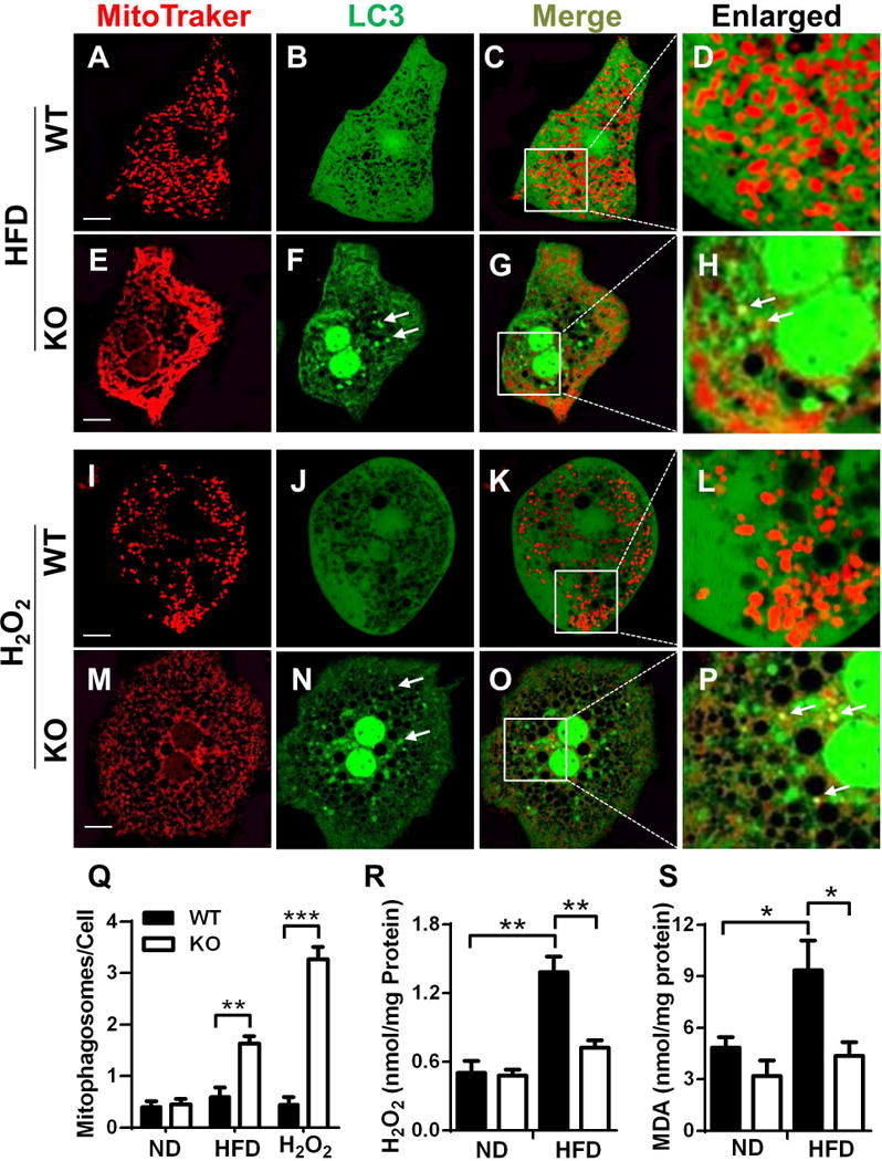
ALCAT1 deficiency promotes autophagosome biogenesis through attenuation of oxidative stress. Primary hepatocytes from ALCAT1−/− mice (KO) and WT controls were infected with recombinant adenoviruses expressing LC3-GFP fusion protein, stained by MitoTracker Red, and followed by confocal imaging analysis of mitochondrial network and autophagosome biogenesis. (A–H) Primary hepatocytes from WT and ALCAT1−/− mice on HFD were analyzed for mitochondrial architecture (A & E), autophagosomes (B & F), and mitophagosomes (C & G, enlarged in D & H). (I–P) Primary hepatocytes from WT and ALCAT1−/− mice on chow diet were treated with 0.5 mM H2O2 for 2 hours, followed analysis of mitochondrial architecture (I & M), autophagosome biogenesis (J & N), and mitophagosomes (K & O, enlarged in L & P). (Q) Quantitative analysis of autophagosomes per hepatocyte (n=100). (R–S) Analysis of oxidative stress and lipid peroxidation in the forms of H2O2 and malondialdehyde (MDA), a byproduct of lipid peroxidation. Mean ± SEM; n=6–8, *P<0.05, **P<0.01, and ***P<0.001. Scale Bar: 10 μm.