


Fig. 1.
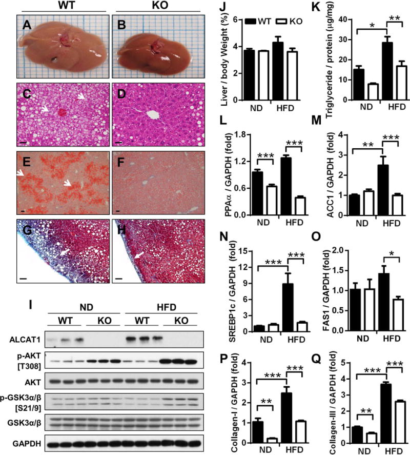
Ablation of ALCAT1 prevents NAFLD and its related metabolic complications. ALCAT1−/− mice and WT controls were fed a high-fat diet (HFD) for 18 consecutive weeks, and were analyzed for: (A–B) gross morphology; (C–D) liver sections by H&E staining; (E–F) hepatosteatosis by Oil Red staining; and (G–H) hepatic fibrosis by trichrome staining. (I) Western blot analysis of ALCAT1 expression and insulin-stimulated phosphorylation of AKT and GSK3α/β in the liver. (J–K) Analysis of liver weight and triglyceride level. (L–O), RT-PCR analysis of genes that promote lipogenesis in the liver, including PPARα, ACC1, SREBP1c and FAS1. (P–Q) RT-PCR analysis of hepatic fibrosis biomarkers, including collagen-I (P) and collagen-III (Q). Data are expressed as mean ± SEM; n = 6–8, *p < 0.05, **p < 0.01 and ***p < 0.001. Scale Bar: 50μm.