


Fig. 5.
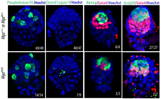
MgaGt/Gt embryos showed more apoptosis, but cell proliferation and differentiation were normal at E4.5. Cell proliferation, as measured by phospho-H3 immunostaining, was similar in MgaGt/Gt compared with Mga+/+ and MgaGt/+ embryos. Confocal Z-stacks of cleaved caspase 9 immunofluorescence showed that more MgaGt/Gt embryos had fragmented nuclei (7/9) than did Mga+/+ and MgaGt/+ embryos (7/47). The pluripotency markers NANOG and POU5F1, as measured by a GFP reporter, are similar in MgaGt/Gt embryos compared with Mga+/+ and MgaGt/+ embryos, as was the PE marker GATA4. Numbers in the panels indicate the number of embryos out to the total with an appearance similar to that shown.