


Figure. 1. Schematic representing how protein folding in the ER modulates mitochondrial ATP and ROS production.
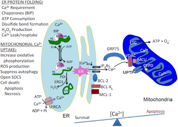
Mitochondria and ER are tethered by the actions of the MFNs, of which MFN2 is localized to the mitochondrial-associated membrane (MAM), that promote efficient Ca2+ transfer from the ER to the mitochondria. Ca2+ loading in the ER is mediated by the abundance of Ca2+-binding proteins, including CNX, CRT, as well as the protein chaperones BIP and PDI. Protein folding in the ER requires Ca2+ and ATP for chaperone function, proper glycosylation, and correct disulfide bond formation. Misfolded proteins may sequester protein chaperones that facilitates opening of Ca2+ channels to initiate Ca2+ transfer to mitochondria to stimulate oxidative phosphorylation. Ca2+ transfer occurs through the activity of several Ca2+ channels that include the ER localized inositol-1,4,5-triphosphate receptors (IP3Rs), as well as the ryanodine receptors (RyRs) and the mitochondrial-localized voltage-dependent anion channel (VDAC) and the mitochondrial Ca2+ uniporter complex MCU (MCU, including MICU1, MICU2, MCUR1 and EMRE). The IP3Rs enriched at the MAMs are linked to VDAC on the OMM by the protein chaperone GRP75. VDAC tightly controls Ca2+ permeation into mitochondria by IP3Rs-mediated Ca2+ signals. Once Ca2+ transverses the OMM it can subsequently cause depolarization of the inner mitochondrial permeability transition pore (MPTP) and induction of apoptotic stimuli. Conditions that prevent Ca2+ transfer from the ER to mitochondria include overexpression of anti-apoptotic proteins such as BCL-2 and BCL-XL and constitute survival signaling. A number of mechanisms have been proposed to cause Ca2+ leak from the ER and are depicted as red identities on the ER membrane (SEC61, SERCA1T, BCL-2, BCL-XL, MCL-1, BI-1 and IP3Rs). As Ca2+ accumulates in mitochondria, cells are predisposed to disruption of the electron transport chain (ETC) to produce ROS, MPTP, mitochondrial swelling, disruption of the OMM, release of cytochrome C and apoptosome components leading to caspase activation and apoptosis. Mechanisms the limit mitochondrial loading of Ca2+ include MPTP itself, and the mitochondrial Ca2+ exchangers NCLX and HCX. In addition to protein synthesis, ATP-utilizing processes include chaperone (BIP)-assisted protein folding in the ER lumen, SERCA-mediated Ca2+ reuptake into the ER and possibly hydrolysis of ATP by the F1/F0 ATP synthase upon collapse of the IMM electrochemical potential. Finally, in addition to superoxide production from the ETC, disulfide bond formation mediated by the protein thiol-disulfide isomerases (PDI, ERP57) and ER oxidase 1 (ERO1) generates hydrogen peroxide upon electron transport to molecular O2 as the acceptor. The balance between the amount of Ca2+ stored in the ER lumen and the amount loaded into the mitochondrial matrix may be a determinant in the decision between survival and death.