


Figure 1. Identification of nanotube structures in resected tumors.
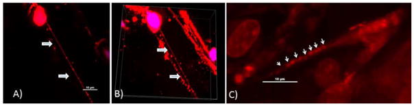
A) Visualization of a long nanotube in a murine orthotopic model of osteosarcoma. Tumors were sectioned using a Vybratome and stained using MitoTracker Red dye to visualize mitochondria. The samples we imaged represent only a very small portion of the entire tumor volume. Nonetheless, for surgically excised murine OS tumors, using confocal microscopy and 3-dimensional reconstruction with Imaris software analysis, we readily identified and visualized nanotubes in 2 of 3 tumors excised from 3 mice. We measured nanotube width in order to differentiate these protrusions from other cellular extensions, and confirmed these nanotubes were well under 1.0 μm in width. These intratumoral nanotubes measured ~80 μm in length, and were consistent with our in vitro findings of OS and other forms of cancer cells culture in vitro (both cell lines and primary).
B) Snapshot of 3-dimensional z-stacked imaging of several nanotubes within the matrix of a murine orthotopic osteosarcoma tumor.
C) Nanotube in a tumor surgically resected from a human patient with ovarian adenocarcinoma. The tumor was stained with fluorescing MitoTracker Orange dye and imaged using confocal microscopy with z-stacking of images.