


Figure 4.
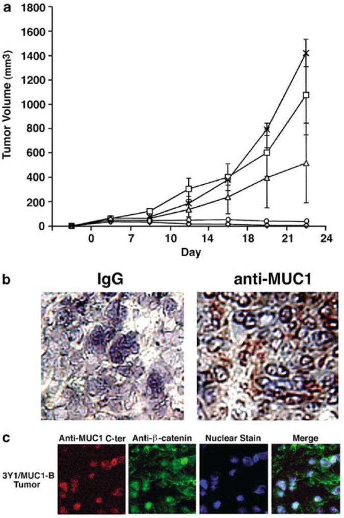
MUC1 induces tumorigenicity of 3Y1 cells in nude mice. (a) 3Y1/Vector-A (◇), 3Y1/Vector-B (○), 3Y1/MUC1-A (□), -B (X) and -C (Δ) cells (1 × 10⁷) were injected subcutaneously into the posterior flank of nude mice. Tumor volumes were calculated from bidimensional measurements at each time point. The results are expressed as the tumor volume (mean±s.d.) obtained from two independent experiments that each included six to eight mice per group. (b) Frozen tumor tissue was sectioned and stained with a control IgG or the DF3 anti-MUC1 antibody. (c) Fixed sections from a 3Y1/MUC1-B tumor were permeabilized and incubated with anti-MUC1 C-ter and anti-β-catenin. Nuclei were stained with TO-PRO-3. Confocal microscopy was performed as described (Li et al., 2003). Little if any staining was obtained when the Texas Red- and fluorescein-conjugated secondary antibodies were used alone as controls (data not shown)