


Figure 5.
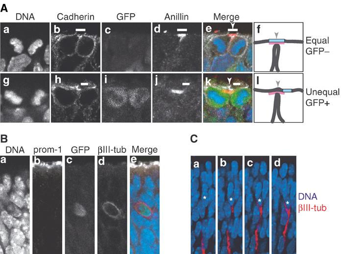
Neurons arising from asymmetric division of NE cells do not inherit apical membrane and prominin-1. (A) Frozen sections of E11.5 hindbrain (a–e) and E11.5 telencephalon (g–k) of heterozygous TIS21-GFP knock-in embryos showing quadruple-labeled dividing NE cells that have almost completed cytokinesis. Single optical sections are shown; DNA, DAPI-stained nuclei (a, e blue, g, k blue). In the NE cell in the top row (a–e), which does not express TIS21-GFP (GFP−) (c, e green) and hence undergoes a proliferative division, the cleavage furrow is about to bisect the apical membrane (i.e. the cadherin hole, white bars), as revealed by immunostaining for cadherin (b, e white) and anillin (d, e red) (which at this stage of cytokinesis indicates the midbody (Oegema et al, 2000)), resulting in its distribution to both daughter cells (equal). In contrast, in the NE cell in the bottom row (g–k), which does express TIS21-GFP (GFP+) (i, k green) and hence undergoes a neurogenic division, the cleavage furrow is about to bypass the apical membrane (i.e., the cadherin hole, white bars), as revealed by immunostaining for cadherin (h, k white) and anillin (j, k red), resulting in its distribution to only one daughter cell (unequal). (f and l) Cartoons summarizing the imminent completion of cytokinesis by plasma membrane fusion in the cell shown in (e) and (k), respectively; black lines, lateral and cleavage furrow plasma membranes; light blue box, apical membrane; red, anillin; arrowheads, site of cleavage. (B) Frozen section of E10.5 telencephalon of heterozygous TIS21-GFP knock-in embryos showing a quadruple-labeled newborn neuron that was identified by immunostaining for βIII-tubulin (βIII-tub, d, e red) and shows GFP fluorescence (c, e green), indicative of its origin from a TIS21-GFP-expressing NE cell (Haubensak et al, 2004). A single optical section is shown; DNA, DAPI-stained nuclei (a, e blue). Note the lack of extension of the neuron to the apical surface immunostained for prominin-1 (prom-1, b, e white). (C) Four consecutive optical sections of a newborn neuron (asterisks), identified by immunostaining for βIII-tubulin (red) in a frozen section of E11.5 hindbrain of heterozygous TIS21-GFP knock-in embryos. Note the extension of the neuron in the basal direction, but not towards the apical side of the neuroepithelium (top). Blue, DAPI-stained nuclei.