


FIG 4.
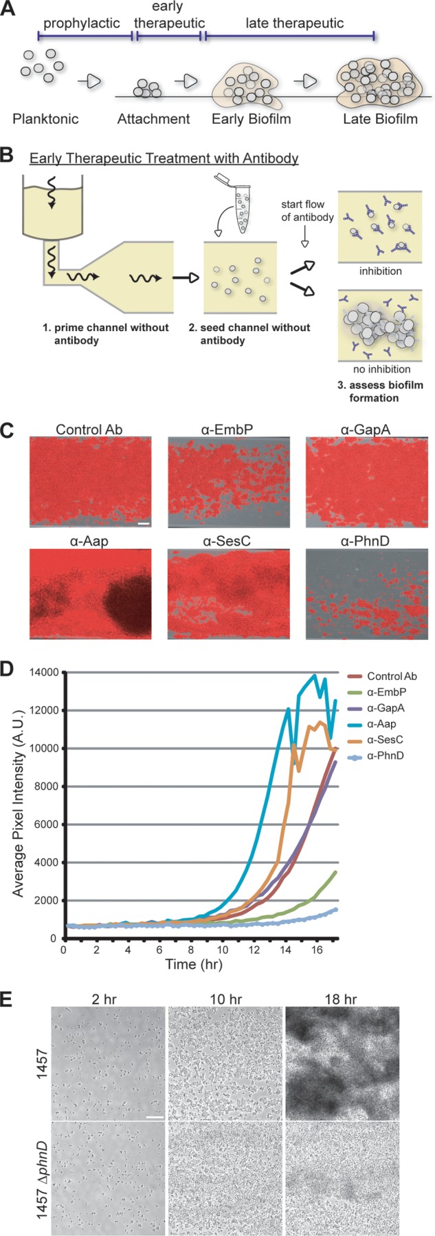
Assessing biofilm inhibition by antibodies added postattachment. (A) Biofilm development cycle. Possible antibody intervention points include the following: prophylactic (prior to bacterial attachment), early therapeutic (before intercellular aggregation), and late therapeutic (after biofilm establishment and maturation). (B) Diagram of the assay for biofilm inhibition, in which antibodies are introduced postattachment (model for early therapeutic intervention). Biofilm formation is monitored for the duration of the experiment. Wavy arrows indicate the direction of flow. (C) Endpoint images at 18 h of 1457-FL grown at 30°C and treated with antibodies postattachment at 100 μg/ml. Scale bar, 30 μm. (D) Longitudinal quantification of the experiment described in the panel C legend using average pixel intensity of images captured at 20-min intervals for 18 h. (E) Deletion of phnD results in defective biofilm formation. Biofilms of S. epidermidis 1457 and its isogenic ΔphnD mutant were grown under conditions of flow at 30°C. Images represent biofilm development after 2, 10, and 18 h. Scale bar, 25 μm.