


Figure 1. Antidepressants reduced the number of α-syn-positive cells in basal ganglia in a transgenic model of MSA.
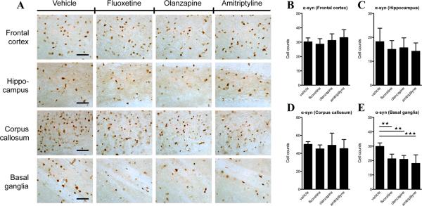
(A) α-syn immunostaining of frontal cortex, hippocampus, corpus callosum and basal ganglia of MBP1-hα-syn transgenic mice treated with vehicle, fluoxetine, olanzapine or amitriptyline. (B) Cell counts per field of α-syn-positive cells in frontal cortex. (C) Cell counts per field of α-syn-positive cells in hippocampus. (D) Cell counts per field of α-syn-positive cells in corpus callosum. (E) Cell counts per field of α-syn immunostaining of basal ganglia. Error bars represent ± SEM. ** p<0.01. *** p<0.001. Scale bar = 50 μm.