


Figure 5.
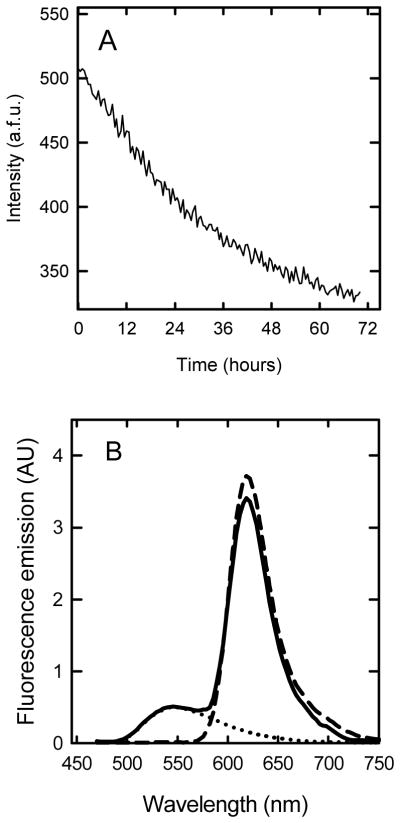
FRET analysis of the interaction between Alexa 594 apoC-II fibrils and NBD-Lyso-12-PC. A. Time course for the change in FRET during apoC-II fibril formation. Alexa 594-labelled apoC-II (33 μM) was incubated with NBD-Lyso-12-PC (60 μM) at 20 °C and the fluorescence emission at 620 nm measured with excitation at 430 nm. B. Fluorescence emission spectra (in arbitrary units) for Alexa 594 apoC-II fibrils alone (5 μM, dashed line), Alexa 594 apoC-II fibrils (5 μM) in the presence of 60 μM NBD-Lyso-12-PC (solid line) and 60 μM NBD-Lyso-12-PC alone (dotted line) were acquired using excitation at 430nm.