


Table 4.
Methods, advantages and disadvantages of nanoparticle attachment techniques to cells.
| Attachment Method | Advantages | Disadvantages | Ideal Cell Type |
|---|---|---|---|
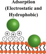
|
No cell preparation Minimal particle requirements: (hydrophobic or positively charged) |
Attachment may not be strong: Particles may detach unpredictably in vivo Positively charged particles can damage cell membranes |
Cells that persist in circulation: Red blood cells and platelets Cells that traverse endothelium may cause unwanted particle detachment during migration |
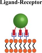
|
Can potentially be used to attach particles to cells in vivo since attachment is mediated by specific cell receptors | Ligand-receptor interaction may signal specific cellular functions | Cells that express sufficient receptors which allow for multivalent binding: CD44-hyaluranon interaction allows for stable multivalent bonding to multiple cell types |
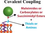
|
Offers the strongest bond between particle and cell Conjugate directly to amine or thiol groups present on all cells (membrane proteins) |
Requires permanent modification to cell surface | Cells that migrate through endothelium: Monocytes, macrophages and T-cells since particle attachment must be strong |
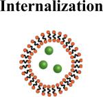
|
Leaves the cell membrane unmodified Can carry particles through endothelium |
Can lead to degradation of internalized biodegradable particles | Cells capable of phagocytosis |