


Figure 2. CQ inhibits PTX-induced autophagy and sensitizes TNBC cell lines to PTX.
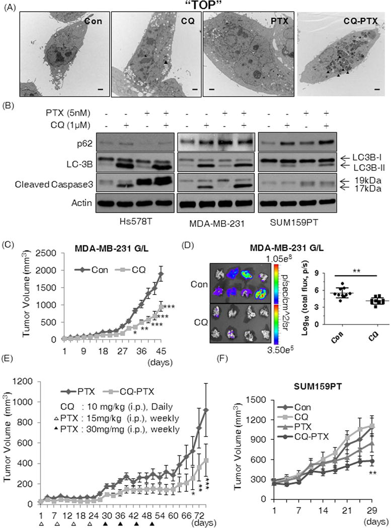
(A) TEM images of autophagosomes in Hs578t cells after 48 hours treatment with Con (DMSO), CQ (1 μM), PTX (5 nM) or combination of CQ and PTX. Solid black arrows indicate autophagosomes. The TEM experiment was performed once and the representative images are shown here. The black bars are 2 μm in length. (B) Western blotting results for p62, LC3B, and cleaved caspase 3 from lysates of Hs578t, MDA-MB-231, and SUM159PT cells after 48-hour treatments as indicated. We confirmed similar effects of CQ in independently repeated experiments. CQ attenuated (C) growth of orthotopic MDA-MB-231 G/L tumors and (D) inhibited spontaneous lung metastasis of the tumors; Con: PBS and CQ: 10 mg/kg i.p. daily (n=10 per group). Total photon counts were normalized by setting the minimum at 3.50 × 10⁵ and the maximum at 1.05 × 10⁸ (p/sec/cmˆ2/sr) and transformed by log10 to analyze the effects of CQ on metastasis and tumor burden in lungs. Two-tailed Student’s t-test was used for the statistical comparison. (E) CQ-PTX attenuated tumor recurrence (MDA-MB-231 G/L) compared to PTX aline. (F) The CQ-PTX combination therapy reduced the growth of orthotopic SUM159PT tumors (n=10). All mice received the scheduled dose of each drug as indicated in the Materials and Methods. Two-way ANOVA (Boneferroni post-tests) was used to compare tumor volumes in all animal experiments (C, E, and F). All control groups received PBS as the vehicle (*p<0.05, **p<0.01, ***p<0.001).