


Figure 2. FoxO deficiency promotes cell proliferation suppression and cell death induction under NVP-BEZ235 treatment.
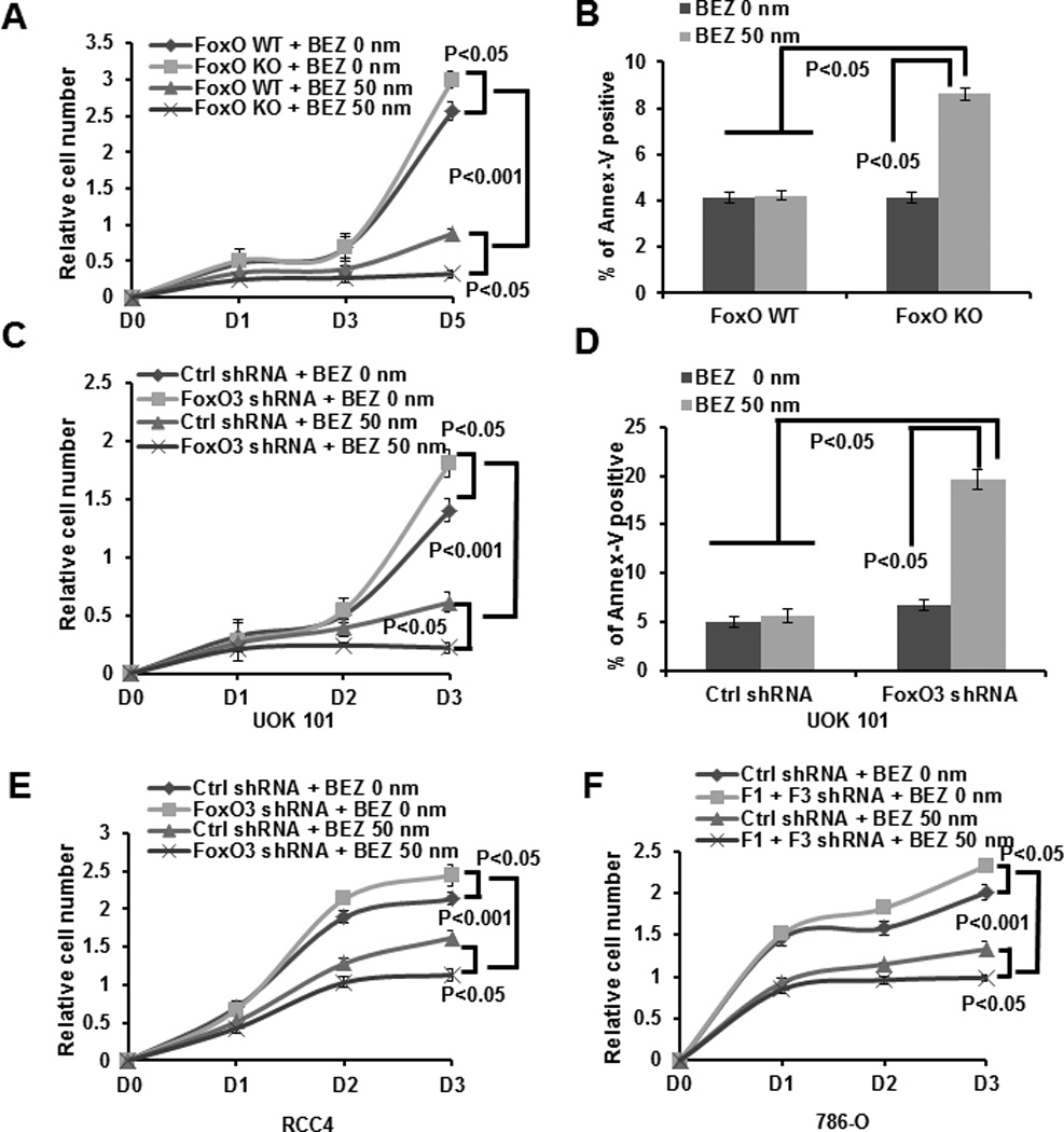
(A) FoxO WT and KO MEFs were treated with 0 (vehicle) or 50 nM NVP-BEZ235 for different days as indicated, and then subjected to cell proliferation analysis. (B) Bar graph showing the percentages of Annexin V staining of FoxO WT and KO MEFs which were treated with 0 or 50 nM NVP-BEZ235 for 24 hours. (C) UOK 101 cells infected with either control shRNA or FoxO3 shRNA were treated with 0 or 50 nM NVP-BEZ235 for different days as indicated, and then subjected to cell proliferation analysis. (D) Bar graph showing the percentages of Annexin V staining of UOK101 cells infected with either control shRNA or FoxO1/FoxO3 shRNA which were treated with 0 or 50 nM NVP-BEZ235 for 24 hours. (E) RCC4 cells infected with either control shRNA or FoxO3 shRNA were treated with 0 or 50 nM NVP-BEZ235 for different days as indicated, and then subjected to cell proliferation analysis. (F) 786-O cells infected with either control shRNA or FoxO1/FoxO3 shRNA were treated with 0 or 50 nM NVP-BEZ235 for different days as indicated, and then subjected to cell proliferation analysis.