


Figure 3. IMQ increased HIF-1α expression at the transcriptional and translational levels.
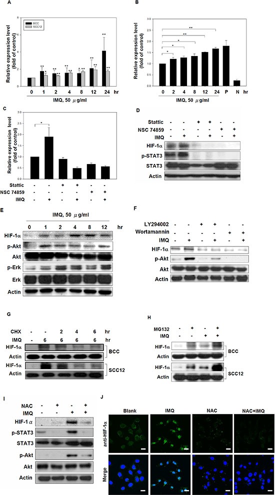
(A) IMQ stimulated HIF-1α mRNA expression in tumor cells. BCC and SCC12 cells were treated with 50 μg/ml IMQ for 0, 1, 2, 4, 8, 12 or 24 hours, and then, the HIF-1α mRNA levels were determined by quantitative real-time PCR. (B) IMQ enhanced STAT3 transcriptional activity in BCC cells. BCC cells were transfected with a STAT3-driven luciferase reporter vector for 48 hours. The cells were treated with 50 μg/ml IMQ for 0, 2, 4, 8, 12 or 24 hours and then analyzed by a luciferase assay. (C and D) The inhibition of STAT3 reduced the HIF-1α mRNA (C) and protein levels (D) in IMQ-treated cells. BCC cells were pre-treated with Stattic (10 μM) or NSC74859 (1 μM) for 1 hour and then treated with or without 50 μg/ml IMQ for 4 hours. Cell lysates were collected, and the mRNA and protein levels were determined by quantitative real-time PCR and immunoblotting with HIF-1α, p-STAT3, STAT3 and β-actin antibodies, respectively. (E) IMQ-induced HIF-1α expression was associated with the activation of the Akt and ERK signaling pathways. BCC cells were incubated with medium containing 50 μg/ml IMQ for 0, 1, 2, 4, 8 or 12 hours, and cell lysates were then harvested for immunoblotting with HIF-1α, phosphorylated-Akt, Akt, phosphorylated-ERK, ERK and β-actin antibodies. (F) Suppression of the PI3K/Akt signaling pathway reduced IMQ-induced HIF-1α expression. BCC cells were incubated for 1 hour with 20 μM LY294002 or 10 μM wortmannin in medium and then treated with or without 50 μg/ml IMQ for 4 hours. Cell lysates were collected for immunoblotting with HIF-1α, p-Akt, Akt and β-actin antibodies. (G) IMQ treatment did not increase the protein stability of HIF-1α. BCC and SCC12 cells were treated with IMQ (50 μg/ml) for 6 hours with or without co-treated with CHX (10 μg/ml) for 0, 2, 4 or 6 hours. Cell lysates were prepared for immunoblotting with HIF-1α and β-actin antibodies. (H) IMQ did not affect the proteasomal degradation of the HIF-1α protein. BCC and SCC12 cells were pre-treated with MG132 (10 μM) for 1 hour, treated with IMQ (50 μg/ml) for 4 hours and then processed for immunoblotting with HIF-1α and β-actin antibodies. (I) The depletion of ROS resulted in the down-regulation of HIF-1α expression with the repression of the STAT3 and PI3K/Akt signaling pathway. BCC cells were incubated with medium containing 2 mM NAC for 30 minutes and then treated with 50 μg/ml IMQ for 4 hours. Cell lysates were collected for immunoblotting with HIF-1α, p-Akt, Akt, p-STAT3, STAT3 and β-actin antibodies. (J) The inhibition of ROS production repressed IMQ-induced HIF-1α nuclear translocation. BCC cells were pre-treated with 2 mM NAC for 30 minutes and then treated with 50 μg/ml IMQ for 4 hours. The subcellular localization of HIF-1α was determined by immunocytochemistry. Scale bars, 20 μm. The data are expressed as the mean ± S.E.M. of at least three independent experiments. * p < 0.05; ** p < 0.01.