



Abstract
The correction of motion artifacts continues to be a significant problem in MRI. In the case of uncooperative patients, such as children, or patients who are unable to remain stationary, the accurate determination and correction of motion artifacts becomes a very important prerequisite for achieving good image quality. The application of conventional motion-correction strategies often produces inconsistencies in k-space data. As a result, significant residual artifacts can persist. In this work a formalism is introduced for parallel imaging in the presence of motion. The proposed method can improve overall image quality because it diminishes k-space inconsistencies by exploiting the complementary image encoding capacity of individual receiver coils. Specifically, an augmented version of an iterative SENSE reconstruction is used as a means of synthesizing the missing data in k-space. Motion is determined from low-resolution navigator images that are coregistered by an automatic registration routine. Navigator data can be derived from self-navigating k-space trajectories or in combination with other navigation schemes that estimate patient motion. This correction method is demonstrated by interleaved spiral images collected from volunteers. Conventional spiral scans and scans corrected with proposed techniques are shown, and the results illustrate the capacity of this new correction approach.
Keywords: MRI, motion, parallel imaging, SENSE, spiral imaging
Involuntary patient motion is still one of the greatest challenges in MRI. Due to the sequential nature of the MR acquisition process, a significant amount of time can elapse between different samples in k-space, leaving many MRI sequences vulnerable to patient motion. In particular, the diagnostic quality of an MR examination can be severely impaired due to motion if k-space is not sampled on the intended grid. Frequently the medical condition of a patient, such as tremor, pain, or mental status, prevents even willing patients from holding still. Previous attempts to address this problem through sequence approaches, such as snapshot imaging or gated acquisition, have had limited success. Therefore, efficient motion-correction strategies are timely and have the potential to significantly improve health care as MRI is used more commonly in geriatric and pediatric patient populations.
The way in which motion affects the final image depends on the magnitude and direction of the motion, as well as its temporal relation to the acquisition of k-space. Overall, the net effect of patient motion during MRI is k-space data inconsistency, which produces artifacts in the final image. If the corrupted k-space data can be identified and the degree of motion is known, one can correct, or at least reduce, image artifacts by 1) rejecting corrupted k-space data (1–3), 2) reacquiring corrupted k-space data, 3) applying retrospective correction schemes that undo the effects of motion (1,4,5), or 4) determining motion on the fly and prospectively adjusting the acquisition for the new location of interest during scanning (6,7).
The simplest form of motion is rigid-body translation without elastic deformation. In k-space, translational motion is manifested by a linear phase shift along the direction of motion (8). Rotational motion produces a slightly more complicated artifact pattern. For a given object, a rotation in image space produces the same rotation in k-space (8). Therefore, during sequential scanning the expected k-space information at a particular position will have been rotated away and will be partially missing or replaced by data that have already been acquired. Consequently, these object rotations can be the source of significant image artifacts.
To correct for rotational motion artifacts, the trajectory and the acquired k-space data must be synchronized with the rotated object. Specifically, the misaligned k-space lines, or interleaves, can be counter-rotated relative to a reference point. However, the rotation procedure usually puts the corrected k-space data at locations that no longer coincide with an equidistant Cartesian grid, resulting in local undersampling and ill conditioning of the reconstruction problem (Fig. 1). Therefore, interpolation methods for image reconstruction that address the ill conditioning have been proposed (4). In this work we present a novel motion-correction approach that avoids some of the aforementioned problems and improves the conditioning of the reconstruction by first putting the motion problem into a matrix formalism and then using a parallel-imaging-based synthesis of the missing k-space information.
FIG. 1.

The effects of rotation are corrected by counter-rotating the individual interleaves. The effect of this counter-rotation on the k-space trajectories is shown for both interleaved spirals (a) and interleaved EPI (b). It is clearly apparent that the counter-rotation causes a variable sampling density in various areas of k-space. Some regions become oversampled compared to the Nyquist sampling rate, whereas other regions become only sparsely sampled. In the case of EPI, it is also worthwhile to note that the sampled trajectory data are no longer on an equidistant grid.
Parallel imaging was introduced several years ago as a complement to conventional gradient encoding (9,10). Since then it has been employed in a broad spectrum of applications ranging from rapid imaging (11–13) to the reduction of image artifacts (5,14,15). The inhomogeneous RF profiles of individual receiver coils can be used to synthesize missing information in k-space (16,17) or remove aliasing (18). The method proposed here retrospectively corrects rigid body rotation and translation, and compensates for the resulting variable density sampling pattern. Specifically, the method utilizes an augmented iterative parallel imaging reconstruction approach to correct for motion regardless of the type of trajectory used. The proposed correction method was tested with a new variant of spiral navigators that can be incorporated in various pulse sequences.
In summary, we introduce a new formalism for parallel imaging reconstruction in the presence of motion for arbitrary k-space data. Specifically, this approach models how motion affects k-space data. By solving the inverse problem under consideration of coil sensitivity and sampling density variations during data acquisition, one can apply this new framework to the field of motion correction.
THEORY
For a better understanding of how motion affects k-space and thus the reconstructed image, we will first describe the formation of an MRI data sample at a particular point in time, ti (i = 1…κ), using discrete sampling in the time and spatial domain, ru (u = 1…N2):
| [1] |
Therefore, the entire acquisition of κ k-space points can be stored in a measurement vector, m = [m(t1) m(t2)…m(tκ)]T, so that the signal formation in matrix form can be expressed as:
| [2] |
where v = [ρ(r1)ρ(r2)…ρ(rN2)]T is the spatial signal distribution in vector form, ki = [kx(ti)ky(ti)kz(ti)]T|i=1…κ is an arbitrary k-space sampling trajectory, and ru = [rx,ury,urz,u]T|u=1…N2 is the position vector. One can then obtain the solution vector v for the image signal distribution by computing the Moore-Penrose inverse of E:
| [3] |
The encoding matrix E can be replaced by the matrix product GF so that Eq. [2] becomes
| [4] |
Here, instead of populating the matrix E with N2 × κ complex exponentials and performing a direct computation of Eq. [2], an FFT F (CN2×N2) is usually performed followed by a resampling of the Cartesian k-space data along the arbitrary k-space waveform using the matrix G (i.e., the inverse gridding matrix). For optimal sinc interpolation, one element in the inverse gridding matrix becomes Gl,i = sinc(k̃l − ki), where k̃l and ki represent the k-space grid locations. These positions, ki, are defined by the arbitrary sampling trajectory. To boost computation efficiency, more compact resampling kernels (e.g., Kaiser-Bessel window) are normally used instead of sinc interpolation (19). The formalism described in Eq. [4] is also known as the inverse gridding operation (20). From a Cartesian image, inverse gridding is able to generate k-space sample points along an arbitrary trajectory defined by ki, and therefore it represents the inverse operation to conventional gridding reconstruction (19).
Most frequently, MR data that are sampled along non-Cartesian k-space trajectories are reconstructed using gridding reconstruction (19); however, Eq. [3] can also be used to form an image from these arbitrarily sampled k-space data. Because of computational complexity, the direct inversion of Eq. [3] to find v is prohibitively inefficient; therefore, a least-squares solution for v,
| [5] |
is normally found in an iterative fashion by means of the conjugate-gradient method (21). This approach has recently gained popularity because it provides the foundation for iterative SENSE reconstruction (22) and nonlinear phase correction in diffusion-weighted imaging (DWI) (23).
In the next step, we demonstrate how motion impacts the acquired k-space data. Specifically, we assume that motion occurs between interleaves but not during interleaves. This assumption is legitimate for all MR applications for which the positional changes during one interleave are negligible. With FFH = I, where I is the identity matrix, let Ω be a warping matrix that operates on an unperturbed image v0 that yields the image v, and let Λ be the transformation rule in k-space that corresponds to Ω in the image domain, so that
| [6] |
Consequently, in the presence of motion, the signal equation for the acquired MR data changes to
| [7] |
Equation [7] clearly demonstrates that the solution of Eq. [4] yields a solution image v that is corrupted by motion. To obtain the true image v0, the spatial warping Ω must be included in the encoding matrix E. An augmented version of Eq. [3] that considers spatial transformation is therefore
| [8] |
The term in parentheses on the left-hand side of Eq. [8] reflects the gridding of the acquired data, correction for motion, and inverse FFT. Equation [8] can be solved for v0 following the same scheme as outlined in Eqs. [3] and [5]. Here, D(D(γκ),(γκ) = k_density(ki)−1) is introduced in Eq. [8] as a sampling density correction matrix exactly as in Pruessmann et al.’s (22) original approach. Also, the rotation of k-space data will affect the sampling density in k-space. Therefore, in this study the true sampling density was determined by a Voronoi tessellation approach (20) after motion correction was applied to the trajectories. Note, however, that accurate knowledge of the sampling density distribution is not so critical for this estimation problem because the iterative conjugate-gradient approach works even without prior knowledge of the sampling density information (22). Nevertheless, the exclusion of the density matrix causes much slower convergence rates because the density correction acts as a preconditioner. Additional preconditioning can be achieved by adding intensity correction, as suggested by Eq. [24] in Ref. 22.
Despite density correction, severe motion can lead to inconsistencies of k-space data (i.e., some areas in k-space are less densely sampled than required by the Nyquist sampling criterion). In other words, depending on the severity of motion, the linear dependency in Eq. [8] increases and therefore the conditioning of the system matrix E′ worsens, yielding considerable reconstruction artifacts. We hypothesize that parallel imaging helps to ameliorate the conditioning of this matrix system by adding additional encoding information using spatially varying coil sensitivities. Following Pruessmann et al.’s (22) seminal work, the k-space data for nc coils can be written as
| [9] |
where , cγ(ru) is the complex coil sensitivity of the γ-th coil at position ru, and m1 … mnc are the k-space sampling data vectors for each of the nc coils. For each coil, one can combine E with cγ on a per-pixel basis using E · diag(cγ), and v can be estimated based on the aforementioned iterative conjugate-gradient method (22). For increased processing speed and without loss of generality, E is again replaced by GF. Instead of the conventional gridding and inverse gridding, we use in our implementation the convolution approach introduced by Wajer and Pruessmann (24). Alternatively, the “fastest Fourier transform in the West” (FFTW) algorithm (25) could be used for non-power-of-2 matrices and non-integer oversampling factors to speed up the gridding and inverse gridding steps during the conjugate-gradient iteration (26).
Assuming that there is an inverse warping operation (i.e., there exists a matrix Ωinv so that Ω Ωinv s0 = s0 (27)), our motion-correction approach in Eq. [7] can be expanded to incorporate the coil sensitivity information by rewriting the signal equation of the γth coil as
| [10] |
so that when all coils are combined, Eq. [10] can be expanded to
| [11] |
In this context, it is important to understand that in the case of patient motion the conventional frame of reference defined by the imaging gradients and the patient frame of reference are not necessarily aligned. Here the matrices G and F and the vector cγ are all defined in the gradient frame of reference, whereas the final image v0 is given in the patient frame of reference. Specifically, the matrix Ω transforms voxels from the patient frame into the gradient frame of reference. Notice that from the perspective of the patient frame of reference for each shot, the coil sensitivities do not remain static and instead “move around the object” (Fig. 2). To transfer the “coil motion” into the patient frame, the warping Ωinv cγ is used. In this study we assumed that the coil elements are static (because we used a rigid eight-channel head array coil). If needed, any true coil motion in the gradient frame of reference (theoretically, even without subject motion) can be modeled by including the motion pattern into the warping matrix Ωinv. For example, coil motion relative to the patient might occur in cardiac or abdominal imaging during respiratory excursions. However, for self-calibrated data, such as variable-density spirals (Fig. 3a) or a preceding spiral-IN navigator (Fig. 3b), from which the coil sensitivity information can be extracted for each shot, the spatial warping of coil sensitivities is not required since these sensitivity maps are inherently registered to the trajectories to form the desired image.
FIG. 2.

a: Axial GRE image acquired with one component coil (dashed line) from a dedicated head array coil. The coil is most sensitive to nearby regions and thus produces a highly nonuniform image. b: If the head is rotated in such a configuration, a different region of the object enters the most sensitive part of the coil, and hence the coil sensitivity has to be counter-rotated for correction purposes if the patient defines the frame of reference.
FIG. 3.

SE interleaved spiral pulse sequence and k-space trajectories used to obtain navigator images. a and b: A variable-density spiral design can be designed to cover enough information around the center of k-space so that a low-resolution navigator image can be produced. Alternatively, a short (~3–5 ms), single-shot, spiral-IN trajectory can be used during the initial SE formation followed by a conventional interleaved spiral trajectory to form the desired image.
In this study we assumed rigid body motion only, which can be modeled by translations and rotations. Specifically, one can apply rotations most efficiently during gridding by applying the appropriate rotation matrix to the initially prescribed k-space trajectory vector, k′ = Rφ,ι,ξ,n k, for each profile/interleave, n, where
| [12] |
is a 3D rotation around the x, y, and z axes using the angles φn, ϕn, and ξn, respectively. For simple rotation, the expression GΛ can be replaced by the gridding matrix G′, which uses k′ instead of k to compute the elements of G′. The direct rotation of the trajectory avoids interpolation errors that would otherwise occur using image (in our case k-space) rotation functions. In a similar fashion, one can achieve subvoxel translational motion, δx = (δx δy δz)T, most efficiently by adding corresponding phase ramps in k-space during gridding by phasing the sampled k-space data:
| [13] |
Theoretically, the order in which rotational and translational motion is applied is irrelevant as long as it follows the order in which these parameters are acquired during the registration process (see next section). For implementation reasons, in this work we applied the rotation first, followed by the translation.
Determination of Translational and Rotational Motion Components
The extent of motion can be determined from an appropriate set of navigator images. These navigator images can either be generated by self-navigating trajectories (e.g., variable-density spiral waveforms (28,29) (Fig. 3a and b) and PROPELLER (2)) or from a separate acquisition (Fig. 3c and d). After a reference navigator image is chosen, one can coregister all navigator images to this reference scan by adjusting the amount of rotation and translation to obtain the highest degree of similarity between the two images (30 –33). In this study we used a multi-grid registration approach to find the global optimum of the cost function (30,31) by automatically traversing parameter space (rotation, translation) using the Nelder-Mead algorithm (a.k.a. the simplex algorithm) (34). During each iteration step, the source image underwent a rotation and translation. Specifically, the Pearson correlation coefficient
| [14] |
is computed from the resulting magnitude image, A, and the target magnitude image, B, over all pixels, i = 1…N × N, to serve as a similarity measure. Here the multi-grid approach reduces the likelihood that the optimizer will be trapped in a local optimum (30,31). A nice feature of many registration approaches, including the one used in this study, is the ability to coregister images in the subpixel range (31,35). Thus, it is possible to detect motion that is much smaller than the navigator voxels.
MATERIALS AND METHODS
Computer Simulation
All simulations and postprocessing steps were performed on a laptop PC (IBM T43p, Pentium M, 2.13GHz, 1GB RAM) running IDL 6.3 (RSI, Boulder, CO, USA). Synthetic scan data for an interleaved spiral and EPI acquisition (eight interleaves) were generated with the use of inverse gridding operations on MR images of a quality phantom (FOV = 22 cm). First, to simulate rigid body motion we generated the same number of synthetic images as interleaves (i.e., N = 8), with differences only in random object rotation (range = ±30°) and translation (range = ± 15 mm). The images were generated by means of the IDL built-in image rotation (rot) function and a Fourier-domain-based continuous image shift operation. Here the IDL rot function uses a bicubic convolution method to resample the rotated voxel on the Cartesian grid (36). Second, each N image was then multiplied by six simulated receiver coils that were assumed to be stationary and located around the circumference of the object. The coil sensitivity information was generated using the Bio-Savart law. These 48 images were then zero-padded by a factor of 2 and underwent 2D-FFT. Thereafter, artificial k-space data were generated by inverse gridding operations (20) to create interleaved spiral and EPI k-space trajectory data (20). On a per-coil basis, one interleaf was taken from each of the N individually distorted images to create a single, motion-corrupted k-space data set that was used to test the proposed correction approach.
To assess the degree of effective image artifacts in an uncorrected scan, we gridded the aforementioned motion-corrupted data set to Cartesian k-space, using the k-space trajectories as they were initially prescribed. Thereafter, we applied the correction for rotation and translation to the simulated, motion-corrupted k-space data and reconstructed them using either conventional gridding reconstruction or our new parallel imaging-based approach. To avoid any bias from inaccuracies of the navigator image coregistration, we input the rotation and translation parameters that were used to generate the corrupted data into our motion-correction scheme. As outlined in the previous section, inconsistencies in k-space resulting from the rotation and translation correction were removed by means of augmented iterative SENSE reconstruction. The normalized absolute squared difference between the original quality phantom image and either the conventionally gridded, motion-corrupted image or the SENSE-reconstructed image was computed. This served as a surrogate measure to determine the efficacy of the accomplished artifact correction. In addition, other commonly used metrics, such as normalized mutual information (37) and normalized entropy (38), were used to assess agreement between images. In contrast to absolute squared differences and normalized entropy, which decreases with increasing coregistration between images, both image correlation and normalized mutual information increase with increasing coregistration between images.
The registration precision is very important for the efficacy of our correction method. It determines how much the measured rotation and translation parameters will fluctuate, and thus how much the k-space trajectories (and sensitivity maps) will be burdened by positional “jitter.” The latter causes ghosting artifacts and adds unwanted blurring to the final image. Registration accuracy, on the other hand, leads to an overall mismatch between the registered, motion-corrected image and the true position, but will not cause any of the aforementioned image artifacts caused by jitter. To estimate the dependence of registration accuracy and precision as a function of navigator resolution (162, 242, 322, 482, and 642), spiral-IN navigator images (for details see next section) were rotated as well as translated along the x and y dimensions in small increments in the presence of spatially varying coil sensitivities. A total number of 200 equidistantly spaced increments were picked, spanning a range of ±15° for rotation and ±10 mm for translation over an FOV of 240 mm. The original navigator image, free of any rotation or translation, served as a reference image. Rice-Nakagami-distributed noise was added to all images, so that the navigator signal-to-noise ratio (SNR) was 5, 10, or 15. Thereafter, each translated and rotated image was coregistered independently to the reference image using the registration method outlined in the Theory section. The observed motion parameters were compared with the true motion applied by linear regression. Here the correlation coefficient and the standard deviation (SD) of the observed rotation and translation from the regression line served as surrogates for the accuracy and precision of our registration method.
MR Experiments
Axial 2D spinecho (SE) scans were performed on two volunteers on a 1.5T whole-body system (Signa CVi; GE, Milwaukee, WI, USA) with a high-performance gradient set (maximum gradient strength = 50 mT/m, maximum slew rate = 150 mT/m/s), using a new, self-navigated spiral imaging sequence (39). MR signal reception was accomplished using a rigid eight-channel head array coil (MRI Devices, Milwaukee, WI, USA), while signal excitation was performed with the integrated body coil. All of the human studies were approved by the review board of our institution. Informed written consent was obtained from the subjects after the nature of the study was fully explained to them.
The scan parameters for the SE spiral scans were as follows: TR/TE = 4000 ms/56 ms or 85 ms, slice thickness/gap = 5/0 mm, 12 slices, FOV = 24 cm, matrix = 256, interleaves = 32, NEX = 1, and receiver bandwidth = ±125 kHz. The self-navigated spiral sequence used a low-resolution, fully sampled spiral-IN readout (24-cm FOV) prior to the formation of the SE (Fig. 3c and d) to generate a navigator image. This was followed by a standard interleaved spiral-OUT acquisition for high-resolution imaging. Using the same acquisition parameters, we also acquired navigated gradient-echo (GRE) spiral images with the same sequence by turning off the refocusing RF pulse and using a TR/TE of 4000 ms/12 ms and 90 ms. All spiral trajectories were initially slew-rate limited and then gradient limited (40). The spiral-IN navigator was a 3.5-ms single-shot spiral that covered an inner k-space radius of 32 points sampled above the Nyquist rate. The first interleaf of the acquisition had a 1-ms longer TE than the remaining acquisitions. By combining this first navigator image with all subsequent matching navigator images, we were able to generate a field map to demodulate data during the gridding process. Synchronization delays between gradient waveform output and data acquisition that could lead to rotations between the spiral-IN and spiral-OUT images were measured and adjusted if necessary at the beginning of each study.
During the experiment the volunteers were either asked to not move at all or to rotate their head at two levels of motion: a rotation of ±10°, which was deemed mild motion, or a rotation of ±20°, which was deemed moderate motion. The relatively large motion was chosen to reflect the range of motion we see in uncooperative patients, such as stroke patients or unsedated pediatric patients, at our institution. Thereafter, further experiments with smaller motion ±5° and ±5 mm were also conducted.
After the raw data were acquired, the navigator images were reconstructed and corrected for receiver coil inhomogeneities using the coil sensitivities of the individual receiver coil elements (using the sum-of-squares (SOS) image as a reference). The coil sensitivity information was directly retrieved from the navigator images and then filtered and extrapolated as described elsewhere (18). Thereafter, the amount of motion relative to the time-average of all navigator images was determined by coregistration as outlined in the Theory section. An average of all navigator images was used because it provides better SNR and greater registration robustness than a single navigator image. It was previously demonstrated that the average template has great utility for correcting eddy-current distortions in diffusion tensor imaging (DTI) (41). After all images were registered to the average image, a new average image was computed and the registration of the original data to the new template was repeated. The convergence of the algorithm was obvious from the diminishing blur seen in the average image. Typically, no more than three iterations were required before the blurring was no longer noticeable. Ultimately, the acquired k-space data were corrected for translational and rotational motion and underwent the SENSE reconstruction as outlined in the Theory section.
RESULTS
Computer Simulation
Figure 4 shows the effects of rotational and translational motion during an eight-interleave spiral acquisition. If the data were gridded along the desired spiral waveform without correction prior to gridding, significant artifacts became apparent in the image (Fig. 4d). After correction for rotational and translational motion, the image quality was improved (Fig. 4g), but considerable variations in k-space sampling density and regional undersampling (Fig 4h and i) still led to noticeable artifacts. After 10 iterations using the augmented SENSE algorithm, the artifacts were barely noticeable in the final reconstruction (Fig. 4j). The efficacy of this method for removing k-space inconsistencies can also be seen in Fig. 4, in which the final k-space has a more homogeneous appearance overall. Figure 5 provides similar evidence for improved image quality after the application of the correction scheme in a simulated interleaved EPI acquisition. However, it is apparent from both the k-space data and the final SENSE reconstruction that interleaved spirals (Fig. 4) have slightly less artifacts than interleaved EPI scans (Fig. 5). This originates from the sparser coverage of the outer portion of interleaved EPI k-space after rotation correction, and the inherent rotational symmetry of spiral trajectories. The degree of mismatch between the original image and the motion-corrupted data with and without correction was quantified by three different similarity measures and is summarized in Table 1. The similarity measures reported in Table 1 confirm the visual impression of significant artifact reduction after the application of the parallel imaging-based motion-correction algorithm. In the presence of motion, the image obtained from conventional gridding reconstruction has the least similarity to the reference image. Gridding with motion correction and (even more so) iterative SENSE with motion correction increases the similarity between the image obtained under motion and the reference image.
FIG. 4.

Assessment of the effect of object rotation during an interleaved spiral data acquisition (eight interleaves) simulated in a quality phantom. Six receiver coils are distributed equally around the circumference of the phantom. For each interleaf, a random object rotation within the range of ±30° and a random object translation within the range of ±15 mm were introduced, with the following results: (a) gridding reconstruction of a quality phantom without rotation, (b) gridded k-space data, and (c) spiral sampling trajectory. Note that for better visualization of the sampling trajectory, only the region ±kmax/4 is plotted. If the acquired data are gridded according to the prescribed trajectory (c and f), inconsistencies in k-space (e) cause severe artifacts in the reconstructed image (d). Some of these distortions can be reduced (g) if the object rotation is used to counter-rotate the k-space acquisition trajectory for that particular interleaf (i), and these corrected orientations are used for gridding which corresponds to the operations on the left side of Eq. [8] (h). Correction for altered sensitivity and an iterative SENSE reconstruction (10 iterations) can remove most of the residual k-space sampling errors (k) and provides an image that is almost free of artifacts (j).
FIG. 5.

Assessment of the effect of object rotation during an interleaved with phase-encoding left-right EPI acquisition (eight interleaves with phase-encoding left-right) simulated in a quality phantom. Six receiver coils were distributed equally around the circumference of the phantom. For each interleaf, a random object rotation within the range of ±30° and a random object translation within the range of ±15 mm were introduced, with the following results: (a) gridding reconstruction of a quality phantom without rotation, (b) gridded k-space data, (c) EPI sampling trajectory, (d) gridding reconstruction of rotationally corrupted data, (e) corrupted k-space, (f) trajectory used for gridding, (g) gridding reconstruction of rotationally corrupted data using a rotationally corrected sampling trajectory, (h) gridded k-space with variable sampling density and regional undersampling, (i) rotationally corrected trajectory used for gridding, (j) resulting image after correction for altered coil sensitivity and iterative SENSE reconstruction (10 iterations), and (k) corresponding k-space.
Table 1.
Amount of Mismatch Between Reference Image and Distorted (Simulated) Image Assessed by Similarity Measures
| Normalized squared difference
|
Normalized entropy
|
Normalized mutual information
|
|||||||
|---|---|---|---|---|---|---|---|---|---|
| Uncorrected | Trajectory corrected | SENSE corrected | Uncorrected | Trajectory corrected | SENSE corrected | Uncorrected | Trajectory corrected | SENSE corrected | |
| EPI | |||||||||
| Rotation | 380.68 | 98.74 | 9.35 | 0.3793 | 0.3485 | 0.2955 | 1.1287 | 1.1359 | 1.1853 |
| Translation | 498.20 | 138.19 | 5.79 | 0.4247 | 0.3939 | 0.2799 | 1.1181 | 1.1392 | 1.2198 |
| Combined | 384.12 | 226.57 | 8.85 | 0.4118 | 0.4062 | 0.3082 | 1.1079 | 1.1358 | 1.1933 |
| Spiral | |||||||||
| Rotation | 119.84 | 52.01 | 11.13 | 0.3546 | 0.3409 | 0.2990 | 1.1318 | 1.1512 | 1.1784 |
| Translation | 157.00 | 19.42 | 5.90 | 0.3889 | 0.3087 | 0.2802 | 1.1222 | 1.1714 | 1.2227 |
| Combined | 232.42 | 71.20 | 15.50 | 0.4000 | 0.3793 | 0.3317 | 1.1097 | 1.1405 | 1.1779 |
Table 2 summarizes the results of the experiment in which we assessed the influence of navigator resolution on the accuracy and precision of our registration procedure. Even with the smallest navigator resolution, the simulated motions were captured very well, which is also reflected by the high correlation coefficient, R. The fluctuation of the positional changes that were determined by the registration procedure and are reflected by the SD, σ, of the differences between the detected rotations and translations and the corresponding regression line was clearly in the submillimeter range. With increasing navigator resolution and increasing SNR, this fluctuation diminished further.
Table 2.
Correlation Between Simulated and Estimated Rotations and Translations
| Navres = 16 | Navres = 24 | Navres = 32 | Navres = 48 | Navres = 64 | ||
|---|---|---|---|---|---|---|
| x | ||||||
| SNR = 5 | R | 0.999433 | 0.999595 | 0.999795 | 0.999905 | 0.999927 |
| σa | 0.177930 | 0.154053 | 0.112422 | 0.0781978 | 0.0692372 | |
| SNR = 10 | R | 0.999692 | 0.999822 | 0.999896 | 0.999952 | 0.999970 |
| σa | 0.130823 | 0.102574 | 0.0799846 | 0.0552091 | 0.0445193 | |
| SNR = 15 | R | 0.999782 | 0.999841 | 0.999925 | 0.999961 | 0.999976 |
| σa | 0.110199 | 0.0968776 | 0.0680726 | 0.0498330 | 0.0397912 | |
| y | ||||||
| SNR = 5 | R | 0.999546 | 0.999758 | 0.999830 | 0.999923 | 0.999959 |
| σa | 0.162598 | 0.121137 | 0.102916 | 0.0704131 | 0.0519895 | |
| SNR = 10 | R | 0.999747 | 0.999881 | 0.999931 | 0.999966 | 0.999977 |
| σa | 0.122041 | 0.0847798 | 0.0654645 | 0.0465636 | 0.0388907 | |
| SNR = 15 | R | 0.999802 | 0.999910 | 0.999948 | 0.999976 | 0.999982 |
| σa | 0.107670 | 0.0738078 | 0.0566544 | 0.0392259 | 0.0341824 | |
| ϕ | ||||||
| SNR = 5 | R | 0.998987 | 0.999643 | 0.999842 | 0.999938 | 0.999954 |
| σb | 0.344842 | 0.220522 | 0.150436 | 0.0958053 | 0.0822952 | |
| SNR = 10 | R | 0.999294 | 0.999770 | 0.999917 | 0.999966 | 0.999979 |
| σb | 0.289213 | 0.176899 | 0.109040 | 0.0711539 | 0.0554753 | |
| SNR = 15 | R | 0.999349 | 0.999807 | 0.999925 | 0.999968 | 0.999984 |
| σb | 0.277557 | 0.162262 | 0.103673 | 0.0684389 | 0.0485496 | |
Standard deviation of differences (measured-regression) of translation in millimeters.
Standard deviation of differences (measured-regression) of rotation in degrees.
MRI Studies
Figure 6 shows the new navigated interleaved SE spiral-OUT images obtained from a volunteer who was asked to either lie still or perform random mild or moderate head movements during the acquisition of navigated interleaved spiral scans. The images in Fig. 6 demonstrate significant motion artifacts when no motion correction was applied. In particular, the image artifacts become increasingly worse and nondiagnostic with increasing levels of motion. In comparison, the correction algorithm was clearly able to reduce the artifact level and diminish the motion-induced blurring. As required, the application of navigator registration and subsequent motion correction to data sets without apparent motion did not lead to any visual changes in image quality or to significant quantitative differences that could be captured by either one of the three similarity measures. For the case of small motion, the quantification of image mismatch between the static and the moving brain with and without correction is summarized in Table 3.
FIG. 6.

In vivo experiment conducted with a fully sampled, low-resolution (32 × 32), single-shot, spiral-IN navigator preceding each conventional spiral interleaf. Shown are motion-corrected SE T2-weighted interleaved spiral-OUT scans with no motion (a), mild motion (b), and moderate motion (c). d–f: The resulting images reconstructed from the same data (i.e., no motion (d), mild motion (e), and moderate motion (f)) without any motion correction applied. e: Without correction, even mild motion causes significant artifacts that are mostly apparent in the frontal brain. This is because the origin of the rotation axis is located occipitally. f: Increased image artifacts can be seen with moderate motion. Without motion, there are no apparent differences between the reconstruction performed with (a) and without (d) motion correction. However, obvious artifact reduction can be achieved with the parallel imaging-based correction scheme in cases of mild (b vs. e) and moderate (c vs. f) motion. c: With moderate motion, some residual artifacts are apparent due to the severity of motion.
Table 3.
Amount of Mismatch Between Reference Image and Distorted Image Assessed by Similarity Measures
| Motion | Normalized squared difference
|
Normalized entropy
|
Normalized mutual information
|
||||||
|---|---|---|---|---|---|---|---|---|---|
| Uncorrected | Trajectory corrected | SENSE corrected | Uncorrected | Trajectory corrected | SENSE corrected | Uncorrected | Trajectory corrected | SENSE corrected | |
| Mild | 3.48 | 3.30 | 2.55 | 0.4085 | 0.4075 | 0.3979 | 1.1354 | 1.1147 | 1.1397 |
| Moderate | 5.33 | 3.53 | 2.36 | 0.4162 | 0.4087 | 0.3972 | 1.1252 | 1.1179 | 1.1414 |
Figure 7 shows further examples of navigated GRE-based interleaved spiral-OUT images from another volunteer. Due to the intentional head movements of the volunteer, considerable k-space sampling density variations occurred, which led to significant artifacts in the conventional image reconstruction. These artifacts are predominantly visible around the frontal aspect of the brain because when the subject lies in the coil, the axis of rotation is positioned at the back of the head. Equal windowing was chosen for all images to better reveal reduction of ghost artifacts. The amount of head rotation and translation was estimated from the navigator images. Correcting the k-space data for only rotational and translational effects clearly reduced image blurring, but the resulting k-space fragmentation led to considerable aliasing artifacts. With the application of the SENSE-based correction scheme, these aliasing artifacts were suppressed quite well. In Fig. 7b, the motion-corrected image using the parallel imaging method appears to be of better quality than the image with supposedly no motion. Despite being asked to remain still, the volunteer moved slightly during the course of the data acquisition. Therefore, this image is a good example of how our method performs with a small amount of movement. However, we did not test this range of motion systematically.
FIG. 7.

In vivo experiment conducted with a fully sampled, low-resolution (32 × 32), single-shot, spiral-IN navigator preceding each interleaf of a GRE-based interleaved spiral-OUT scan with TE = (a) 12 ms and (b) 90 ms. Top left: The reference sum-of-squares interleaved spiral image is acquired with no subject motion. Top right: Significant artifacts are apparent when the volunteer performs moderate head motion during data acquisition and no motion correction is applied. Bottom left: Correction of k-space data for translational and rotation motion results in sharper object contours, but the image quality is still corrupted by residual ghosting from local undersampling and sampling density variations. Bottom right: Image quality improves after the application of the augmented SENSE reconstruction, especially for the TE = 90 ms case (Fig. 7b). The SENSE-reconstructed image shows less artifacts than the conventionally reconstructed (i.e., gridding reconstruction) image with supposedly no motion.
Finally, Fig. 8 demonstrates an example of the efficacy of the registration algorithm that provided us with the amount of rotation and translation that had to be applied to each individual interleaf. Any spatial misregistration is typically reflected in the difference images (i.e., navigator images or reference image) by a hyperintense rim around the edge of the brain or other structures, with rapid contrast gradients. As expected, after the application of the registration algorithm, this rim was not apparent in the difference images. Occasionally we found that certain image frames demonstrated global hyperintensities despite successful coregistration. This is most likely due to through-plane motion. Spins that are either partially saturated or fully relaxed enter the slice and change the overall signal intensity within the section. These spin history effects can also lead to baseline intensity changes of a particular interleave, and therefore are potentially responsible for residual artifacts. This is a known problem for 2D retrospective motion-correction schemes and is independent of the method proposed herein. In the current implementation, no measures were taken to account for this confounder; however, in the case of redundant sampling, it has been suggested that data sets with significant spin history problems should be rejected (42).
FIG. 8.
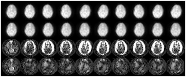
Ten out of 32 navigator images obtained from consecutive acquisitions (32 interleaves). Top row: Navigator images before coregistration. Second row: Aligned navigator images after each image was registered to an average image computed from all 32 navigators. Third row: Absolute-squared-differences images (individual navigator images before registration vs. reference image). Spatial misregistration is clearly apparent from the hyperintense rim at the edge of the brain. The intensity scaling is increased 10-fold compared to the two rows to better highlight the differences. Bottom row: Absolute-squared-differences images (individual navigator images after registration vs. reference image) clearly demonstrate a better alignment. The scaling is identical to that of the images in the third row.
DISCUSSION
Patient motion can significantly reduce the diagnostic quality of MR studies. Often the condition of the patient precludes him or her from being cooperative or remaining still for the duration of the examination. This is especially true for longer examination periods, and the likelihood that the patient will become uncomfortable and begin to move increases as the scan progresses. This in turn can require repeated examinations and lead to overly prolonged studies. In the pediatric setting, often sedation or anesthesia is needed to avoid motion artifacts, which leads to increased logistic problems and higher examination costs. It is therefore of great relevance to reduce the degree of motion artifacts in MRI scans. In the past several years, several attempts have been made to minimize artifacts from patient motion. These correction approaches have been carried out either prospectively or retrospectively (6,7,42,43).
In this study we propose a variant of parallel imaging reconstruction for arbitrary k-space acquisitions that allows for efficient retrospective in-plane motion correction. An excellent review of the potential of parallel imaging to correct for motion was presented recently by Larkman et al. (1). The current work is somewhat similar to the studies of Atkinson and Hill (4), Batchelor et al. (27), and Pipe et al. (2). The novelty of our approach is that it provides 1) a framework for motion correction that can be applied to arbitrarily sampled k-space data; 2) the capacity to overcome local k-space undersampling and different sampling densities by improving the conditioning of the inverse problem by means of additional spatial encoding information from coil sensitivities; 3) a rapid translation correction by the proper phasing of each k-space sample point and rotation correction by applying a rotation matrix to each k-space vector k(ti) that is part of the gridding step; and 4) the extraction of motion information from either a navigator scheme built into the trajectory itself or an additional navigator image. This new method relies heavily on the pioneering work of Pruessmann et al. (22) and Kannengiesser et al. (44) regarding the fast reconstruction of undersampled, non-Cartesian k-space data. A related iterative approach for motion correction was recently proposed by Batchelor et al. (27), although they did not capitalize on the additional encoding capabilities from coil sensitivities to improve the conditioning of the inverse problem. Potential future developments include finding alternative ways to reconstruct the data by synthesizing missing k-space parts using information from adjacent points in k-space, such as GRAPPA-based methods (17,45).
A comparative evaluation of image quality for motion-corrupted data before and after correction for rigid body motion was performed both in vivo and using computer simulations. The proposed rigid body motion correction (i.e., correcting k-space data for rotation and translation) with conventional gridding reconstruction was able to reduce artifacts to some extent; however, considerable residual artifacts were still apparent, due primarily to local violation of the Nyquist sampling criterion in k-space. With the use of augmented iterative parallel imaging reconstruction, the level of residual artifacts was significantly reduced. The amount of motion simulated in this study was fairly large (±30° rotation and ±15 mm translation), but was selected to cover the typical amounts of motion encountered in clinical studies.
Exact knowledge of the rotation and translation components is crucial for any motion-correction scheme to be able to fully correct image artifacts. Like many other correction schemes, the efficacy of our method strongly depends on the ability of the navigator image and the coregistration technique to pick up on small amounts of motion in vivo. However, for the navigated interleaved spiral trajectories that we tested in vivo, the small residual motion errors did not cause any significant residual artifacts. If the residual motion error increases, mismatch will first be noticeable as a slight blurring in the corrected image and/or by “swirl artifacts.” Conversely, the great advantage of low-resolution navigator images over higher resolution images is that the coregistration is much more robust (i.e., the parameter search is not so easily trapped in the local optima of the cost function, and the smaller image size allows very rapid registration). In assessing the influence of navigator resolution on the registration quality, we found that the fluctuations resulting from the registration were in the submillimeter range. A potential difficulty for the registration step is the variable coil sensitivity modulation that affects the image intensity. These additional signal intensity modulations may reduce the effectiveness of traditional similarity measures, and this effect needs to be investigated further. To minimize this confounder, we first intensity-corrected the navigators. In future studies, similarity measures that are known to be more insensitive to differences in image contrast, such as (normalized) mutual information (30,31,37), may be more suitable than the Pearson correlation coefficient. However, in this study we did not find that the choice of the similarity measure had any influence on the registration, regardless of whether Pearson correlation or normalized mutual information was used.
The motion-correction scheme presented here can also be used in conjunction with parallel imaging for scan acceleration. There is essentially no difference between undersampled k-space resulting from conventional parallel imaging and that resulting from local sampling density variations caused by reorganizing k-space for motion correction. The degree to which aliasing can be avoided is determined by the amount of net regional undersampling in k-space. In other words, with increasing data reduction as a result of parallel imaging, the method’s ability to correct for motion diminishes. If more emphasis is to be placed on reducing severe motion, the parallel imaging reduction factor has to be derated accordingly. The ability of the reconstruction ultimately depends on the number of coils used, the geometry factor, and the general conditioning of the design matrix.
One of the shortcomings of the proposed method is its inability to correct for through-plane motion. We found that spin history effects can reduce the quality of the reconstruction, although the final outcome is still significantly better than that achieved without any correction. Ultimately, spin history effects will always challenge 2D acquisition techniques that are exposed to potential movement with higher degrees of motional freedom. Nevertheless, in this work our goal was to investigate the possibility of correcting motion and spatiotemporal coil sensitivity variations using an augmented generalized parallel imaging reconstruction method. This method, in its general form, is expressed as a 3D problem with six degrees of freedom. The application of this technique to 3D acquisitions should also improve the quality of 3D scans, and further work on retrieving 3D motion information in a rapid fashion is warranted. It is important to note that even with prospective motion correction, and regardless of whether Cartesian or arbitrary k-space acquisitions are used, modifications to the original sensitivity information (i.e., translation and rotation) might be required because the patient will be exposed to different coil sensitivities during the course of motion. Therefore, there is a chance that conventional Cartesian parallel imaging algorithms or standard iterative SENSE algorithms do not work properly even when the scan plane can follow the movement of the object.
Although we initially demonstrated the navigator image technique in this study by using a SE approach, we also applied the navigator scheme to regular GRE scans. The limitation is that the minimum TE for the pulse sequence is delayed by a few milliseconds to accommodate the short spiral-IN navigator image. If necessary, and if T2* decay permits, the navigator signal can also be acquired at the end of the spiral-OUT train (i.e., after the interleaf is acquired that contributes to the high-resolution image). For SEs the spiral-IN navigator is particularly useful because it takes advantage of the high SNR available as the signal increases prior to the SE formation. As shown in Fig. 3a and b, a variable-density spiral-out scan can be used as well. The image quality of variable-density spiral scans is affected somewhat more by off-resonance effects and signal decay compared to conventional, constant-pitch spirals, and it depends on the pitch factor and overall readout length. Nevertheless, this trajectory has great potential because of its inherent navigation capabilities, and is a viable alternative that has been used successfully for self-navigated DTI (23,29). Because a spiral readout is used, the overall image quality of our sequence is slightly impaired by the T2* decay during the readout, but one could easily improve this by increasing the number of interleaves. The spiral-IN navigator and the parallel imaging-based correction scheme can be also combined with conventional FSE readouts.
CONCLUSIONS
We have proposed a new motion-correction scheme and pulse sequence that takes advantage of the new parallel imaging techniques. Our motion-correction scheme capitalizes on the ability of parallel imaging methods to correct for missing data in k-space. The amount of motion-induced image displacement and rotation is determined by means of registration algorithms applied to navigator images, and can be included in the iterative parallel imaging reconstruction. An appropriate navigator image can be produced with either a variable-density spiral waveform or an additional low-resolution, single-shot spiral scan prior to regular image formation. This new correction approach may avoid the well-known shortcomings of regular Cartesian encoding in the presence of patient motion, and may become a helpful asset for correcting such artifacts, especially in studies of vulnerable subjects. A variant of this method could also be used to allow parallel imaging in prospectively motion-corrected studies.
Acknowledgments
Grant sponsor: National Institutes of Health; Grant numbers: 1R01EB002711; 1R01NS047607; 5R01NS34866; Grant sponsor: Center of Advanced MR Technology at Stanford; Grant number: P41RR09784; Grant sponsors: Lucas Foundation; Oak Foundation.
We thank Dr. Gary Glover for providing us with his spiral pulse sequence as a basis for our experiments, and for his tireless support and mentorship. We also thank Marcus T. Alley, Ph.D., and Julie Ruiz, Ph.D., for editing the manuscript.
Footnotes
Presented in part at the 14th Annual Meeting of ISMRM, Seattle, WA, USA, 2006.
References
- 1.Larkman DJ, Atkinson D, Hajnal JV. Artifact reduction using parallel imaging methods. Top Magn Reson Imaging. 2004;15:267–275. doi: 10.1097/01.rmr.0000143782.39690.8a. [DOI] [PubMed] [Google Scholar]
- 2.Pipe JG, Farthing VG, Forbes KP. Multishot diffusion-weighted FSE using PROPELLER MRI. Magn Reson Med. 2002;47:42–52. doi: 10.1002/mrm.10014. [DOI] [PubMed] [Google Scholar]
- 3.Bydder M, Larkman DJ, Hajnal JV. Detection and elimination of motion artifacts by regeneration of k-space. Magn Reson Med. 2002;47:677– 686. doi: 10.1002/mrm.10093. [DOI] [PubMed] [Google Scholar]
- 4.Atkinson D, Hill DL. Reconstruction after rotational motion. Magn Reson Med. 2003;49:183–187. doi: 10.1002/mrm.10333. [DOI] [PubMed] [Google Scholar]
- 5.Bydder M, Atkinson D, Larkman DJ, Hill DL, Hajnal JV. SMASH navigators. Magn Reson Med. 2003;49:493–500. doi: 10.1002/mrm.10388. [DOI] [PubMed] [Google Scholar]
- 6.Thesen S, Heid O, Mueller E, Schad LR. Prospective acquisition correction for head motion with image-based tracking for real-time fMRI. Magn Reson Med. 2000;44:457– 465. doi: 10.1002/1522-2594(200009)44:3<457::aid-mrm17>3.0.co;2-r. [DOI] [PubMed] [Google Scholar]
- 7.Ward HA, Riederer SJ, Grimm RC, Ehman RL, Felmlee JP, Jack CR., Jr Prospective multiaxial motion correction for fMRI. Magn Reson Med. 2000;43:459– 469. doi: 10.1002/(sici)1522-2594(200003)43:3<459::aid-mrm19>3.0.co;2-1. [DOI] [PubMed] [Google Scholar]
- 8.Gonzales R, Woods R. Digital image processing. Reading, MA: Addison-Wesley Publishing Company; 1993. [Google Scholar]
- 9.Hutchinson M, Raff U. Fast MRI data acquisition using multiple detectors. Magn Reson Med. 1988;6:87–91. doi: 10.1002/mrm.1910060110. [DOI] [PubMed] [Google Scholar]
- 10.Ra JB, Rim CY. Fast imaging using subencoding data sets from multiple detectors. Magn Reson Med. 1993;30:142–145. doi: 10.1002/mrm.1910300123. [DOI] [PubMed] [Google Scholar]
- 11.Zhu Y, Hardy CJ, Sodickson DK, Giaquinto RO, Dumoulin CL, Ken-wood G, Niendorf T, Lejay H, McKenzie CA, Ohliger MA, Rofsky NM. Highly parallel volumetric imaging with a 32-element RF coil array. Magn Reson Med. 2004;52:869– 877. doi: 10.1002/mrm.20209. [DOI] [PMC free article] [PubMed] [Google Scholar]
- 12.Weiger M, Pruessmann KP, Boesiger P. 2D SENSE for faster 3D MRI. MAGMA. 2002;14:10–19. doi: 10.1007/BF02668182. [DOI] [PubMed] [Google Scholar]
- 13.Jakob PM, Griswold MA, Edelman RR, Manning WJ, Sodickson DK. Accelerated cardiac imaging using the SMASH technique. J Cardiovasc Magn Reson. 1999:1. doi: 10.3109/10976649909080844. [DOI] [PubMed] [Google Scholar]
- 14.Griswold MA, Jakob PM, Chen Q, Goldfarb JW, Manning WJ, Edelman RR, Sodickson DK. Resolution enhancement in single-shot imaging using simultaneous acquisition of spatial harmonics (SMASH) Magn Reson Med. 1999;41:1236–1245. doi: 10.1002/(sici)1522-2594(199906)41:6<1236::aid-mrm21>3.0.co;2-t. [DOI] [PubMed] [Google Scholar]
- 15.Bammer R, Auer M, Keeling SL, Augustin M, Stables LA, Prokesch RW, Stollberger R, Moseley ME, Fazekas F. Diffusion tensor imaging using single-shot SENSE-EPI. Magn Reson Med. 2002;48:128–136. doi: 10.1002/mrm.10184. [DOI] [PubMed] [Google Scholar]
- 16.Sodickson DK, Manning WJ. Simultaneous acquisition of spatial harmonics (SMASH): fast imaging with radiofrequency coil arrays. Magn Reson Med. 1997;38:591– 603. doi: 10.1002/mrm.1910380414. [DOI] [PubMed] [Google Scholar]
- 17.Griswold MA, Jakob PM, Heidemann RM, Nittka M, Jellus V, Wang J, Kiefer B, Haase A. Generalized autocalibrating partially parallel acquisitions (GRAPPA) Magn Reson Med. 2002;47:1202–1210. doi: 10.1002/mrm.10171. [DOI] [PubMed] [Google Scholar]
- 18.Pruessmann KP, Weiger M, Scheidegger MB, Boesiger P. SENSE: sensitivity encoding for fast MRI. Magn Reson Med. 1999;42:952–962. [PubMed] [Google Scholar]
- 19.Jackson JI, Meyer CH, Nishimura DH, Macovski A. Selection of a convolution function for fourier inversion using gridding. IEEE Trans Med Imaging. 1991;10:473– 478. doi: 10.1109/42.97598. [DOI] [PubMed] [Google Scholar]
- 20.Rasche V, Proksa R, Sinkus R, Bornert P, Eggers H. Resampling of data between arbitrary grids using convolution interpolation. IEEE Trans Med Imaging. 1999;18:385–392. doi: 10.1109/42.774166. [DOI] [PubMed] [Google Scholar]
- 21.Golub GH, Van Loan CF. Matrix computations. Baltimore, MD: Johns Hopkins University Press; 1983. [Google Scholar]
- 22.Pruessmann KP, Weiger M, Bornert P, Boesiger P. Advances in sensitivity encoding with arbitrary k-space trajectories. Magn Reson Med. 2001;46:638– 651. doi: 10.1002/mrm.1241. [DOI] [PubMed] [Google Scholar]
- 23.Liu C, Moseley ME, Bammer R. Simultaneous phase correction and SENSE reconstruction for navigated multi-shot DWI with non-Cartesian k-space sampling. Magn Reson Med. 2005;54:1412–1422. doi: 10.1002/mrm.20706. [DOI] [PubMed] [Google Scholar]
- 24.Wajer FTA, Pruessmann KP. Major speedup of reconstruction for sensitivity encoding with arbitrary trajectories. Proceedings of the 9th Annual Meeting of ISMRM; Glasgow, Scotland. 2001. p. Abstract 767. [Google Scholar]
- 25.Frigo M, Johnson S. The design and implementation of FFTW3. 2005. pp. 216–231. [Google Scholar]
- 26.Eggers H, Boernert P, Boesiger P. Comparison of gridding- and convolution-based iterative reconstruction algorithms for sensitivity-encoded non-Cartesian acquisitions. Proceedings of the 10th Annual Meeting of ISMRM; Honolulu, HI, USA. 2002. p. Abstract 743. [Google Scholar]
- 27.Batchelor PG, Atkinson D, Irarrazaval P, Hill DL, Hajnal J, Larkman D. Matrix description of general motion correction applied to multishot images. Magn Reson Med. 2005;54:1273–1280. doi: 10.1002/mrm.20656. [DOI] [PubMed] [Google Scholar]
- 28.Kim DH, Adalsteinsson E, Spielman DM. Simple analytic variable density spiral design. Magn Reson Med. 2003;50:214–219. doi: 10.1002/mrm.10493. [DOI] [PubMed] [Google Scholar]
- 29.Liu C, Bammer R, Kim DH, Moseley ME. Self-navigated interleaved spiral (SNAILS): application to high-resolution diffusion tensor imaging. Magn Reson Med. 2004;52:1388–1396. doi: 10.1002/mrm.20288. [DOI] [PubMed] [Google Scholar]
- 30.Wells WM, 3rd, Viola P, Atsumi H, Nakajima S, Kikinis R. Multi-modal volume registration by maximization of mutual information. Med Image Anal. 1996;1:35–51. doi: 10.1016/s1361-8415(01)80004-9. [DOI] [PubMed] [Google Scholar]
- 31.Studholme C, Constable RT, Duncan JS. Accurate alignment of functional EPI data to anatomical MRI using a physics-based distortion model. IEEE Trans Med Imaging. 2000;19:1115–1127. doi: 10.1109/42.896788. [DOI] [PubMed] [Google Scholar]
- 32.Woods RP, Grafton ST, Holmes CJ, Cherry SR, Mazziotta JC. Automated image registration: I. General methods and intrasubject, intramodality validation. J Comput Assist Tomogr. 1998;22:139–152. doi: 10.1097/00004728-199801000-00027. [DOI] [PubMed] [Google Scholar]
- 33.Ashburner J, Friston KJ. Voxel-based morphometry—the methods. Neuroimage. 2000;11(Pt 1):805–821. doi: 10.1006/nimg.2000.0582. [DOI] [PubMed] [Google Scholar]
- 34.Press W, Teukolsky S, Vetterling W, Flannery B. Numerical recipes in C. Cambridge, MA: Cambridge University Press; 1992. [Google Scholar]
- 35.Friston KJ, Ashburner J, Frith C, Poline JB, Heather JD, Frackowiak R. Spatial registration and normalization of images. Hum Brain Mapp. 1995;2:165–189. [Google Scholar]
- 36.Park S, Schowengerdt R. Image reconstruction by parametric cubic convolution. Comput Vis Graph Image Process. 1983;23:258–272. [Google Scholar]
- 37.Pluim JP, Maintz JB, Viergever MA. Mutual-information-based registration of medical images: a survey. IEEE Trans Med Imaging. 2003;22:986–1004. doi: 10.1109/TMI.2003.815867. [DOI] [PubMed] [Google Scholar]
- 38.Lempel A, Ziv J. On the complexity of finite sequences. IEEE Trans Inform Theory. 1976;22:75– 81. [Google Scholar]
- 39.Aksoy M, Liu C, Moseley ME, Bammer R. A self-navigated spiral in & out pulse sequence design for retrospective motion correction. Proceedings of the 14th Annual Meeting of ISMRM; Seattle, WA, USA. p. Abstract 3197. [Google Scholar]
- 40.Glover GH, Lai S. Self-navigated spiral fMRI: interleaved versus single-shot. Magn Reson Med. 1998;39:361–368. doi: 10.1002/mrm.1910390305. [DOI] [PubMed] [Google Scholar]
- 41.Bammer R, Auer M. Correction of eddy-current induced image warping in diffusion-weighted single-shot EPI using constrained non-rigid mutual information image registration. Proceedings of the 9th Annual Meeting of ISMRM; Glasgow, Scotland. 2001. p. Abstract 508. [Google Scholar]
- 42.Pipe JG. Motion correction with PROPELLER MRI: application to head motion and free-breathing cardiac imaging. Magn Reson Med. 1999;42:963–969. doi: 10.1002/(sici)1522-2594(199911)42:5<963::aid-mrm17>3.0.co;2-l. [DOI] [PubMed] [Google Scholar]
- 43.Zech CJ, Herrmann KA, Huber A, Dietrich O, Stemmer A, Herzog P, Reiser MF, Schoenberg SO. High-resolution MR-imaging of the liver with T2-weighted sequences using integrated parallel imaging: comparison of prospective motion correction and respiratory triggering. J Magn Reson Imaging. 2004;20:443– 450. doi: 10.1002/jmri.20127. [DOI] [PubMed] [Google Scholar]
- 44.Kannengiesser SAR, Brenner AR, Noll TG. Accelerated image reconstruction for sensitivity encoded imaging with arbitrary k-space trajectories. Proceedings of the 8th Annual Meeting of ISMRM; Denver, CO, USA. 2000. p. Abstract 155. [Google Scholar]
- 45.Griswold M, Heidemann R, Jakob P. Direct parallel imaging reconstruction of radially sampled data using GRAPPA with relative shifts. Proceedings of the 11th Annual Meeting of ISMRM; Toronto, Canada. 2003. p. Abstract 2349. [Google Scholar]