


Figure 6. Ubiquitination of PSD-95 Regulates Endocytosis of AMPA Receptors.
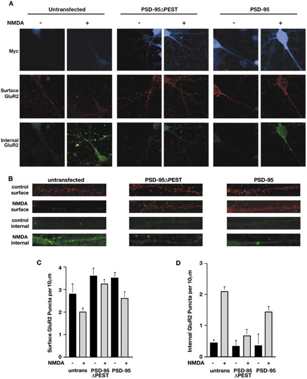
(A) (Top row) An antibody feeding technique was used to visualize endocytosed GluR2 in untransfected neurons (blue; left panels), neurons transfected with myc-PSD-95ΔPEST (blue; middle panels), or myc-tagged PSD-95 (blue; right panels). (Middle row) In untransfected neurons, NMDA treatment causes a loss of surface GluR2 staining (red; +; left panels). NMDA-induced loss of surface GluR2 is largely blocked in PSD-95ΔPEST-expressing neurons (red; middle panels) and blunted in neurons expressing wild-type PSD-95 (red; right panels). (Bottom row) In untransfected neurons (−; left panels), there is scant intracellular GluR2 staining (green). NMDA treatment (20 μM, 3 min) causes internalization of GluR2 (green; +; left panels). NMDA-induced GluR2 endocytosis was blocked in neurons expressing PSD-95ΔPEST (green; middle panels). NMDA-induced endocytosis of GluR2 was partially inhibited in neurons expressing wild-type PSD-95 (green; right panels). Background levels of myc antigen are faintly detected in the control neurons.
(B) Higher-magnification view of representative dendritic regions shows that in untransfected neurons (left column), NMDA treatment causes a loss of surface GluR2 (red) and a concomitant increase in intracellular GluR2 (green). In contrast, in neurons expressing PSD-95ΔPEST (middle column), the NMDA-induced loss of surface GluR2 (red) and increase in intracellular GluR2 (green) is largely attenuated. Both NMDA effects are blunted in neurons expressing wild-type PSD-95 (right columns).
(C) Quantification of the density of surface GluR2 puncta revealed untransfected, untreated control neurons had 2.6 ± 0.6 GluR2 puncta/10 μm (n = 16). Following NMDA treatment, untransfected neurons exhibited 1.9 ± 0.3 puncta per 10 μm (p < 0.02 versus control, n = 16). In neurons expressing PSD-95ΔPEST, GluR2 puncta density was 3.6 ± 0.4/10 μm (n = 15). NMDA treatment did not cause a significant decrease in density of surface GluR2 puncta (3.25 ± 0.2/10 μm, p > 0.09, n = 15). Neurons expressing wild-type PSD-95 had 3.5 ± 0.26 puncta/10 μm (n = 16). NMDA treatment reduced GluR2 puncta density to 2.6 ± 0.3/10 μm(p < 0.05, n = 16). Quanitatnion of data were performed using a double blind protocol.
(D) In untransfected neurons, NMDA treatment increased the density of internalized GluR2 puncta from 0.44 ± 0.1 puncta/10 μm to 2.1 ± 0.15 puncta/10 μm within 30 min (n = 16, p < 0.01). This NMDA-induced increase in internal GluR2 was blocked by transfection with PSD-95ΔPEST (control: internal GluR2 puncta was 0.33 ± 0.2 puncta/10 μm; n = 15; NMDA internal GluR2 density was 0.68 ± 0.17/10 μm; n = 15, p > 0.15). In neurons expressing wild-type PSD-95, NMDA had a reduced effect compared to controls (0.36 ± 0.37 puncta/10 μm in untreated cells versus 1.44 ± 0.16 puncta/10 μm in NMDA treated cells; n = 16, p < 0.05). Quantitation of data were performed using a double blind protocol.