


Fig. 4.
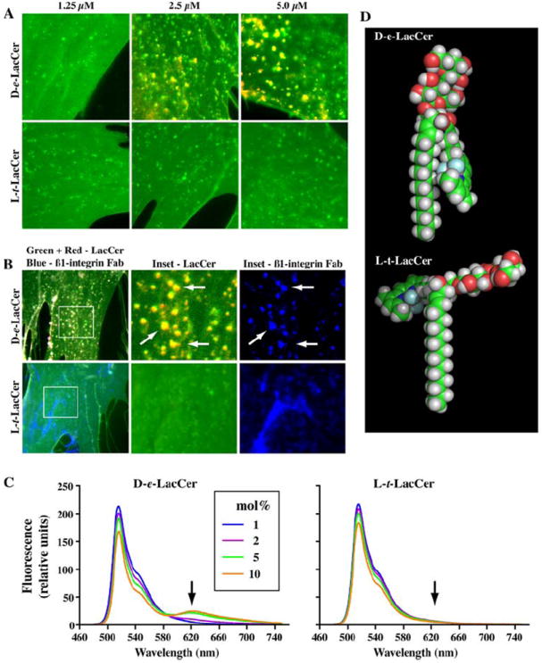
Comparison of d-erythro and l-threo stereoisomers of Bodipy-LacCer. (a) Human skin fibroblasts were incubated with the indicated concentration of Bodipy-d-erythro (d-e) or l-threo- (l-t) LacCer for 30 min at 10°C, washed, and observed under the fluorescence microscope at low temperature to inhibit endocytosis. Images were acquired simultaneously at green and red wavelengths, pseudocolored and digitally merged. Note the presence of yellow/orange micron size patches when Bodipy-d-erythro-LacCer was used at high concentrations. No such domains were observed using Bodipy-l-threo-LacCer. b Mapping endocytic markers to PM domains. HSFs were co-incubated with 5 μM Bodipy-d-erythro- or l-threo-LacCer and AF647-labeled anti-β1 integrin Fab. c Fluorescence emission spectra of small unilamellar vesicles formed from POPC and containing 1, 2, 5, or 10 mol % Bodipy-d-erythro-LacCer or -l-threo-LacCer. Note the presence of monomer (515 nm) and excimer (620 nm) fluorescence emission peaks when the d-erythro isomer was used at high mol % fractions, similar to Bodipy-Cer in Fig. 1b. In contrast, no excimer fluorescence was seen under the same conditions using the l-threo isomer. d Molecular models of Bodipy-LacCer stereoisomers created using the Chem3D program (Ultra 9.0, CambridgeSoft Corp., Cambridge, MA, USa). All data are from (Singh et al. 2006), reproduced with permission