



Summary
Objectives
A common experimental model of status epilepticus (SE) utilizes intraperitoneal administration of the cholinergic agonist pilocarpine preceded by methyl-scopolamine treatment. Currently, activation of cholinergic neurons is recognized as the only factor triggering pilocarpine SE. However, cholinergic receptors are also widely distributed systemically and pretreatment with methyl-scopolamine may not be sufficient to counteract the effects of systemically injected pilocarpine. The extent of such peripheral events and the contribution to SE are unknown and the possibility that pilocarpine also induces SE by peripheral actions is yet untested.
Methods
We measured in vivo at onset of SE: brain and blood pilocarpine levels, blood–brain barrier (BBB) permeability, T-lymphocyte activation and serum levels of IL-1β and TNF-α. The effects of pilocarpine on neuronal excitability was assessed in vitro on hippocampal slices or whole guinea pig brain preparations in presence of physiologic or elevated [K+]out.
Results
Pilocarpine blood and brain levels at SE were 1400 ± 200 μM and 200 ± 80 μM, respectively. In vivo, after pilocarpine injection, increased serum IL-1β, decreased CD4:CD8 T-lymphocyte ratios and focal BBB leakage were observed. In vitro, pilocarpine failed to exert significant synchronized epileptiform activity when applied at concentrations identical or higher to levels measured in vivo. Intense electrographic seizure-like events occurred only in the copresence of levels of K+ (6 mM) mimicking BBB leakage.
Conclusions
Early systemic events increasing BBB permeability may promote entry of cofactors (e. g. K+) into the brain leading to pilocarpine-induced SE. Disturbance of brain homeostasis represents an etiological factor contributing to pilocarpine seizures.
Keywords: Status epilepticus, Inflammation, Muscarinic receptors, Blood–brain barrier, Electrophysiology
The importance of cholinergic mechanisms in epilepsy has been demonstrated as early as in the nineteenth century (Cavalheiro et al., 2006). The cholinergic agonist pilocarpine has been extensively used experimentally to mimic status epilepticus (SE). Pilocarpine-treated animals are not only useful to study epileptogenesis but are also models of multiple drug resistance to antiepileptic drugs (Hoffmann et al., 2006). Pilocarpine acts on muscarinic receptors; both M1 and M2 receptors appear to be involved, but M1 receptors mediate most of the proepileptogenic actions (Bymaster et al., 2003). This CNS activation is currently believed to be the only factor initiating a cascade of events leading to hyperexcitability, apoptosis, and remodeling of the limbic system (Cavalheiro et al., 2006).
It is accepted as true that systemic effects induced by pilocarpine (300–350 mg/Kg, i.p.) are sufficiently antagonized by pretreatment with the peripherally-acting muscarinic antagonist methyl-scopolamine (0.5–1 mg/kg, also given i.p.) (Cavalheiro et al., 2006). This approach was meant to minimize the contribution of systemic cholinergic activation in favor of CNS effects. However, scarce information is available in support of this assumption. For example, at the concentrations of pilocarpine used to induce SE, a significant muscarinic blockade by methyl-scopolamine is virtually impossible (Dehaye et al., 1984; Wang et al., 1999). Understanding whether early peripheral events take place in spite of methyl-scopolamine pre-treatment may be futile if one only wishes to produce a valid animal model of limbic epilepsy. Nevertheless, it is important to fully understand how a peripherally administered cholinergic agonist may lead to SE, particularly in view of the fact that muscarinic receptors are widely distributed on heart muscle and sinoatrial node, blood vessels, white blood cells (WBC), and blood–brain barrier (BBB) endothelium.
Pilocarpine can potently affect peripheral organs, vessels, and circulating blood cells. Non-CNS effects of pilocarpine are observed at dosages much lower (1/20th) than those used to induce SE (Cook and Persinger, 1999). Very low dosages of peripherally administered pilocarpine (4 mg/kg) caused a substantial decrease in arterial pressure (Saad et al., 2003). Pilocarpine can also directly activate WBC, therefore decreasing the CD4/CD8 T-lymphocyte ratio (Arzt et al., 1989; Prync et al., 1992). These results show that at least in principle a possible extra-CNS target of peripherally administered pilocarpine may be the white blood cell muscarinic receptor. A peripheral cholinergic activation may be of importance when considering that a periphery-CNS interplay may favor the development of brain pathologies including seizures (Simard and Rivest, 2005).
Pilocarpine can be administered either peripherally or directly into the brain. In spite of the relatively high affinity of pilocarpine for CNS muscarinic receptors (5 nM, 0.2 μM and 30 μM for the high, intermediate and low affinity sites (Hedlund and Bartfai, 1981; Hedlund et al., 1982)), dosages well in excess of the predicted Km need to be administered to obtain a reliable development of seizures (Hamani and Mello, 1997). Direct intrahippocampal injections require 10 mM pilocarpine boluses, with the affinity constant for M1 receptors being several hundredfold less. In comparison, kainic acid induces seizures at concentrations that are close to the Km value for the target receptors (Tauck and Nadler, 1985; Rodrigues et al., 2005). The fact that activation of CNS receptors is not fully consistent with the dosage administered is also confounded by the fact that in vitro, pilocarpine can elicit electrographic seizure only if applied together with other epileptogenic factors (e. g., high extracellular K+ or low Mg++) (Rutecki and Yang, 1998; Avoli et al., 2002).
Given these premises and the overall relevance of pilocarpine as a model of SE and spontaneous seizures, we decided to further investigate whether early peripheral events occur in the time frame “pilocarpine injection–onset of SE”. The endpoints we studied include WBC activation, blood IL-1β and TNF-α levels, and BBB integrity. We also evaluated the brain permeation of pilocarpine and the efficacy of pilocarpine to elicit seizure-like activity when applied to naïve hippocampal neurons, under condition of physiological or high of extracellular K+.
Methods
EEG recording and video monitoring
With the exception of whole brain guinea pig experiments that were performed at the Neurological Institute Besta (Milan), all the experiments were carried out at the Cleveland Clinic, Lerner Research Institute. Stereotactic electrode implantation was performed under pentobarbital anaesthesia (45 mg/kg i.p.), using the Kopf stereotactic frame and a stereotactic atlas of the rat brain. For intrahippocampal recordings, bipolar twisted stainless wire electrodes (0.1 mm diameter, 0.5 mm vertical tip separation, Medwire, New York, New York, U.S.A.) are placed bilaterally in the dorsal hippocampus (LH: left hippocampus, RH: right hippocampus, A: −3.9 mm, L: ± 3.0 mm, D: −2.5 mm from bregma). Stainless steel screws (MX-0090–2, Small Parts Inc., Miami, FI, U.S.A.) were placed bilaterally on the dura mater of the frontal cortex (LC: left frontal cortex, RC: right frontal cortex, A: 1.0 mm, L: ± 2.5 mm from bregma). An additional screw electrode is placed in the frontal sinus and as a referential recording electrode. Rats were left unrestrained for 72 h for recovery from surgery before prolonged EEG recordings are performed. Each rat was kept in separate cage, under 12-h dark–light cycles, with free access to food and water. Digital EEG recordings (five channels per rat) were performed using two Vangard systems (Lamont, Madison, WI, U.S.A.). Each system consists of a recording Hewlett-Packard workstation and a similar review system connected to two 21-inch monitors. EEG data were sampled a rate of 100 Hz.
Animals and tissue sample preparation
Rats were housed in a controlled environment (21 ± 1°C; humidity 60%; lights on 08:00 a.m. – 8:00 p.m.; food and water available ad libitum). Procedures involving animals and their care were conducted in conformity with the institutional guidelines that are in compliance with international laws and policies (EEC Council Directive 86/609, OJ L 358, 1, Dec. 12, 1987; Guide for the Care and Use of Laboratory Animals, U. S. National Research Council, 1996). Rats (male Sprague-Dawley 200–250 g) were injected with methylscopolamine (1 mg/kg, i.p., Sigma-Aldrich, St. Louis, Mo, U.S.A.) 30 min before pilocarpine (350 mg/kg i.p.) to prevent peripheral cholinergic side effects. Animals (n = 6) were anesthetized (isofluorane) during recurrent motor seizures and at onset of SE as assessed by behavioral observation (stages 4 and 5 of Racine’s scale) and EEG recordings. Blood was withdrawn from the right heart ventricles; brain areas (hippocampus and entorhinal cortex) were resected and frozen. Serum samples were obtained after centrifugation at 1500×g for 10 min at 4 °C.
HPLC
Chemicals and reagents: pilocarpine hydrochloride was purchased from Sigma Aldrich. HPLC grade acetonitrile was purchased from Merck (Darmstadt, Germany). Milli-Q water (Millipore, Bedford, MA, U.S.A.) was used throughout. The other chemicals used were of analytical reagent grade. Chromatographic system and conditions: The chromatographic analysis was carried out according to a method previously described by Kuks et al. (1990). The HPLC system used was an Agilent 1100 s HPLC (Agilent, Santa Clara, CA, U.S.A.) consisting of a quaternary pump and a Diode Array Detector (DAD) set at wavelength of 220 nm. The analytical column was a microbondapak C18 3.9*300 mm from Waters (Milford, MA, U.S.A.). The system was run on isocratic gradient utilizing a mobile phase of 0.6% triethylamine in water (adjusted to pH 2.3 with 85% phosphoric acid) with a run time of 20 min. The chromatographic analysis was performed at room temperature with flow-rate of 1.0 ml/min and injection volume of 5 μl. Data collection and integration were performed by Agilent Chemstation Software.
Standard samples and calibration curves
Stock solutions (10 mM) of pilocarpine hydrochloride were freshly prepared in water and working solutions (0.1 nM–10 mM) were prepared by diluting stock solutions with water.
FACS and cytokine ELISA
Blood samples were withdrawn before development of SE (n = 6). For fluorescent-activated cell sorting (FACS) analysis, 150 μL of whole blood (or brain homogenized) were incubated with a combination of specific antibodies recognizing different population of WBC. In particular mouse anti-rat CD8 a-FITC, CD3-R-PE and CD4-PE-Cy5 (BD Pharmingen, San Diego, CA, U.S.A.) were added (2 μL, 5 μL, and 5 μL, respectively) to blood samples. Mouse anti-rat CD11b-FITC, CD3-R-PE, and CD45-PE-Cy5 were added (2μL, 5μL, and 5μL, respectively) for the determination of neutrophils/monocytes. Blood samples were analyzed by the FACS-Core facility at LRI. IL-1β, TNF-α, and IL-6 ELISA–tests (rats n = 6 control, n = 6 Stage IV, n = 6 SE) were purchased from Pierce Biotechnology Inc (Rockford, IL, U.S.A.) and performed as described by the vendor.
Slice electrophysiology
We used a total of 40 viable slices obtained from 10 rats. Slices are judged morphologically by looking at swollen cells and recovery of the same over time, and by detecting extracellularly evoked field potential recordings in response to stimulation of white matter. In most of the experiments performed for the purposes described here, CA1 activation was used (Janigro et al., 1997; D’Ambrosio et al., 1999). Rats were deeply anesthetized with isoflurane and then decapitated. The brain was quickly removed from the skull and immediately placed into ice-cold oxygenated (95% O2; 5% CO2) cutting solution, composed in mM of 2.5 KCl, 28 NaHCO3, 1.25 NaHPO4, 7 Dextrose, 7 MgCl2, 0.5 CaCl2, 235 sucrose, 1 ascorbic acid, 3 Na-Pyruvate. The brainstem and cerebellum were dissected out. The central section of the cerebral hemisphere containing somatosensory cortical regions and dorsal or dorsoventral hippocampi was subsequently cut in the coronal plane into slices of 450 μm thickness using a Vibratome 3000 (The Vibratome Co., St. Louis, MO, U.S.A.). Prior to recording, slices were kept at room temperature and pH 7.4 in a preincubation chamber containing oxygenated artificial cerebrospinal fluid (aCSF) of the composition (in mM) 120 NaCl, 3.1 KCl, 3 MgCl2, 26 NaHCO3, 1.25 KH2PO4, 10 Dextrose, 1 CaCl2, 1 ascorbic acid, 3 Na-Pyruvate. After 1 h in recovery, the slices were transferred to a chamber containing recording aCSF [(in mM) 120 NaCl, 3.1 KCl, 1 MgCl2, 26 NaHCO3, 1.25 KH2PO4, 10 Dextrose, 2 CaCl2 and allowed to equilibrate to the temperature achieved in the recording chamber. In the experiment at 6 mM K+ the aCSF had the following composition: [(in mM) 120 NaCl, 4.75 KCl, 1 MgCl2, 26 NaHCO3, 1.25 KH2PO4, 10 Dextrose, 2 CaCl2. For electrophysiological recordings, a slice was placed in a submerged recording chamber and constantly superfused with oxygenated recording aCSF at a flow rate of 2 ml/min at 32°C. Concentrations ranging from 10–500 μM of pilocarpine were dissolved into the recording aCSF and were, thus, applied to the slices. Extracellular field potentials (FP) were recorded using borosilicate glass micropipettes (0.5–2 MΩ) filled with aCSF.
In vitro guinea pig brain preparation
Brains were isolated from young adult Hartley guinea pigs (150–200 g; Charles River Laboratories, Calco, Italy) according to the standard technique described in detail elsewhere (de Curtis et al., 1998b; de Curtis et al., 1991). After induction of barbiturate anesthesia, the brain was carefully isolated and transferred to the incubation chamber. The basilary artery was cannulated with a polyethylene cannula to ensure arterial perfusion with a complex saline solution (composition: NaCl 126 mM, KCl 3 mM, KH2PO4 1.2 mM, MgSO4 1.3 mM, CaCl2 2.4 mM, NaHCO3 26 mM, glucose 15 mM, 3% dextran MW 70,000) oxygenated with a 95% O2; 5% CO2 gas mixture, pH 7.3. The arterial perfusion rate was 5.5 ml/min. Brain isolation was performed at low temperature (15 °C), and brain temperature was slowly increased to and maintained at 32 °C during the experiments. Adequate measures were taken to minimize animal pain and discomfort, in accordance with the International guidelines on animal research. The experimental protocol was approved by the Ethical Committee of the Istituto Nazionale Neurologico Besta in Milano. Extracellular recordings were simultaneously performed in the piriform cortex (PC), in the lateral and medial entorhinal cortex (EC) and in the CA1 region of the hippocampus with glass micropipettes filled with a 0.9% NaCl solution (2–5 MOhm input resistance). The functional viability of these cortical regions and the preservation of the BBB in the isolated brain preparation has been extensively demonstrated (de Curtis et al., 1998), (Librizzi et al., 2001, 2006). Bipolar silver stimulating electrodes positioned on the lateral olfactory tract were utilized to evoke potentials. Data were digitized and stored on a personal computer. The software for data acquisition and analysis (ELPHO) was developed by Dr. Vadym Gnatkovsky.
FITC-albumin solution composition
A recently developed microangiographic technique based on the injection of fluorescent probes enables confocal visualization of the entire brain micro- and macrovasculature (Cavaglia et al., 2001). Simultaneous observation of functional changes in BBB permeability is also possible if appropriate microangiographic molecules are employed. To this end, fluorescent albumin was used, since albumin does not extravasate abluminally if the BBB is intact (Cavaglia et al., 2001). The fluorescent albumin solution was prepared by reconstituting 500 mg of bovine desiccate albumin-fluorescein isothiocyanate (FITC-albumin, MW 69 KD; 12 mol/mol albumin; Sigma, St. Louis, MO, U.S.A.) in 50 ml of phosphate buffered saline (0.1M PBS) lacking magnesium and calcium ions. The FITC solution was stirred at room temperature in the dark for 5 min prior to injection. FITC albumin was directly infused in the left ventricle (Cavaglia et al., 2001). 10 ml of solution were injected at rate of 1 ml/min under general anesthesia. Presence of vascular leakage in pilocarpine-treated and control animals was evaluated by visual inspection and by confocal-microscopy (Carvey et al., 2005; Serduc et al., 2006; Yuan et al., 2006).
BBB permeability to radiotracers
A concentrated bolus of [14C] Diazepam and [3H] Sucrose (2 μC each) also containing 1 μM pilocarpine was injected into the left ventricle of an anesthetized animal that had been prepared as for FITC-albumin injection. The diffusion into the brain was monitored 15 min after the injection. Blood and brain samples containing samples of [14C] sucrose or [3 H] Diazepam were introduced into vials with 4 ml of Ready Protein Beckman scintillation cocktail (Packard Ultima Gold, ECN, Costa Mesa, CA, U.S.A.). Radioactivity was counted with an LS 6500 scintillation counter (Beckman).
Endothelial cells
The isolation and culture of rat brain microvascular endothelial cells has been described (Stanness et al., 1997). Brain tissue was obtained from 2 week old male Sprague-Dawley rats (20 g; six rats/experiment) anesthetized with 1% isoflurane (Abbott Labs, Chicago, IL, U.S.A.). Brain cortical tissue was homogenized, filtered through a 147-μm filter (Sefar America, Kansas City, MO, U.S.A.) and enzymaticaly processed with protease (50 μg/ml for 30 min) (Roche Diagnostics Corp., Indianapolis, IN, U.S.A.). Dextran (15%) (Sigma U.S.A.) was used to remove fat from the tissue prior to additional 2 h digestion with collagenase (100 μg/ml). Brain microvascular endothelial cells, separated by a Percoll gradient (1,580 g) were cultured in 75-cm2 tissue culture flasks precoated with fibronectin (3 μg/cm2) (Biomedical Technologies, Stoughton, MA, U.S.A.). Brain microvascular endothelial cells were characterized by the uptake of acetylated low-density lipoprotein labeled with DiI Ac-L-DL (Biomedical Technologies) and by von Willebrand factor staining, visualized by the Vector Stain Elite kit (Vector Labs, Burlingame, CA, U.S.A.). On day-7, when flasks were confluent, more than 96% of the cells were positive for DiI Ac-L-DL and von Willebrand factor. WBC were isolated as described (Krizanac-Bengez et al., 2006a, 2006b). Approximately 106 cells were added to each well.
Statistical analysis was performed with aid of Origin 7.0 (MicroCal, Northampton MA, U.S.A.); data were considered to be significantly different when p < 0.05 (by ANOVA).
Results
Seizures induced by pilocarpine are staged by the methods developed by Racine and modified by Gilbert (Bawden and Racine, 1979; McIntyre and Racine 1986; Gilbert, 1995). For the experiments described herein, staging of limbic seizures was performed by video EEG monitoring. The EEG patterns recorded prior to drug treatment, before SE (Stage IV) and during SE are shown in Fig. 1. Gilbert’s stage IV is characterized by prolonged myoclonic jerks and pronounced automatisms. At this stage, which usually occurred 20 min after the injection of pilocarpine, the EEG was characterized by spikes corresponding to myoclonic jerks, and progressive spread of activity from the hippocampal formation to the cortex (Fig. 1). This was followed by a transition period (indicated by Stage IV in the figure) followed by SE. We used a total of 41 pilocarpine-treated rats. Note that approximately 20 animals in the pilocarpine group were not used for further data analysis because they (1) did not develop EEG and behavioral changes consistent with Stage IV, or (2) they reached preconvulsive stages after more than 30 min. Thus, we report data from 21 animals experiencing Stage IV within 30 min after i.p. pilocarpine.
Figure 1.

Relationship between postpilocarpine stages and the experimental end points. EEG recordings from frontal cortex (LF–RF) and hippocampus (LH–RH) in rats exposed to 350 mg/kg of pilocarpine after pretreatment with methyl-scopolamine (1 mg/kg). Note the progressive synchronization and spread of epileptiform activity until Stage IV. Behavioral and electrographic seizures were observed approximately 30 min after pilocarpine (bottom trace). Note the different time scales in the bottom panels. Also note that all the data presented in this paper were obtained from either naive, or Stage I, or Stage IV animals, to emphasize progressive changes that always preceded SE.
Penetration of pilocarpine across the blood–brain barrier
We measured pilocarpine levels in rat serum and brain tissue following a systemic injection of 350 mg/kg. This was done either acutely (e. g., 2–3 min after the injection and in absence of behavioral modifications) or in correspondence of behavioral and EEG changes typical of Stage IV. The details of the HPLC procedure and calibration methods are described in the methods. In the acutely exposed group, rats were sacrificed 3 to 5 min after pilocarpine administration and labeled as “naive” in Fig. 2A. We evaluated the permeability of the BBB to the hydrophilic molecule 14C Sucrose (m. w. 324), the lipophilic tracer 3 H-diazepam (m. w. 284), and pilocarpine (m. w. 208). Brain pilocarpine levels were significantly lower than predicted by the lipophilicity of the drug (Fig. 2A). Unexpectedly, pilocarpine’s brain levels were on average similar to sucrose, and significantly lower than those for diazepam (n = 6) in spite of the significant difference in oil:water partition coefficients (Log P: −5.19, and 1.02 for sucrose and pilocarpine, respectively); Diazepam (Log P = 2.98) was, as expected, significantly more permeant than sucrose. Note that at approximately stage IV, brain levels of pilocarpine were significantly lower than those measured in serum (Fig. 2B), suggesting low BBB permeability even in presence of obvious behavioral and EEG changes. In conclusion, these results show that pilocarpine’s penetration across the BBB was significantly lower than expected based on its structural properties (lipophilicity and low molecular weight).
Figure 2.
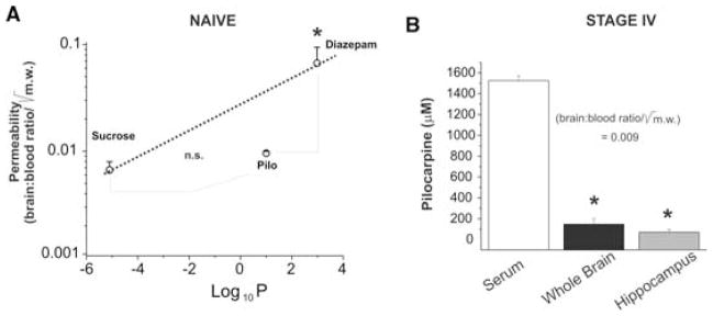
Modest pilocarpine permeability in naïve or Stage IV rats. (A) In naive animals (n = 3), pilocarpine permeability was significantly lower than predicted by its oil:water coefficient. Thus, pilocarpine permeability was similar to the paracellular pathway tracer sucrose. (B) Similar trans-BBB values were obtained in Stage IV animals in the regions indicated (n = 6). The asterisks indicate p < 0.05 by ANOVA; n. s., not significantly different.
In vitro effects of pilocarpine
The concentrations of pilocarpine found in rat brain at around stage IV were used in rat hippocampal slice experiments to assess their effects on CA1 pyramidal cell excitability (Fig. 3A). After establishment of a baseline field potential recording (usually 20–30 min), slices were exposed either individually or sequentially to pilocarpine concentrations ranging from 10 to 500 μM (n = 30 slices from seven rats). Specifically, 10 slices were exposed to increasing concentrations of pilocarpine (Fig. 3A). In an additional set of experiments, each slice was exposed to only one concentration of pilocarpine (Fig. 3B). A total of 20 slices (four slices each concentration) was used. Overall, in the 87% of slices tested, pilocarpine failed to exert any significant effect on spontaneous FP (e. g., traces in Figs. 3A and B). Seizure-like activity characterized by direct current (DC) shifts or repetitive bursting, interictal-like bursts, or depolarizing shifts were never observed regardless of the concentration of pilocarpine used. Modest increases in firing rate were observed in the remaining 13% slices. Individual slices were exposed to a single concentration of pilocarpine to rule out desensitization or habituation. Initial responses to pilocarpine were again minimal, but burst activity became more pronounced at steady state (usually 30 min of exposure, Fig. 3B). Again, no seizure-like events were detected in these experiments.
Figure 3.

In vitro electrophysiological recordings of pilocarpine’s action on rat hippocampal slices (A–B). C. Effect of 4 μM kainic acid perfused for 3 min via the arterial system in the guinea pig whole brain preparation (n = 5). Recordings were performed in the anterior and posterior piriform cortex (APC and PPC), in the medial and lateral entorhinal cortex (MERC and LERC) and in the CA1 subfield of the hippocampus. In C2, the effects of 800 μM pilocarpine for 30 min is shown. Note the different voltage calibrations in C and D. See text for details.
A possible explanation for pilocarpine’s lack of effect on CA1 firing may have depended on the deafferented nature of the slice preparation itself (see, for example, Avoli et al., 2000). To address this, we performed field potential recordings in the isolated guinea pig brain preparation perfused with high concentrations of pilocarpine (100–1000 μM). The effects of kainic acid, applied at 4 μM, were used as a positive control (Fig. 3C1). Note that while kainic acid produced the expected epileptiform effects on the brain, high dosages of pilocarpine produce low-voltage oscillations in the gamma range that resembled those seen in limbic structures after local application of carbachol (Fig. 3C2; (Dickson et al., 2003)). Seizure-like events were never observer under these conditions. Evoking seizures required millimolar concentrations of pilocarpine or manipulation of BBB permeability (data not shown; see (Uva et al., 2006)).
Peripheral actions of pilocarpine
Expression of muscarinic receptors in WBC (Costa et al., 1990; Rinner and Schauenstein, 1993; Cook and Persinger, 1999) may also have constituted a target for pilocarpine’s action after systemic administration. This was investigated by fluorescence-based analysis of immune cells collected from blood samples immediately after pilocarpine injection, before or during SE (Fig. 4). Note that, a significant decrease of CD4-expressing cells was observed shortly (within 30 min) after pilocarpine injection and prior to SE. At the same time point, we also measured the serum levels of the proinflammatory cytokine Il-1β (Fig. 5A). These were greatly elevated at early time points after pilocarpine injection and remained elevated throughout the experiment. TNF-α levels remained unaffected (Fig. 5B).
Figure 4.

Expression of muscarinic receptors in white blood cells may be an early event after pilocarpine exposure. The results shown were obtained by fluorescence-based analysis of immune cells collected from blood samples immediately after pilocarpine injection, before or during SE. Note that, a significant decrease of CD4-expressing cells was observed shortly (20–30 min) after pilocarpine injection and prior to SE (n = 6).
Figure 5.
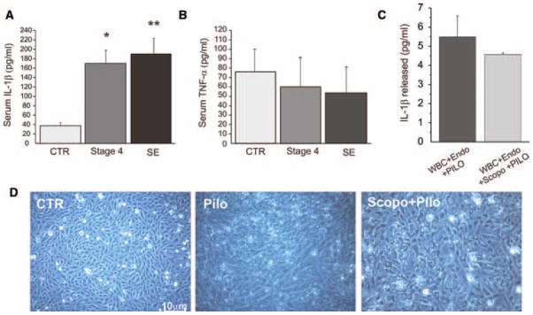
The proinflammatory mediator Il-1β is greatly elevated prior to pilocarpine-induced status epilepticus (A). In contrast, TNF-α levels were unaffected (B). The asterisk indicates p < 0.05, while the double asterisk indicates p < 0.02 (n = 6). Methyl-scopolamine fails to counterbalance the effects of pilocarpine in vitro. The concentration of pilocarpine used in these experiments was 1.5 mM to match the levels measured in rat serum prior to SE (shown in Fig. 2B). The concentration of methyl-scopolamine was 4 μM, again chosen to match the dosage of in vivo experiments. We allowed 45 min between exposure to the blocker and the pilocarpine challenge. The muscarinic antagonist methyl-scopolamine failed to prevent release of Il-1β by endothelial cells in presence of white blood cells (C). Similarly, the morphological alterations induced by pilocarpine were not affected by pretreatment with methyl-scopolamine. N = 6 wells per experimental data point.
In vitro effects of methyl-scopolamine
Since the experiments described so far were performed in animals pretreated with methyl-scopolamine, we tested its efficacy in preventing peripheral activation of muscarinic receptors in two separate experimental paradigms. For this purpose, we used the levels of pilocarpine measured in serum of animals (see Fig. 2B) and a concentration of scopolamine that was comparable to the one used in vivo (i.e., a 1/350 ratio against pilocarpine). We first exposed rat brain microvascular endothelial cells (Krizanac-Bengez et al., 2006a, 2006b) to pilocarpine in the presence or absence of methyl-scopolamine (Fig. 5C). These experiments were complemented by a set of trials where WBC were also added to the culture system (Fig. 5C). Regardless of the presence or absence of methyl-scopolamine, pilocarpine caused a significant release of Il-1β comparable in time course to what observed in vivo (Fig. 5A). We also measured the effects of methyl-scopolamine on cell morphology. To this end, we challenged endothelial cell cultures with pilocarpine exposures and compared the results to naive, time-matched cultures. Note that methyl-scopolamine failed to prevent the profound changes in cell morphology and membrane integrity induced by pilocarpine (Fig. 5D).
Pilocarpine’s effect on the blood-brain barrier
Since the data so far presented suggested that the CNS levels of pilocarpine that can be achieved after peripheral administration are not sufficient to cause seizures, we tested the hypothesis that other systemic changes may trigger neuronal hyperexcitability. We first tested the intactness of the endothelial cell BBB, a crucial determinant of homeostatic control of the neuronal microenvironment. At pilocarpine stage IV, that is, at the time serum and brain levels of pilocarpine were measured, and before the development of SE, BBB integrity was measured by comparing 14C-Sucrose permeability in naive versus pilocarpine treated rats. On average, 14C-Sucrose penetrated pilocarpine-treated rat brains in a fashion comparable to naïve brain (Fig. 6A). The permeability to sucrose was only slightly increased, suggesting little damage to the BBB at this stage. However, FITC extravasation analysis demonstrated a reproducible pattern of patchy leaks in pilocarpine-treated hippocampi before SE (Fig. 6B–D and Figs. 7 & 8). BBB leakage was observed in the frontal neocortex, hippocampus, EC, and dentate gyrus. Interestingly, neurons in pyramidal and granule cell layers were filled with FITC-albumin, suggesting avid uptake by these neurons (e. g., Fig. 7B1–2 and 8C).
Figure 6.

(A) Blood–brain barrier permeability for the hydrophilic low molecular weight tracer 14C-Sucrose is preserved after pilocarpine injection, but prior to development of SE. Note that, the brain:serum ratio in animals at stage IV was similar to the ratio measured in scopolamine-treated controls. In addition, numerous areas of hypoperfusion were seen (B), as demonstrated by the poor filling of regional vascular districts; these regions are highlighted by dashed lines. The blue fluorescent images were obtained by DAPI nuclear staining to show that the corresponding images were taken from approximately the same locations. As term of paragon, compare these micrographs with ischemia-reperfusion data presented elsewhere (Cavaglia et al., 2001). (C, D) The fluorescent protein-based microangiographic technique (in green) used to visualize regional breakdown of the blood–brain barrier revealed multiple small leaky spots in all limbic and parietal cortex regions measured.
Figure 7.

(A) BBB integrity is altered by pilocarpine. FITC extravasation was photographed in selected areas to demonstrate patchy leakage spots in pilocarpine-treated animals. The blue fluorescent images were obtained by DAPI nuclear staining to show that the corresponding images were taken from approximately the same locations. The bar corresponds to 750 μm. Neuronal uptake of FITC-albumin was observed in correspondence of area of leaky vessels (B1–B2).
Figure 8.

BBB integrity is altered by pilocarpine: neuronal cells take up the fluorescent albumin used for this study. (A) Examples of leaky spots in pre-SE animals. The left panel refers to a naive control. (B) Montage to show the entire hippocampal formation and the perihippocampal cortex. (C) Uptake of FITC-albumin was observed in pyramidal cell and granular cell layers. Note the overall of DAPI staining and the uptake of FITC signal.
A possible explanation of the differences found between Psucrose (little change) and extravasation of plasma protein may be due to the fact that while Psucrose was measured in whole brain homogenates, FITC leakage was assessed histologically. In other words, it is possible that hemodynamic changes may have accounted for a deficient perfusion of radioactive tracer (14C-Sucrose) thus leading to an underestimate of actual permeability. This was tested by reexamining FITC perfusion patterns in naïve versus stage IV animals (Fig. 6). Note that, the hippocampal formation and adjacent areas were dramatically hypoperfused, as demonstrated by lack of visible, FITC-albumin filled vessels. Taken together, these results suggest that pilocarpine caused two different cerebrovascular changes, one consisting of increased BBB permeability and the other related to hemodynamic changes (Saad et al., 2003).
Synergism between [K]out and pilocarpine
Since the results so far presented suggested little direct effects of pilocarpine on hippocampal neurons but increased BBB permeability, we tested the hypothesis that slightly elevated [K]out, as one may expect in the proximity of focal cerebrovascular leaks, may unveil proepileptogenic actions of muscarinic agonists (Fig. 9). As expected based on the work by Avoli et al. (2000) and Rutecki and Yang (1998), we found that even slight elevation of [K]out (to 6 mM) unmasked a powerful proepileptogenic effect of low concentrations of pilocarpine (50 μM). This effect was remarkable compared to what seen in low (4.35 mM) [K]out under identical experimental conditions (Fig. 9A versus B, 10 slices obtained from three rats).
Figure 9.

The epileptogenic effects of pilocarpine are revealed using hippocampal slice (n = 10) by concomitant increases of ACSF potassium to serum levels. (A) Under low (4.35 mM) [K]out, pilocarpine exerts little excitatory synchronous field effects at concentrations comparable to those measured in vivo brain (see Fig. 2B). However, when the same, or lower, concentrations are applied in presence of slightly elevated [K]out, pilocarpine acts as a potent generator of epileptiform discharges.
Discussion
The main finding of our study is that the mechanism of pilocarpine’s action as an epileptogenic agent may also include a peripheral site of action. Our findings suggest that activation of WBC leading to a cascade of events implicating BBB permeability may synergistically potentate direct CNS action of the drug leading to seizures. A novel finding of this work is that brain levels of pilocarpine found in rats at the time of initiation of electrographic seizures are significantly lower than those required for induction of hyperexcitability/synchronization in vitro under physiologic concentrations of [K]out. There are two more general implications that derive from these findings: (1) At least in principle, peripheral activation of the immune system may lead to changes in neuronal excitability; and (2) cholinergic agonists or inhibitors of acetylcholine cleavage may cause seizures in spite of poor penetration across the BBB.
Specifically, we found that increased serum levels of IL-1β, a decrease in CD4:CD8 T-lymphocyte ratio, and focal BBB leakage occur in vivo after pilocarpine injection and despite of methyl-scopolamine pretreatment. All these effects were observed prior to development of SE and could therefore not be due to generalized seizures. Pilocarpine blood and brain levels before SE were 1,400 ± 200 μM and 200 ± 80 μM, respectively, suggesting a poor CNS penetration. These results prompted us to formulate a hypothesis whereby pilocarpine exerts its epileptogenic effects also by peripheral activation of lymphocytes. This was supported by the finding that, in vitro, pilocarpine failed to induce any significant synchronized epileptiform activity at concentrations identical or higher to those measured in vivo. Intense electrographic seizure-like events in vitro occurred only in presence of levels of K+ (6 mM), mimicking BBB leakage.
Pilocarpine’s permeability across the blood–brain barrier
Our experiments show that pilocarpine does not efficiently permeate the BBB. Direct comparison with an impermeant and permeant compound (sucrose and diazepam) revealed that pilocarpine belongs to the low-permeation lipophilic agents, which are commonly extruded by multiple drug resistance proteins (Marroni et al., 2003). Comparison between pilocarpine and sucrose/diazepam was performed in control animals, leaving open the possibility that BBB permeability changes occurring after exposure of pilocarpine may have caused increased drug transfer into the brain. This was clearly not the case since pilocarpine did not significantly enter the brain at EEG Stage IV. It therefore appears that pilocarpine is a poor permeant across the BBB as also suggested by previous radiotracer studies (Omori et al., 2004).
Patchy leaky spots could be seen in the brain by FITC extravasation. It is thus possible that local concentrations of pilocarpine were slightly higher in those regions. However, while the brain levels of pilocarpine achieved appear to be low compared to dose initially injected intraperitoneally, our results cannot rule out the hypothesis of focally increased pilocarpine (Egorov et al., 2003). Under this scenario, initiation of seizures depends on BBB loss in regions highly sensitized to cholinergic agonists. Concomitantly and perhaps synergistically, failure of BBB protection could determine an alteration of the brain homeostasis thus facilitating the entry of blood-borne molecules/ions (e. g., K+) (Janigro, 1999; Grant and Janigro, 2004; Oby and Janigro, 2006). We have recently demonstrated that focal motor seizures can be triggered by BBB openings induced by intracarotid injection of a hyperosmolar bolus of mannitol (Marchi et al., 2007). In this study, BBB opening was achieved immediately after the first brain passage of the 1.4 M bolus of mannitol injected directly in the cerebral circulation. The probability of seizures occurrence was directly correlated with extent of BBB opening and seizures occurred minutes after a successful BBB opening. Recent evidences points to the same etiological role of BBB dysfunction in contributing to seizures occurrence (Ivens et al., 2007; van Vliet et al., 2007). However these results cannot be directly compared. For example, van Vliet and colleagues investigated the role of BBB integrity in the progression of temporal lobe epilepsy using a different experimental paradigm consisting of a systemic (and not intracarotid) administration of mannitol. Mannitol was injected at concentrations that are commonly used to prevent post-traumatic events including epileptogenesis. Ivens and colleagues identified a selective glial uptake of FITC-albumin after a chronic opening of the BBB. In our acute experiments, as well as in other animal models including the aforementioned van Vliet paper, albumin accumulated in neurons and glia. In spite of these minor discrepancies, our data suggest that early focal BBB damage observed in the pilocarpine model may contribute to the development of seizures allowing entrance into the brain of serum-derived factors.
Lack of efficacy with methyl-scopolamine pretreatment
Previous results by others (Dehaye et al., 1984; Wang et al., 1999) question the usefulness of methyl-scopolamine as blocker of peripheral actions by pilocarpine. In fact, the levels of pilocarpine used to induced SE, as well as those we measured in blood, are well in excess of those capable of fully displacing antagonist’s binding. This was confirmed by our in vitro studies that demonstrated that pilocarpine’s effects on WBC or endothelial cells are not significantly antagonized by methyl-scopolamine. This was observed in spite of a 1–2 h interval between administration of the antagonist and that of pilocarpine. This insufficient blockade by methyl-scopolamine may suggest that this pretreatment, while useful in decreasing mortality and in diminishing a number of behavioral events (e. g., body tremors, salivation, chromodacryorrhea, and diarrhea), may not be capable of fully counteracting the systemic effects of pilocarpine.
In vitro action of pilocarpine
Our electrophysiological evidence demonstrates that concentrations of pilocarpine close to the range of activation of muscarinic receptors or to those measured in vivo do not cause any considerable epileptiform activity at physiologic [K]out. In vitro seizure-like activity was obtained only in condition of elevated extracellular K+ (6 mM). Previous findings by others have indirectly implied the same, since seizure-like activity in vitro was obtained only when cofactors, such as high K+ were applied (Rutecki and Yang, 1998). In similar experiments but using the EC-hippocampal slice, low Mg++ was used to trigger seizure-like activity with pilocarpine (Massimo Avoli, personal communication and Avoli et al., 2000). The induction of epileptiform discharges by intraparenchymal, intraventricular, or intrahippocampal application of pilocarpine also required much greater amounts of the drug in view of the high affinity for muscarinic receptors (Dehaye et al., 1984; Leite et al., 2002; Cavalheiro et al., 2006).
Work by Alonso et al. demonstrated that the cholinergic agonist carbachol induces seizure-like in slices (Dickson and Alonso, 1997). For several reasons, these findings are not comparable to ours because a different agent has been used. For example, carbachol appeared to specifically target interneurons versus principal cells. Paulsen and colleagues (Fisahn et al., 1998) failed to reproduce these findings, and at the same concentration carbachol did not cause seizure-like activity in vitro. Interestingly, and consistent with our hypothesis, the only major difference between the two approaches is the fact that Alonso and colleagues used a slightly elevated concentration of potassium (5 mM) while when no epileptic activity was seen, the concentration of K+ was 3 mM. The latter concentration is comparable to the potassium present in CSF while the former is expected in the brain after BBB opening. Egorov et al., (Egorov et al., 2003) observed localized depolarizing effects of carbachol, but focal injections required 10 mM of the drug, clearly a much higher concentration than what was used by the authors in the other papers. Finally, Magistretti and colleagues did not observe significant field potential changes suggestive of epileptiform activity after exposure of brain slices to muscarinic agents in spite of elevated potassium concentrations (Magistretti et al., 2004). Thus, previously published data and our presented results strongly suggest that cofactors are required to produce cholinergic seizures.
A limitation of the slice approach for pharmacological studies is the use of largely nonphysiological conditions (Schwartzkroin and Prince, 1976). We thus expanded our studies to the in vitro whole brain preparation. This preparation allows the investigation of drug or circuitry effects under controlled conditions and functional preservation of the BBB that closely mimic what the in vivo/in situ condition (de Curtis et al., 1991; Muhlethaler et al., 1993; Librizzi et al., 1999, 2001, 2006). Application of extreme concentrations of pilocarpine (up to 1 mM) did not cause epileptiform activity, but rather induced fast oscillations in the gamma range that were comparable to those induced by activation of cholinergic synapses by carbachol (Fisahn et al., 1998; van Der et al., 1999; Dickson et al., 2003). The effect of pilocarpine on FP recorded in CA1 and entorhinal cortices following stimulation of the lateral olfactory tract in the isolated guinea pig brain were evaluated to overcome the limitations of the hippocampal slice preparation. Pilocarpine reduced the conditioned response in a pairing test and reduced the propagation of lateral olfactory tract (LOT)-evoke activity into CA1. This suggests that the muscarinic effect of pilocarpine is inhibitory on field activity. In addition, pilocarpine’s effect in the brain was limited to gamma rhythm, while seizure-like events were never observed at concentrations similar to those seen prior to SE in vivo (Uva et al., 2006).
A complementary mechanism of action of pilocarpine?
The fact that pilocarpine-induced activity can be triggered in condition of high extracellular K+ has been already demonstrated by others (Rutecki and Yang, 1998; Avoli et al., 2000). Increased extracellular K+ mimics the consequences of in vivo opening of the BBB and subsequent loss of brain homeostasis. What remains to be elucidated is the extent of K+ increase in the brain parenchyma after opening of the BBB. These changes are difficult to predict or model considering the buffering capacity of glial cells. We previously demonstrated that small, but significant changes in [K]out are sufficient to account for post-traumatic excitability changes (D’Ambrosio et al., 1998, 1999). Thus, it is possible that a similar change in [K]out occurred in pilocarpine brain since both traumatic brain injury and pilocarpine have a profound effect on BBB integrity (data presented herein and Schmidt and Grady, 1993).
Under nonphysiological conditions, the delicate balance between large depolarizing and hyperpolarizing potentials occurring in the hippocampus is lost, and synchronous, repetitive neuronal depolarizations may occur. While most studies have concentrated on epileptogenesis following failure of inhibitory potentials or exaggerated excitation, nonsynaptic contributions to epileptogenesis have been relatively neglected despite the fact that several features of abnormal neuronal hyperexcitability and synchronization can be understood by models which focus on inadequate K+ clearance, excessive accumulation mechanisms (Lux et al., 1986; Traynelis and Dingledine, 1988; Gabriel et al., 1998) and/or alterations in the size of the extracellular space. Elevated potassium, as seen after loss of brain homeostasis, is a potent convulsant (Traynelis and Dingledine, 1988) and also acts as a vasoactive agent capable of both dilation (at low [K]out) or constriction (Nguyen et al., 1998). There is a significant difference between brain and plasma [K]out, favoring potassium entry into the CNS when the BBB is breached (Keep et al., 1993; Grant and Janigro, 2004). Thus, if a secondary effect of pilocarpine is an inflammatory change in BBB permeability, even focal changes in [K]out may be responsible in part for the initiation of seizures. Of course other mechanisms can be involved, since many epileptogenic agents are more concentrated in serum than brain (e. g., glutamate (O’Kane et al., 1999; Grant et al., 2003)).
A less well-understood possibility is a direct action of inflammatory mediators, such as Il-1β, on neurons. Admittedly, attempts to tie interleukins to ion channels is in its infancy, but several reports have suggested that elevation of Il-1β may cause sudden rapid changes in excitability of both inhibitory and excitatory neurons (Plata-Salaman and Ffrench-Mullen, 1992; Sawada et al., 1992; Yang et al., 2005). Thus, focal entry of Il-1β via the para-cellular way left open by the initial proinflammatory insult by pilocarpine may constitute a possible contributor for pilocarpine seizures after systemic injection. If a peripheral inflammatory process contributes to pilocarpine-induced seizures then pretreatment with antiinflammatory drugs should protect from the onset of SE. The effect of non-steroidal antiinflammatory drugs on seizures elicited by pilocarpine was investigated (Ikonomidou-Turski et al., 1988). Interestingly, only mefenamic acid was able to reduce the severity of seizures while sodium salicylate and phenylbutazone exacerbated the effects of pilocarpine. These controversial results demonstrate the complexity of pilocarpine’s action but point to an inflammatory component.
Conclusions
Our results have demonstrated that pilocarpine may act by both peripheral activation of proinflammatory mechanisms and by direct action on CNS neurons. The former seems to be synergistically required to induce SE.
Acknowledgments
This work was supported by NIH 2RO1 HL51614, NIH RO1 NS43284, NIH RO1 NS38195, and NIH NS149514, and by a Mariani Foundation grant R 06–50.
References
- Arzt ES, Fernandez-Castelo S, Diaz A, Finkielman S, Nahmod VE. The muscarinic agonist pilocarpine inhibits DNA and interferon-gamma synthesis in peripheral blood mononuclear cells. Int J Immunopharmacol. 1989;11:275–281. doi: 10.1016/0192-0561(89)90165-3. [DOI] [PubMed] [Google Scholar]
- Avoli M, D’Antuono M, Louvel J, Kohling R, Biagini G, Pumain R, D’Arcangelo G, Tancredi V. Network and pharmacological mechanisms leading to epileptiform synchronization in the limbic system in vitro. Prog Neurobiol. 2002;68:167–207. doi: 10.1016/s0301-0082(02)00077-1. [DOI] [PubMed] [Google Scholar]
- Bawden HN, Racine RJ. Effects of bilateral kindling or bilateral sub-threshold stimulation of the amygdala or septum on muricide, ranacide, intraspecific aggression and passive avoidance in the rat. Physiol Behav. 1979;22:115–123. doi: 10.1016/0031-9384(79)90412-8. [DOI] [PubMed] [Google Scholar]
- Bymaster FP, Carter PA, Yamada M, Gomeza J, Wess J, Hamilton SE, Nathanson NM, McKinzie DL, Felder CC. Role of specific muscarinic receptor subtypes in cholinergic parasympathomimetic responses, in vivo phosphoinositide hydrolysis, and pilocarpine-induced seizure activity. Eur J Neurosci. 2003;17:1403–1410. doi: 10.1046/j.1460-9568.2003.02588.x. [DOI] [PubMed] [Google Scholar]
- Carvey PM, Zhao CH, Hendey B, Lum H, Trachtenberg J, Desai BS, Snyder J, Zhu YG, Ling ZD. 6-Hydroxydopamine-induced alterations in blood-brain barrier permeability. Eur J Neurosci. 2005;22:1158–1168. doi: 10.1111/j.1460-9568.2005.04281.x. [DOI] [PubMed] [Google Scholar]
- Cavaglia M, Dombrowski SM, Drazba J, Vasanji A, Bokesch PM, Janigro D. Regional variation in brain capillary density and vascular response to ischemia. Brain Res. 2001;910:81–93. doi: 10.1016/s0006-8993(01)02637-3. [DOI] [PubMed] [Google Scholar]
- Cavalheiro EA, Mello ML, Leite JP. The pilocarpine model of seizures. In: Pitkanen A, Schwartzkroin PA, Moshe SL, editors. Models of seizures and epilepsy. Elsevier; Burlington, MA: 2006. pp. 433–448. [Google Scholar]
- Cook LL, Persinger MA. Infiltration of lymphocytes in the limbic brain following stimulation of subclinical cellular immunity and low dosages of lithium and a cholinergic agent. Toxicol Lett. 1999;109:77–85. doi: 10.1016/s0378-4274(99)00123-x. [DOI] [PubMed] [Google Scholar]
- Costa LG, Kaylor G, Murphy SD. In vitro and in vivo modulation of cholinergic muscarinic receptors in rat lymphocytes and brain by cholinergic agents. Int J Immunopharmacol. 1990;12:67–75. doi: 10.1016/0192-0561(90)90069-y. [DOI] [PubMed] [Google Scholar]
- D’Ambrosio R, Maris DO, Grady MS, Winn HR, Janigro D. Selective loss of hippocampal long-term potentiation, but not depression, following fluid percussion injury. Brain Res. 1998;786:64–79. doi: 10.1016/s0006-8993(97)01412-1. [DOI] [PubMed] [Google Scholar]
- D’Ambrosio R, Maris DO, Grady MS, Winn HR, Janigro D. Impaired K homeostasis and altered electrophysiological properties of post-traumatic hippocampal glia. J Neurosci. 1999;19:8152–8162. doi: 10.1523/JNEUROSCI.19-18-08152.1999. [DOI] [PMC free article] [PubMed] [Google Scholar]
- de Curtis M, Pare D, Llinas RR. The electrophysiology of the olfactory-hippocampal circuit in the isolated and perfused adult mammalian brain in vitro. Hippocampus. 1991;1:341–354. doi: 10.1002/hipo.450010402. [DOI] [PubMed] [Google Scholar]
- de Curtis M, Biella G, Buccellati C, Folco G. Simultaneous investigation of the neuronal and vascular compartments in the guinea pig brain isolated in vitro. Brain Res Brain Res Protoc. 1998;3:221–228. doi: 10.1016/s1385-299x(98)00044-0. [DOI] [PubMed] [Google Scholar]
- Dehaye JP, Winand J, Poloczek P, Christophe J. Characterization of muscarinic cholinergic receptors on rat pancreatic acini by N-[3 H]methylscopolamine binding. Their relationship with calcium 45 efflux and amylase secretion. J Biol Chem. 1984;259:294–300. [PubMed] [Google Scholar]
- Dickson CT, Alonso A. Muscarinic induction of synchronous population activity in the entorhinal cortex. J Neurosci. 1997;17:6729–6744. doi: 10.1523/JNEUROSCI.17-17-06729.1997. [DOI] [PMC free article] [PubMed] [Google Scholar]
- Dickson CT, Biella G, de Curtis M. Slow periodic events and their transition to gamma oscillations in the entorhinal cortex of the isolated Guinea pig brain. J Neurophysiol. 2003;90:39–46. doi: 10.1152/jn.01063.2002. [DOI] [PubMed] [Google Scholar]
- Egorov AV, Angelova PR, Heinemann U, Muller W. Ca2+-independent muscarinic excitation of rat medial entorhinal cortex layer V neurons. Eur J Neurosci. 2003;18:3343–3351. doi: 10.1111/j.1460-9568.2003.03050.x. [DOI] [PubMed] [Google Scholar]
- Fisahn A, Pike FG, Buhl EH, Paulsen O. Cholinergic induction of network oscillations at 40 Hz in the hippocampus in vitro. Nature. 1998;394:186–189. doi: 10.1038/28179. [DOI] [PubMed] [Google Scholar]
- Gabriel S, Eilers A, Kivi A, Kovacs R, Schulze K, Lehmann TN, Heinemann U. Effects of barium on stimulus induced changes in extracellular potassium concentration in area CA1 of hippocampal slices from normal and pilocarpine-treated epileptic rats. Neurosci Lett. 1998;242:9–12. doi: 10.1016/s0304-3940(98)00012-3. [DOI] [PubMed] [Google Scholar]
- Gilbert ME. Repeated exposure to lindane leads to behavioral sensitization and facilitates electrical kindling. Neurotoxicol Teratol. 1995;17:131–141. doi: 10.1016/0892-0362(94)00064-k. [DOI] [PubMed] [Google Scholar]
- Grant GA, Janigro D. The blood-brain barrier. In: Winn HR, editor. Youmans Neurological Surgery. Saunders; Philadelphia, PA: 2004. pp. 153–174. [Google Scholar]
- Grant GA, Meno JR, Nguyen TS, Stanness KA, Janigro D, Winn RH. Adenosine-induced modulation of excitatory amino acid transport across isolated brain arterioles. J Neurosurg. 2003;98:554–560. doi: 10.3171/jns.2003.98.3.0554. [DOI] [PubMed] [Google Scholar]
- Hamani C, Mello LE. Status epilepticus induced by pilocarpine and picrotoxin. Epilepsy Res. 1997;28:73–82. doi: 10.1016/s0920-1211(97)00030-2. [DOI] [PubMed] [Google Scholar]
- Hedlund B, Bartfai T. Binding of [3 H]-pilocarpine to membranes from rat cerebral cortex. Naunyn Schmiedebergs Arch Pharmacol. 1981;317:126–130. doi: 10.1007/BF00500067. [DOI] [PubMed] [Google Scholar]
- Hedlund B, Grynfarb M, Bartfai T. Two ligands may bind simultaneously to the muscarine receptor. Naunyn Schmiedebergs Arch Pharmacol. 1982;320:3–13. doi: 10.1007/BF00499064. [DOI] [PubMed] [Google Scholar]
- Hoffmann K, Gastens AM, Volk HA, Loscher W. Expression of the multidrug transporter MRP2 in the blood-brain barrier after pilocarpine-induced seizures in rats. Epilepsy Res. 2006 doi: 10.1016/j.eplepsyres.2005.12.005. [DOI] [PubMed] [Google Scholar]
- Ikonomidou-Turski C, Cavalheiro EA, Turski L, Bortolotto ZA, Kleinrok Z, Calderazzo-Filho LS, Turski WA. Differential effects of non-steroidal anti-inflammatory drugs on seizures produced by pilocarpine in rats. Brain Res. 1988;462:275–285. doi: 10.1016/0006-8993(88)90556-2. [DOI] [PubMed] [Google Scholar]
- Ivens S, Kaufer D, Flores LP, Bechmann I, Zumsteg D, Tomkins O, Seiffert E, Heinemann U, Friedman A. TGF-beta receptor-mediated albumin uptake into astrocytes is involved in neocortical epileptogenesis. Brain. 2007;130:535–547. doi: 10.1093/brain/awl317. [DOI] [PubMed] [Google Scholar]
- Janigro D. Blood-brain barrier, ion homeostasis and epilepsy: possible implications towards the understanding of ketogenic diet mechanisms. Epilepsy Res. 1999;37:223–232. doi: 10.1016/s0920-1211(99)00074-1. [DOI] [PubMed] [Google Scholar]
- Janigro D, Gasparini S, D’Ambrosio R, McKhann G, DiFrancesco D. Reduction of K+ uptake in glia prevents long-term depression maintenance and causes epileptiform activity. J Neurosci. 1997;17:2813–2824. doi: 10.1523/JNEUROSCI.17-08-02813.1997. [DOI] [PMC free article] [PubMed] [Google Scholar]
- Keep RF, Xiang J, Betz AL. Potassium transport at the blood-brain and blood-CSF barriers. Adv Exp Med Biol. 1993;331:43–54. doi: 10.1007/978-1-4615-2920-0_8. [DOI] [PubMed] [Google Scholar]
- Krizanac-Bengez L, Hossain M, Fazio V, Mayberg M, Janigro D. Loss of flow induces leukocyte-mediated MMP/TIMP imbalance in dynamic in vitro blood-brain barrier model: role of pro-inflammatory cytokines. Am J Physiol Cell Physiol. 2006a;291:C740–C749. doi: 10.1152/ajpcell.00516.2005. [DOI] [PubMed] [Google Scholar]
- Krizanac-Bengez L, Mayberg MR, Cunningham E, Hossain M, Ponnampalam S, Parkinson FE, Janigro D. Loss of shear stress induces leukocyte-mediated cytokine release and blood-brain barrier failure in dynamic in vitro blood-brain barrier model. J Cell Physiol. 2006b;206:68–77. doi: 10.1002/jcp.20429. [DOI] [PubMed] [Google Scholar]
- Kuks PF, Weekers LE, Goldhoorn PB. Decomposition of pilocarpine eye drops assessed by a highly efficient high pressure liquid chromotographic method. Pharm Weekbl Sci. 1990;48:732–742. doi: 10.1007/BF01980046. [DOI] [PubMed] [Google Scholar]
- Leite JP, Garcia-Cairasco N, Cavalheiro EA. New insights from the use of pilocarpine and kainate models. Epilepsy Res. 2002;50:93–103. doi: 10.1016/s0920-1211(02)00072-4. [DOI] [PubMed] [Google Scholar]
- Librizzi L, Biella G, Cimino C, de Curtis M. Arterial supply of limbic structures in the guinea pig. J Comp Neurol. 1999;411:674–682. doi: 10.1002/(sici)1096-9861(19990906)411:4<674::aid-cne11>3.0.co;2-o. [DOI] [PubMed] [Google Scholar]
- Librizzi L, Janigro D, De Biasi S, de Curtis M. Blood-brain barrier preservation in the in vitro isolated guinea pig brain preparation. J Neurosci Res. 2001;66:289–297. doi: 10.1002/jnr.1223. [DOI] [PubMed] [Google Scholar]
- Librizzi L, Mazzetti S, Pastori C, Frigerio S, Salmaggi A, Buccellati C, Di Gennaro A, Folco G, Vitellaro-Zuccarello L, de Curtis M. Activation of cerebral endothelium is required for mononuclear cell recruitment in a novel in vitro model of brain inflammation. Neuroscience. 2006;137:1211–1219. doi: 10.1016/j.neuroscience.2005.10.041. [DOI] [PubMed] [Google Scholar]
- Lux HD, Heinemann U, Dietzel I. Ionic changes and alterations in the size of extracellular space during epileptic activity. In: Delgado-Escueta AV, Ward AA, editors. Advances in Neurology. Raven Press; New York: 1986. pp. 619–639. [PubMed] [Google Scholar]
- Magistretti J, Ma L, Shalinsky MH, Lin W, Klink R, Alonso A. Spike patterning by Ca2+-dependent regulation of a muscarinic cation current in entorhinal cortex layer II neurons. J Neurophysiol. 2004;92:1644–1657. doi: 10.1152/jn.00036.2004. [DOI] [PubMed] [Google Scholar]
- Marchi N, Angelov L, Masaryk T, Fazio V, Granata T, Hernandez N, Hallene K, Diglaw T, Franic L, Najm I, Janigro D. Seizure-promoting effect of blood-brain barrier disruption. Epilepsia. 2007 doi: 10.1111/j.1528-1167.2007.00988.x. [DOI] [PMC free article] [PubMed] [Google Scholar]
- Marroni M, Marchi N, Cucullo L, Abbott NJ, Signorelli K, Janigro D. Vascular and parenchymal mechanisms in multiple drug resistance: a lesson from human epilepsy. Curr Drug Targets. 2003;4:297–304. doi: 10.2174/1389450033491109. [DOI] [PubMed] [Google Scholar]
- McIntyre DC, Racine RJ. Kindling mechanisms: current progress on an experimental epilepsy model. Prog Neurobiol. 1986;27:1–12. doi: 10.1016/0301-0082(86)90010-9. [DOI] [PubMed] [Google Scholar]
- Muhlethaler M, de Curtis M, Walton K, Llin’as R. The isolated and perfused brain of the guinea-pig in vitro. European Journal of Neuroscience. 1993;5:915–926. doi: 10.1111/j.1460-9568.1993.tb00942.x. [DOI] [PubMed] [Google Scholar]
- O’Kane Rl, Martinez-Lopez I, DeJoseph MR, Vina JR, Hawkins RD. Na+-dependent Glutamate Transporters (EAAT1, EAAT2, and EAAT3) of the blood-brain barrier. J Biol Chem. 1999;274:31891–31985. doi: 10.1074/jbc.274.45.31891. [DOI] [PubMed] [Google Scholar]
- Oby E, Janigro D. The blood-brain barrier and epilepsy. Epilepsia. 2006;47:1761–1774. doi: 10.1111/j.1528-1167.2006.00817.x. [DOI] [PubMed] [Google Scholar]
- Omori Y, Endo T, Hara Y, Nishiyama M, Midgley I, Smart CI, John AJ, Chasseaud LF, McBurney A, John BA. Absorption, distribution and excretion of 14 C-pilocarpine following oral administration to rats. Arzneimittelforschung. 2004;54:171–178. doi: 10.1055/s-0031-1296955. [DOI] [PubMed] [Google Scholar]
- Plata-Salaman CR, Ffrench-Mullen JM. Interleukin-1 beta depresses calcium currents in CA1 hippocampal neurons at pathophysiological concentrations. Brain Res Bull. 1992;29:221–223. doi: 10.1016/0361-9230(92)90029-w. [DOI] [PubMed] [Google Scholar]
- Prync AE, Arzt E, Fernandez CS, Finkielman S, Nahmod V. The inhibitory effect of the muscarinic agonist pilocarpine on lymphocyte activation involves the IL-2 pathway and the increase in suppressor cell function. Int J Neurosci. 1992;62:277–285. doi: 10.3109/00207459108999780. [DOI] [PubMed] [Google Scholar]
- Rinner I, Schauenstein K. Detection of choline-acetyltransferase activity in lymphocytes. J Neurosci Res. 1993;35:188–191. doi: 10.1002/jnr.490350209. [DOI] [PubMed] [Google Scholar]
- Rodrigues MC, Rossetti F, Foresti ML, Arisi GM, Furtado MA, Dal-Col ML, Bertti P, Fernandes A, Santos FL, Del Vecchio F, Garcia-Cairasco N. Correlation between shaking behaviors and seizure severity in five animal models of convulsive seizures. Epilepsy Behav. 2005;6:328–336. doi: 10.1016/j.yebeh.2005.02.005. [DOI] [PubMed] [Google Scholar]
- Rutecki PA, Yang Y. Ictal epileptiform activity in the CA3 region of hippocampal slices produced by pilocarpine. Am Physiol Soc. 1998:3019–3029. doi: 10.1152/jn.1998.79.6.3019. [DOI] [PubMed] [Google Scholar]
- Saad WA, Guarda IF, Camargo LA, Santos TA, Guarda RS, Saad WA, Simoes S, Rodrigues JA. Role of nitric oxide of the median preoptic nucleus (MnPO) in the alterations of salivary flow, arterial pressure and heart rate induced by injection of pilocarpine into the MnPO and intraperitoneally. Braz J Med Biol Res. 2003;36:897–905. doi: 10.1590/s0100-879x2003000700010. [DOI] [PubMed] [Google Scholar]
- Sawada M, Hara N, Maeno T. Reduction of the acetylcholine-induced K+ current in identified Aplysia neurons by human interleukin-1 and interleukin-2. Cell Mol Neurobiol. 1992;12:439–445. doi: 10.1007/BF00711544. [DOI] [PMC free article] [PubMed] [Google Scholar]
- Schmidt RH, Grady MS. Regional patterns of blood-brain barrier breakdown following central and lateral fluid percussion injury in rodents. Journal of Neurotrauma. 1993;10:415–430. doi: 10.1089/neu.1993.10.415. [DOI] [PubMed] [Google Scholar]
- Schwartzkroin PA, Prince DA. Epileptogenesis in mammalian cortex studied in vitro. Trans Am Neurol Assoc. 1976;101:P288–P291. [PubMed] [Google Scholar]
- Serduc R, Verant P, Vial JC, Farion R, Rocas L, Remy C, Fadlallah T, Brauer E, Bravin A, Laissue J, Blattmann H, Van Der Sanden B. In vivo two-photon microscopy study of short-term effects of microbeam irradiation on normal mouse brain microvasculature. Int J Radiat Oncol Biol Phys. 2006;64:1519–1527. doi: 10.1016/j.ijrobp.2005.11.047. [DOI] [PubMed] [Google Scholar]
- Simard AR, Rivest S. Do pathogen exposure and innate immunity cause brain diseases? Neurol Res. 2005;27:717–725. doi: 10.1179/016164105X49526. [DOI] [PubMed] [Google Scholar]
- Stanness KA, Westrum LE, Fornaciari E, Mascagni P, Nelson JA, Stenglein SG, Myers T, Janigro D. Morphological and functional characterization of an in vitro blood-brain barrier model. Brain Res. 1997;771:329–342. doi: 10.1016/s0006-8993(97)00829-9. [DOI] [PubMed] [Google Scholar]
- Tauck DL, Nadler JV. Evidence of functional mossy fiber sprouting in hippocampal formation of kainic acid-treated rats. J Neurosci. 1985;5:1016–1022. doi: 10.1523/JNEUROSCI.05-04-01016.1985. [DOI] [PMC free article] [PubMed] [Google Scholar]
- Traynelis SF, Dingledine R. Potassium-induced spontaneous electrographic seizures in rat hippocampal slices. J Neurophysiol. 1988;59:259–276. doi: 10.1152/jn.1988.59.1.259. [DOI] [PubMed] [Google Scholar]
- Uva L, Janigro D, de Curtis M. Submillimolar and supramillimolar concentrations of pilocarpine induce, respectively, gamma oscillations and epileptiform activity in the in vitro isolated guinea pig brain. Soc for Neuroscience Annual Meeting; Atlanta, GA. October 14–18, 2006; 2006. abstract #796. 3-D35. [Google Scholar]
- Van Der LS, Panzica F, de Curtis M. Carbachol induces fast oscillations in the medial but not in the lateral entorhinal cortex of the isolated guinea pig brain. J Neurophysiol. 1999;82:2441–2450. doi: 10.1152/jn.1999.82.5.2441. [DOI] [PubMed] [Google Scholar]
- van Vliet EA, da Costa AS, Redeker S, van Schaik R, Aronica E, Gorter JA. Blood-brain barrier leakage may lead to progression of temporal lobe epilepsy. Brain. 2007;130:521–534. doi: 10.1093/brain/awl318. [DOI] [PubMed] [Google Scholar]
- Wang H, Shi H, Lu Y, Yang B, Wang Z. Pilocarpine modulates the cellular electrical properties of mammalian hearts by activating a cardiac M3 receptor and a K+ current. Br J Pharmacol. 1999;126:1725–1734. doi: 10.1038/sj.bjp.0702486. [DOI] [PMC free article] [PubMed] [Google Scholar]
- Yang S, Liu ZW, Wen L, Qiao HF, Zhou WX, Zhang YX. Interleukin-1beta enhances NMDA receptor-mediated current but inhibits excitatory synaptic transmission. Brain Res. 2005;1034:172–179. doi: 10.1016/j.brainres.2004.11.018. [DOI] [PubMed] [Google Scholar]
- Yuan H, Gaber MW, Boyd K, Wilson CM, Kiani MF, Merchant TE. Effects of fractionated radiation on the brain vasculature in a murine model: blood-brain barrier permeability, astrocyte proliferation, and ultrastructural changes. Int J Radiat Oncol Biol Phys. 2006;66:860–866. doi: 10.1016/j.ijrobp.2006.06.043. [DOI] [PubMed] [Google Scholar]