


Figure 8. Confocal microscopy to show the co-localization of syndecan-1 with FITC-labeled MSNP-PEI in differentiated cells.
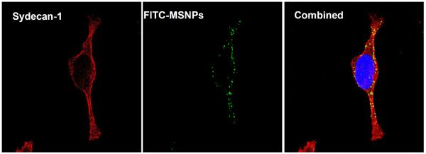
Syndecan-1 was detected by an antibody coupled to Alexa Fluor 594 (red fluorescence). FITC-labeled MSNP-PEI 10 kD (25 μg/mL) was added to the cells for 3 hr prior to protein and nuclear staining as in Fig. 7A. The combined panel on the right hand side demonstrates particle co-localizing with syndecan-1. The extent of the co-localization was 64.3% as determined by Image J software.