


Figure 2.
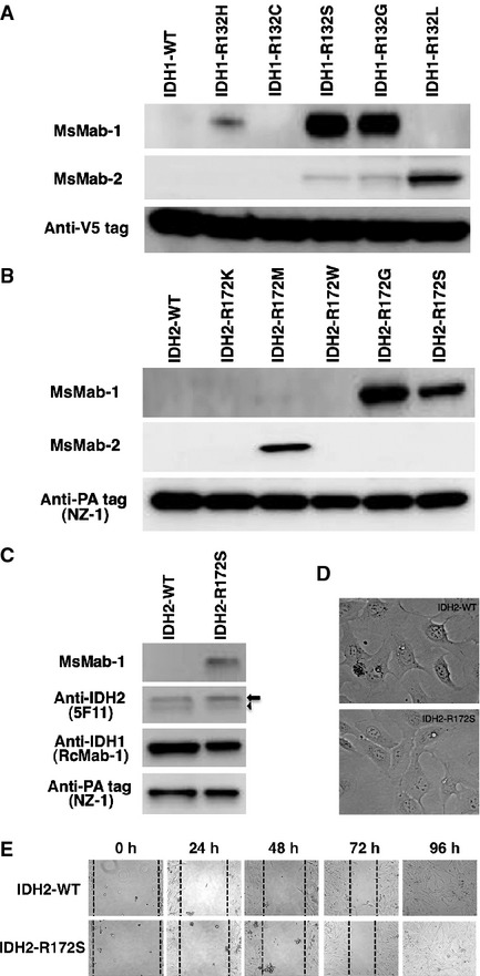
(A) Western blot analyses by MsMab-1 against mutated IDH1-expressing CHO cells. Total cell lysate from CHO cells expressing IDH1 wild type (WT, lane 1) and IDH1 mutants (lane 2, IDH1-R132H; lane 3, IDH1-R132C; lane 4, IDH1-R132S; lane 5, IDH1-R132G; lane 6, IDH1-R132L) were electrophoresed under reducing conditions using 5–20% gels, and were Western blotted with MsMab-1, MsMab-2, and anti-V5 tag. (B) Western blot analyses by MsMab-1 against mutated IDH2-expressing CHO cells. Total cell lysate from CHO cells expressing IDH2-WT (lane 1), IDH2 mutants (lane 2, IDH2-R172K; lane 3, IDH2-R172M; lane 4, IDH2-R172W; lane 5, IDH2-R172G; lane 6, IDH2-R172S) were electrophoresed under reducing conditions using 5–20% gels, and were Western blotted with MsMab-1, MsMab-2, and anti-PA tag (NZ-1). (C) Western blot analyses by MsMab-1 against mutated IDH2-expressing U-2 OS cells. Total cell lysate from U-2 OS cells expressing IDH2-WT (lane 1) and IDH2-R172S (lane 2) were electrophoresed under reducing conditions using 5–20% gels, and were Western blotted with MsMab-1, anti-IDH2 (5F11), anti-IDH1 (RcMab-1), and anti-PA tag (NZ-1). Arrow and arrowhead indicate transfected and endogenous IDH2, respectively. (D) The morphology of transfected U-2 OS cells (IDH2-WT and IDH2-R172S) was investigated 2 days after transfection. No morphological or growth difference was observed among those cells: 6.41 × 10⁵ cells and 6.56 × 10⁵ cells in 10-cm dish, respectively. (E) Measurement of cell migration by in vitro scratch assay. U-2 OS cells were transiently transfected with IDH2-WT or IDH2-R172S. The migration ability was assessed 24, 48, 72, and 96 h from the scratch. The figure shows the cells at 0, 24, 48, 72, and 96 h after the scratch. Magnification is 20×. IDH, isocitrate dehydrogenase; MsMab, multi-specific anti-mutated IDH1/2 mAb; CHO, Chinese hamster ovary; OS, osteosarcoma.