



Abstract
Shal K+ (Kv4) channels in mammalian neurons have been shown to be localized exclusively to somato-dendritic regions of neurons, where they function as key determinants of dendritic excitability. To gain insight into the mechanisms underlying dendritic localization of Kv4 channels, we use Drosophila melanogaster as our model system. We show that Shal K+ channels display a conserved somato-dendritic localization in vivo in Drosophila. From a yeast-2-hybrid screen, we identify the novel interactor, SIDL (for Shal Interactor of Di-Leucine Motif), as the first target protein reported to bind the highly conserved di-leucine motif (LL-motif) implicated in dendritic targeting. We show that SIDL is expressed primarily in the nervous system, co-localizes with GFP-Shal channels in neurons, and interacts specifically with the LL-motif of Drosophila and mouse Shal channels. We disrupt the Shal-SIDL interaction by mutating the LL-motif on Shal channels, and show that Shal K+ channels are then mis-localized to some, but not all, axons in vivo. These results suggest that there are multiple mechanisms underlying Shal K+ channel targeting, one of which depends on the LL-motif. The identification of SIDL may provide the first step for future investigation into the molecular machinery regulating the LL-motif-dependent targeting of K+ channels.
Keywords: Kv4, Shal, Potassium Channels, Drosophila, Dendrites, Trafficking
Introduction
Voltage-gated Shal K+ (Kv4) channels are highly conserved in amino acid sequence and underlie transient A-type K+ currents from jellyfish to human (reviewed in: Birnbaum et al., 2004; Jerng et al., 2004). Recent work has highlighted that voltage-dependent Shal K+ channels function as key determinants of dendritic excitability. Accordingly, Kv4 channels in mammalian neurons display a strictly somato-dendritic distribution (Baro et al., 2000; Maletic-Savatic et al., 1995; Misonou and Trimmer, 2004; Sheng et al., 1992). Kv4.2 channels in hippocampal dendrites act at subthreshold potentials, regulating the integration of high-frequency trains of synaptic input (Ramakers and Storm, 2002), the shape of mEPSCs (Kim et al., 2007), backpropagating action potentials (Cai et al., 2004; Chen et al., 2006; Kim et al., 2005), and induction of long-term potentiation (LTP) (Chen et al., 2006; Kim et al., 2007). Although the function of Shal channels is clearly dependent on their somato-dendritic localization, little is known about how Shal channels actually achieve this polarized distribution in neurons.
As a first clue to the molecular mechanisms underlying dendritic targeting of Shal K+ channels in neurons, Rivera et al. (2003) reported that a highly conserved 16 amino acid di-leucine motif (LL-motif) on the C-terminus of Shal channels is required and sufficient for dendritic targeting of Kv4.2 channels in cultured cortical pyramidal neurons (Rivera et al., 2003). The investigators showed that a deletion of the LL-motif resulted in loss of the polarized distribution of Kv4.2 channels in neurons. Conversely, the LL-motif itself was able to target both the non-channel protein CD8, and the normally axonal Kv1.3 and Kv1.4 (Shaker) channels, to dendrites (Rivera et al., 2003). Interestingly, another di-leucine motif on the C-terminus of the Na+ channel Nav1.2 has been implicated in axonal targeting (Garrido et al., 2001). Di-leucine motifs identified in other cell types have been shown to be involved in polarized trafficking by binding to adaptor proteins that organize target proteins into clathrin coated vesicles for transport from the trans-Golgi network, plasma membrane, or endosomal compartments (reviewed in: (Horton and Ehlers, 2003). The LL-motif of Shal channels may interact with proteins directing somato-dendritic localization.
In this report, we examine the subcellular localization of Drosophila Shal K+ channels and the mechanisms underlying this distribution. We show for the first time that Shal channels in Drosophila also display a somato-dendritic distribution. Since 14 of the 16 amino acids of the LL-motif are identical between Drosophila and mouse Shal channels, we suspect that mechanisms underlying the dendritic localization of Shal channels are similar across species. Here, we identify SIDL as the first interactor of this LL-motif. We show that SIDL is primarily expressed in the nervous system and co-localizes with GFP-Shal channels in neurons. Disruption of the LL-motif, and interaction with SIDL, results in mislocalization of Shal K+ channels to axons in a subset of neurons, suggesting that SIDL may function in one of several mechanisms underlying dendritic localization of Shal channels.
Materials and Methods
Yeast-Two-Hybrid (Y2H) Assays for Mapping Interaction Sites
The C-terminus of Shal1 (Shal1C; amino acids 411-571) was used as “bait” in a Y2H screen of pGADT7 “prey” constructs containing cDNAs reverse transcribed from RNA isolated from Drosophila embryos (aged 0-24 hrs; Clontech Laboraties Inc., Palo Alto, CA), as previously described (Diao et al., 2009).
For direct Y2H assays testing the specificity of the SIDL-Shal interaction, the C-termini of Shaker and Shaw were amplified from cDNA clones of ShA1 and Shaw2 by PCR, the C-terminus of Shab-RB was amplified by RT-PCR, the N-terminus of Shal and C-terminus of mouse Kv4.2 (mKv4.2C) were amplified from cDNA clones by PCR; amplified products were subcloned into the pGBKT7 bait vector. For mapping the SIDL-Shal interaction, deletion constructs of SIDL and Shal were amplified and cloned into pGADT7 and pGBKT7 vectors, respectively. Shal2C(LL→AA) and Shal2C(LL→VV) constructs were generated using primers that incorporated the corresponding leucines substitutions. The LL-motif construct was obtained by extending two overlapping primers. All constructs were confirmed by DNA sequencing, co-transformed into AH109 yeast, and assayed for interaction by the expression of reporter genes (HIS3, Ade2, and MEL1).
GST-Pulldown Assays
Shal1C, Shal2C and Shal2CΔ9 were amplified from Shal cDNA clones using appropriately engineered primers. The C-terminus of SIDL (SIDLC) was amplified from the clone identified in our Y2H screen; PCR amplification rounds were designed to fuse the epitope c-myc to the N-terminus of SIDL. Shal and myc-SIDLC products were then subcloned into pGEX-2TK and transformed into BL21 cells to express GST-fusion proteins. GST and GST-fusion proteins were purified using glutathione-agarose beads (Sigma Aldrich, St. Louis, MO). myc-SIDLC protein was cut with biotinylated thrombin (Novagen Inc, San Diego, CA) from GST-myc-SIDLC, which contains an engineered thrombin site. Thrombin was removed using streptavidin-agarose beads. GST proteins were then incubated with purified myc-SIDLC or Drosophila embryo membrane extracts. Glutathione-agarose beads were washed, boiled in 4×SDS loading buffer, and run by standard SDS-PAGE techniques. Proteins were transferred to nitrocellulose and immunoblots were probed with designated antibodies.
In Situ Hybridization
Wild type (w1118) embryos aged 12 hours (at 25°C) were collected, washed with water, dechorionated with bleach, and fixed with 4% paraformaldehyde in PBS. After rehydration, embryos were treated with protease K and then hybridized with probe at 55°C. Single-stranded digioxygenin-labeled (DIG-11-UTP, DIG DNA Labeling and Detection Kit, Roche, Basel, Switzerland) probes were amplified by PCR. The linearized SIDLC clone from the Y2H screen was used as template. The 3′activation domain primer was used to amplify the SIDLC probe. After hybridization and wash, embryos were incubated with anti-DIG antibody and developed by NBT/BCIP.
Antibodies
The anti-Shal antibody was generated by our lab, using the peptide sequence CGIELDDNYRD, as previously described (Diao et al., 2009). Monoclonal anti-syntaxin (8C3), anti-myc (9E10), anti-elav (Elav-9F8A9), and anti-β-galactosidase (40-1a) were obtained from the Developmental Studies Hybridoma Bank (DSHB, University of Iowa). Anti-GST was obtained from Invitrogen Molecular Probes (Eugene, OR).
Immunostaining Embryonic Cell Cultures
Embryos aged 5-6 hours (25°C; stage 9-10) were dissociated into culture media (Schneider’s Drosophila media supplemented with 18% fetal bovine serum, 100 units/mL penicillin, 100 ug/mL streptomysin) using a glass micropipet, as previously described (Tsunoda and Salkoff, 1995a). Cell cultures grown for 2 to 7 days were immunostained, as previously described (Diao et al., 2009). Briefly, cultures were fixed (4% formaldehyde), incubated with primary antibody, washed (0.1% sapponin in PBS), incubated with rhodamine or FITC-conjugated secondary antibody (Jackson Immunoresearch Laboratories, West Grove, PA; 2 hours, room temperature), washed again, and mounted in 90% glycerol with p-phenylenediamine (Sigma Aldrich, St. Louis, MO).
Transgenic UAS-myc-SIDL, UAS-GFP-Shal, UAS-GFP-ShalΔLL-motif, and UAS-UAS-GFP-Shal(LL→AA) Lines
The Gateway System was used to generate pUAST-6×myc-SIDL (referred to as UAS-myc-SIDL), pUAST-GFP-Shal2 (referred to as UAS-GFP-Shal), and pUAST-GFP-Shal2ΔLL-motif (referred to as UAS-GFP-ShalΔLL-motif) clones. SIDL was amplified by PCR from an available cDNA clone (Drosophila Genomics Resource Center, DGRC). Shal2 and Shal2ΔLL-motif were amplified by PCR from a cDNA clone of Shal2 using appropriate primers. SIDL, Shal2 and Shal2ΔLL-motif were then subcloned into the “entry vector”, pENTR1A (Invitrogen, Carlsbad, CA), such that sequences were flanked with attL1 and attL2 recombination sites. SIDL, Shal2 and Shal2ΔLL-motif were then recombined in vitro using lambda integrase into “destination” vectors (Drosophila Gateway Vector collection, DGRC), containing the attR1 and attR2 recombination sites. pUAST Drosophila transformation destination vectors have been designed to fuse 6 sequential myc epitopes (6xmyc) (pTMW) or GFP (pTGW) to the N-termini of the corresponding sequences, under the control of the upstream-activating-sequence (UAS). For UAS-GFP-Shal(LL→AA), the resulting Shal2 recombined into pTGW vector was altered so that nucleotides (1441-1446) corresponding to the two conserved leucines in the LL-motif were changed to encode two alanines; this site-directed change was performed by GenScript (Piscataway, NJ). All transgenic lines were generated by microinjection of constructs into w1118 embryos and standard P-element mediated germline transformation.
Fly Brain Imaging
Adult brains were dissected in 0.1M Phosphate buffer, fixed for 20 minutes in 4% paraformaldehyde and washed in 0.1M phosphate buffer for 30 minutes. Images were captured using Zeiss LSM 510 confocal microscope and processed using Adobe Photoshop. Images are of a single frontal section approximately 60um from the antennal lobe, into the brain. For quantification in Figure 5, GFP intensity was calculated by first measuring the mean signal intensity along a line drawn over the medial antennocerebral tract (mACT) using ImageJ software (Rasband, W.S., ImageJ, U. S. National Institutes of Health; http://rsb.info.nih.gov/ij/, 1997-2009.). Background fluorescence was then measured by placing the same line over the neighboring neuropil for each antennal lobe. Mean GFP signal intensity was adjusted by subtracting the background, and then expressed as a percentage above background fluorescence.
Figure 5. GFP-ShalΔLL-motif and GFP-Shal(LL→AA) Channels are Mis-Targeted to mACT Axons.

(A) GAL4-GH146 was used to drive expression of UAS-DsRed (right column) and either UAS-GFP-Shal, UAS-GFP-ShalΔLL-motif, or UAS-GFP-Shal(LL→AA) (left column) in vivo. Shown are single plane confocal images focused on mACT axons (arrowheads) which project to the lateral horn (LH). The proximal segment of axons in the iACT can also be seen in this focal plane (see arrows). GFP-Shal is observed in the proximal segment of the iACT, but not in the mACT. GFP-ShalΔLL-motif and GFP-Shal(LL→AA) are found in both the proximal segment of iACT axons (arrows) and the axons of the mACT (arrowheads); images are representative of 11 and 8 brains examined for GFP-ShalΔLL-motif and GFP-Shal(LL→AA), respectively. (B) Quantification of GFP signal intensity along the mACT, expressed as the percentage over background fluorescence. There is significantly more GFP-ShalΔLL-motif (78.93% ± 0.54, N=20 antennal lobes) and GFP-Shal(LL→AA) (68.44% ± 0.35, N=15 antennal lobes) localized to the mACT when compared to GFP-Shal (3.16 ± 0.05, N= 10 antennal lobes).
Results
Somato-Dendritic Localization of Shal K+ Channels is Conserved in Drosophila
To determine if the somato-dendritic localization of Shal K+ channels seen in mammalian systems is also conserved in Drosophila, we used the tissue-specific UAS-GAL4 expression system (Brand and Perrimon, 1993) to examine the localization of GFP-tagged Shal channels in an identified cell type. We chose to use the projection neurons (PNs) of the antennal lobe of the adult Drosophila brain because axonal and dendritic processes are easily discernable in vivo. PN cell bodies are localized in three major clusters (anterodorsal, lateral, and ventral) around the periphery of the antennal lobe. Like many insect neurons, PNs are unipolar and this single process extends into the antennal lobe where it forms dendritic arbors that receive input from olfactory receptor neurons, as well as an axonal projection that extends in the opposite direction to higher centers in the brain (Stocker et al., 1997; Stocker et al., 1990; Wong et al., 2002). We used the GAL4-GH146 transgene to drive expression of UAS-transgenes specifically in PNs (Stocker et al., 1997; Wong et al., 2002). For orientation, we first used GAL4-GH146 to drive expression of three different UAS-transgenes previously shown to display expression in dendrites only (Dscam[17.1]-GFP; Wang et al., 2004), axons and dendrites (Dscam[17.2]-GFP;(Wang et al., 2004), or nuclei (ms2::GFP-nls; Ashraf et al., 2006). Adult brains were dissected, fixed, and subjected to confocal microscopy to examine GFP localization in PNs. As expected, Dscam [17.1]-GFP signal was observed exclusively in the dendrite enriched glomeruli within the antennal lobe, while Dscam [17.2]-GFP was localized to axons in the antennocerebral tract as well as dendrites (Figure 1A). The ms2::GFP-nls nuclear signal illustrated the position of PN cell bodies (Figure 1A).
Figure 1. Somato-Dendritic Localization of GFP-Shal is Conserved in Drosophila and Dependent on the Conserved LL-motif.
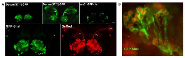
(A) Top panels, GAL4-GH146 was used to drive expression of three UAS-transgenes shown previously to localize to distinct subcellular domains. Adult brains were dissected, fixed, and subjected to confocal microscopy to examine GFP localization in PNs. As expected, Dscam[17.1]-GFP (top left) is restricted to the dendrite rich interior of the antennal lobe, while Dscam[17.2]-GFP (top middle) is strongly expressed in axons present in the inner antennocerebral tract (iACT, see arrows) and dendrites. GFP fused to the nuclear localization signal (ms2::GFP-nls, top right) indicates the location of PN cell body nuclei present in clusters around the periphery of the antennal lobe. Bottom panels, GAL4-GH146 was used to drive expression of UAS-GFP-Shal (left) and UAS-DsRed (right). GFP-Shal signal was detected in PN cell bodies and dendrites, but not in axons. DsRed expressed in the same brain is shown (right) with arrows indicating the axonal iACT projecting to the lateral horn (LH). Shown are representative images, composed of collapsed confocal projections in order to display labeling in all cellular compartments. Note that collapsed images reveal more background, especially evident in the mushroom bodies. Scale bar represent 50 μm. (B) Under higher magnification (630X) it is evident that GFP-Shal channels are present in cell bodies as well as in dendritic tracts of the AL. These results suggest that somato-dendritic localization of GFP-Shal is conserved in Drosophila.
We then generated a GFP-tagged Shal2 construct under the control of UAS (UAS-GFP-Shal). As a control for cell morphology, we used an available transgenic line expressing UAS-DsRed. We then made transgenic lines in which GAL4-GH146 was used to drive simultaneous expression of both UAS-GFP-Shal and UAS-DsRed in the same brains. We found that GFP-Shal channels were indeed localized to dendrites and cell bodies, but not at all to axons; figure 1A shows collapsed confocal planes in order to display cell bodies, the antennal lobe (dendrites), and axonal tracts in the same image. Ds-Red in the same PNs displayed expression throughout the cell bodies, axons, and dendrites, suggesting that the localization pattern of GFP-Shal was specific, and not due to any structural abnormality. Note that collapsed confocal images also amplified background, especially in the mushroom bodies and can be seen in both GFP and DsRed images. The major axonal tract of PNs, the inner antennocerebral tract (iACT) is indicated (arrows) for clarity (Figure 1A). Figure 1B shows a higher magnification overlay of GFP-Shal and DsRed in a cluster of PN cell bodies and their proximal dendritic tracts extending into the antennal lobe. These results suggest that somato-dendritic localization of GFP-Shal is conserved in Drosophila, making Drosophila, and specifically the PNs, a viable model for investigating mechanisms underlying dendritic targeting of Shal channels.
Identification of SIDL as a Novel Interactor of the Highly Conserved Di-Leucine Motif of Shal K+ Channels
The Shal LL-motif is likely to bind component(s) involved in dendritic localization of Shal channels. Such an interactor may regulate the release of Shal channel subunits from the ER/trans-Golgi network, or function in the targeting, transport, retention, or recycling of Shal channels in dendrites. To take an unbiased approach to identifying protein interactors of the LL-motif, we performed a yeast-two-hybrid (Y2H) screen for candidate interactors. We used the entire 161 residue C-terminal tail of the Drosophila Shal1 isoform (Shal1C), containing the LL-motif, as “bait” and screened for interacting “prey” from a cDNA library (Diao et al., 2009). One of the interactors identified was the C-terminal 382 residues of a protein we now call SIDL (for Shal Interactor of Di-Leucine Motif). To examine the specificity of the SIDL-Shal1 interaction, we used direct Y2H assays to test whether the C-terminus of SIDL (SIDLC) was also able to interact with the C-termini of three other Drosophila voltage-gated K+ channels, Shaker (Kv1), Shab (Kv2), and Shaw (Kv3), and/or the C-terminus of the mouse Shal K+ channel, mKv4.2. We found that SIDLC interacted with Drosophila Shal1 and mKv4.2, but not the C-termini of the other Drosophila K+ channels tested (Figure 2A). These results suggest that SIDL interacts specifically, even across species, with Shal K+ channels.
Figure 2. SIDL Interacts Specifically with the Highly Conserved Di-Leucine Motif of Shal Channels.

(A) Shown are direct Y2H assay results testing interaction of the C-terminal 382 residues of SIDL (SIDLC) with full-length and deletion constructs of the C-terminal tails of Shal, Kv4.2, Shaker, Shaw, and Shab channel subunits (amino acid numbers indicated). Interaction strength, determined by reporter gene expression, is indicated by number of “+” symbols; “−” indicates no reporter gene expression. Note that the 16-residue dileucine motif (red) of Shal channels is required and sufficient for binding SIDLC. (B-C) Shown are immunoblots from GST-pull down assays using GST-fusion proteins indicated above each lane, incubated with purified myc-SIDLC protein (B), or embryonic membrane extracts (C). (B) Significant myc-SIDLC protein was pulled-down by GST-Shal1C and GST-Shal2C, but not by GST alone or GST-Shal2CΔ9. Immunoblots were probed with anti-GST (B) or Ponceau S (C) to show the presence of GST proteins in each assay. (C) Note that GST-SIDLC pulled-down endogenous Shal protein (Shal(bound)) from embryonic membrane extracts, even with far less GST-SIDLC protein present than GST alone. Immunoblot of unbound fractions is also shown using anti-syntaxin (syx(unbound)) as a control for the presence of membrane extract proteins
The interaction of SIDLC with mKv4.2C suggested that SIDL interacts with a conserved binding site on Shal channels. Using direct Y2H assays, we tested whether the binding site is the highly conserved LL-motif required for dendritic targeting. We first tested whether SIDLC was able to bind the shorter Shal2C isoform, which also contains the LL-motif. SIDLC also showed strong interaction with Shal2C. We then made deletion constructs of Shal2C as illustrated in Figure 2A. We found that deletions affecting the 16 amino acid LL-motif resulted in a loss of interaction between SIDLC and Shal. This was first seen in larger deletions of up to 40 residues of the C-terminal end of Shal2C, and then in progressively smaller deletions. A deletion of the entire LL-motif (Shal2CΔ16), or the last 9 residues of the LL-motif (Shal2CΔ9), including the two conserved leucines, resulted in a complete loss of interaction (Figure 2A). Substitution of the two leucines to alanines (Shal2C(AA)) also significantly reduced interaction with SIDLC, while substitution to more similar valine residues (Shal2C(VV)) did not. Finally, we tested whether the LL-motif alone was sufficient to bind SIDLC. We found that SIDLC displayed equally strong interaction with the 16-residue LL-motif as with the entire Shal2C (Figure 2A). Thus, our results show that the conserved LL-motif on the C-terminus of Shal is both required and sufficient for binding SIDL.
To validate the SIDL-Shal interaction using another independent approach, we used the GST-pull down assay. We generated constructs to express GST fused to Shal1C (GST-Shal1C), Shal2C (GST-Shal2C), and Shal2CΔ9 (GST-Shal2CΔ9). GST-fusion proteins were expressed and incubated with the C-terminal 382 amino acids of SIDL tagged with myc (myc-SIDLC). We found that GST-Shal1C and GST-Shal2C indeed pulled-down myc-SIDLC, while GST alone and GST-Shal2CΔ9 did not (Figure 2B). To test the GST pull-down assay in the opposite configuration, we fused SIDLC to GST (GST-SIDLC) and incubated GST and GST-SIDLC with embryonic membrane extracts this time. We found that GST-SIDLC indeed pulled-down native Shal protein, while GST alone did not (Figure 2C).
SIDL is Primarily Expressed in the Nervous System
SIDL, which has not previously been described, contains 1146 amino acids, with no consensus protein binding or enzymatic domains. Sequence analysis using multiple transmembrane prediction programs resulted in conflicting predictions. Some programs predicted SIDL to be a soluble protein while others predicted an integral membrane protein. Two of the Kyte-Doolittle based prediction programs (waveTM, Liakopoulos et al., 2001), available at http://athina.biol.uoa.gr/bioinformatics/waveTM/, and TopPred2, (Claros and von Heijne, 1994), available at http://bioweb.pasteur.fr/seqanal/interfaces/toppred.html) predicted SIDL to contain one to two transmembrane domains at 787-807 (amino acids) and possibly also 1079-1099 (amino acids; see Figure 3A).
Figure 3. SIDL is Primarily Expressed in the Nervous System and Co-Localizes with GFP-Shal in Dendrites.

(A) SIDL protein illustration showing Shal binding region identified in Y2H (765-1146 amino acids; green) and the two putative transmembrane domains predicted at residues 787-807 and 1079-1099 (see text). (B) RT-PCR for SIDL and actin in RNA isolated from Drosophila at different developmental stages: embryo (hours AEL at 25°C indicated), larvae (instar stage indicated), pupae (days aged indicated), and adult (heads (H) or bodies (B)). (C) RNA in situ hybridization for SIDL in representative 12-hour embryos; immunostaining for neuron-specific elav protein is shown for reference. SIDL appears to be expressed primarily in the nervous system. (D) Small clusters and single neurons with processes shown for primary cultures grown from transgenic lines expressing GFP-Shal (left) or CD8-GFP (right). Note that GFP-Shal is present in puncta along neuronal processes (see arrowheads for examples), while CD8-GFP is expressed throughout processes. (E) Shown is a single representative neuron from primary cultures made from a transgenic line expressing GFP-Shal (green) and nod-β-Gal (red). GFP-Shal positive processes all show immunostaining for the dendritic marker, nod-β-Gal. (F) A single representative neuron in primary culture from a transgenic strain expressing GFP-Shal and myc-SIDL in neurons. Immunostaining shows that GFP-Shal (left-green) and myc-SIDL (middle-red) co-localize (right-yellow) in cell bodies and in puncta along dendritic processes. Scale bars represent 5 μm for (D,E) and 10 μm for (F).
To examine the expression of SIDL at different developmental stages, we performed RT-PCR for SIDL from embryos, at 5, 12, and 17 hours after egg laying (AEL), larvae in all three instar stages, pupae at 1 day and 4 days post-pupariation, and adult fly heads and bodies. We found that SIDL is expressed at all of these developmental stages (Figure 3B). To examine whether SIDL is expressed in the nervous system, we examined the expression of SIDL in the developing embryo, where Shal channels have been shown to underlie the transient A-type K+ current present in virtually all neurons (Tsunoda and Salkoff, 1995a). We performed RNA in situ hybridizations in wild-type embryos (12 hours AEL) using a single-stranded digoxygenin-labeled probe for SIDL. We found that SIDL displayed expression primarily in the nervous system, similar to the nervous system specific elav protein (Figure 3C).
SIDL Co-Localizes with Shal K+ Channels in Neurons
To determine if SIDL co-localizes with Shal channels at distinct subcellular sites, we examined neurons in primary cultures from transgenic lines. We first used the neuron-specific elav-GAL4 transgene to drive expression of UAS-GFP-Shal alone. We found that GFP-Shal protein was localized in neuronal cell bodies and in distinct puncta along neuronal processes (Figure 3D, 3F; Diao et al., 2009). As a control, we examined a transgenic line expressing the membrane targeted CD8-GFP fusion protein under the control of the same GAL4 neuronal driver. In contrast to GFP-Shal, CD8-GFP was localized uniformly throughout neuronal processes (Figure 3D), suggesting that GFP-Shal puncta in neuronal processes are indeed distinct sites of localization. To confirm that GFP-Shal puncta are indeed localized in dendrites, we prepared primary cultures from embryos expressing GFP-Shal and the fusion protein, nod-ß-Gal, which localizes only in dendrites (Andersen et al., 2005; Clark et al., 1997; Lee et al., 2000; Rolls et al., 2007; Yamamoto et al., 2006). Indeed, GFP-Shal positive processes were also positive for nod-ß-Gal (Figure 3E).
We next examined whether SIDL co-localizes with GFP-Shal in these dendritic puncta. We generated transgenic lines expressing both UAS-GFP-Shal and UAS-6xmyc-tagged SIDL (UAS-myc-SIDL) in neurons, using the elav-Gal4 driver. We found that GFP-Shal and myc-SIDL co-localized both in neuronal cell bodies and in puncta along neuronal processes (Figure 3F). Nearly all GFP-Shal positive puncta were also positive for myc-SIDL; there were a minority of myc-SIDL positive puncta that did not appear to contain GFP-Shal. One possibility is that SIDL may function in clustering Shal channels at these sites. The co-localization of GFP-Shal and myc-SIDL in dendritic puncta further implicated a role for SIDL in Shal K+ channel regulation.
Disruption of the Shal-SIDL Interaction Results in Axonal Localization of Shal Channels in a Subset of Projection Neurons in the Adult Brain
To test whether the Shal-SIDL interaction is required for somato-dendritic localization of Shal K+ channels, we replicated mutations in Shal that resulted in a loss of the Shal-SIDL interaction in Y2H assays (Figure 2) and created transgenic lines expressing these Shal variants tagged with GFP. The two transgenic lines we generated included one expressing UAS-GFP-Shal with a deletion of the 16-residue LL-motif (UAS-GFP-ShalΔLL-motif), and another with the two central leucines of the LL-motif changed to alanines (UAS-GFP-Shal(LL→AA)). We then used GAL4-GH146 to drive expression of UAS-GFP-ShalΔLL-motif and UAS-GFP-Shal(LL→AA) in PNs. In case any observed effects might be due to gross morphological changes in the PNs, we simultaneously expressed UAS-DsRed in the same fly brains with each of the UAS-GFP-Shal variants. Whole brains were dissected, fixed and imaged by confocal microscopy. Analysis of confocal planes appropriate to examine PN clusters and dendrites in the antennal lobe, showed that GFP-Shal was localized in both cell bodies and dendrites, as described earlier. Figure 4 shows confocal images collapsed to display PN cell body clusters, the antennal lobe with PN dendrites, and the major iACT PN axonal tract. Interestingly, however, GFP-ShalΔLL-motif and GFP-Shal(LL→AA) channels displayed a distinctly punctate localization pattern in both cell bodies and in the antennal lobe (Figure 4). It is unlikely that this was due to a defect in PN morphology since DsRed was present diffusely throughout PNs in GFP-Shal, GFP-ShalΔLL-motif, and GFP-Shal(LL→AA) expressing transformants (Figure 4). The clustering of GFP-ShalΔLL-motif and GFP-Shal(LL→AA) channels may indicate a role for the Shal-SIDL interaction in maintaining channel distribution.
Figure 4. GFP-ShalΔLL-motif and GFP-Shal(LL→AA) Channels are Not Present in iACT Axons.

GAL4-GH146 was used to drive expression of UAS-DsRed and either UAS-GFP-Shal, UAS-GFP-ShalΔLL-motif, or UAS-GFP-Shal(LL→AA) in vivo. DsRed expression (right column) shows diffuse localization in PN cell bodies in clusters around the antennal lobe, in dendrites in the antennal lobe, and axons projecting in the iACT (arrows) to the lateral horn (LH). All GFP-Shal variants (left column) appear restricted to cell bodies and dendrites, and absent from the iACT. Images are representative of a total of 29 brains for GFP-Shal, 11 brains for GFP-ShalΔLL-motif, and 8 brains for GFP-Shal(LL→AA). Images shown are collapsed Z-series confocal projections to display PN cell bodies, dendrites, and iACT axons together. Note the more punctate signal evident for GFP-ShalΔLL-motif and GFP-Shal(LL→AA) channels, compared with GFP-Shal. Scale bars represent 50um.
We were surprised that GFP-ShalΔLL-motif and GFP-Shal(LL→AA) channels were excluded from axons in the iACT. To investigate whether these GFP-Shal mutants were excluded from all PN axons, we examined single confocal planes through the brain that captured PN axons projecting in the middle ACT (mACT). PNs extend axons to higher centers in the brain in three antennocerebral tracts (ACTs): inner (iACT), middle (mACT), and outer (oACT). The iACT axons project from cell bodies that reside in anterodorsal and lateral PN clusters to the calyx of the mushroom body and the lateral horn (LH) in the protocerbrum (Figures 1,4,5; see arrows), while mACT axons project from cell bodies that reside in the ventral PN cluster directly to the LH (Figure 5, see arrowheads; Okada et al., 2009; Stocker, 1994; Stocker et al., 1990). oACT axons project from anterodorsal and lateral cluster PNs to the LH and calyx by way of an outer tract. We did not observe expression in the oACT with the GH146-Gal4 driver, most likely because the GH146-Gal4 only expresses Gal4 in a single PN that projects via the oACT (Wong et al., 2002) and it was not detectable in our hands. GH146-Gal4 clearly promoted expression in PNs with axons in the iACT, as shown in Figures 1 and 4, and the mACT, as shown in Figure 5. GFP-Shal, however, was never seen to extend into either tract (Figures 1,5), consistent with an exclusively somato-dendritic localization pattern. Interestingly, in confocal planes used to image the mACT, we also observed GFP-Shal in the very proximal segment of the iACT axons just where they bundle to leave the antennal lobe (Figure 5, see arrows). One possibility is that Shal channels are excluded from axons by a diffusion barrier present in this proximal segment, similar to mechanisms proposed in mammalian neurons (Winckler et al., 1999).
When we examined the localization of GFP-ShalΔLL-motif and GFP-Shal(LL→AA) channels in PNs, we found that although these mutant channels were not present in the distal iACT (Figure 4), they were present in the proximal segment of the iACT, similar to GFP-Shal (Figure 5). In contrast, GFP-ShalΔLL-motif and GFP-Shal(LL→AA) channels were consistently found throughout the mACT (Figure 5, see arrowheads). Thus, mislocalization to axons appeared to occur only in those PNs with axons that project into the mACT, but not into the iACT. Quantification of GFP signal intensity along the mACT, expressed as percent above background, showed that GFP-ShalΔLL-motif and GFP-Shal(LL→AA) displayed 78.9% and 68.4% signal in mACT over background, respectively, compared with only 3.2% over background for GFP-Shal (Figure 5C). Our studies show that disruption of the LL-motif, and therefore the Shal-SIDL interaction, result in axonal targeting in the subset of PN neurons, those that reside in the ventral cluster, and project axons in the mACT tract. These results suggest then that there are likely to be multiple mechanisms for targeting Shal K+ channels in different neurons. The role of other potential Shal interactors, such as SKIP3 (Diao et al., 2009) and a putative KChIP homolog, in trafficking will need to be explored in the future as well.
Mislocalization of GFP-ShalΔLL-motif Channels in Sensory Neurons
To test whether the localization patterns of GFP-Shal variants showed any differences in another system, we used the ppk-Gal4 transgene to drive expression of GFP-Shal variants in identified neurons in the peripheral nervous system (PNS). In the Drosophila PNS, the dendritic arborization (da) neurons are present in four clusters (dorsal (d), lateral (l), ventral’ (v’), and ventral (v)) and project multiple dendrites along the epidermis. The ppk-Gal4 transgene drives expression in class IV da neurons (Grueber et al., 2003; Grueber et al., 2007). Class IV da neurons display the most extensive array of dendritic arborization of the da neurons (Grueber et al, 2002), and three class IV da neurons, named ddaC, v’ada, vdaB, have been identified in each abdominal hemisegment (A2-A6), present in the dorsal, ventral’, and ventral clusters, respectively. da neurons are easily examined in the late embryo through larval stages, where their cell bodies and dendritic projections are found just beneath the surface of a rather transparent cuticle. In contrast, da axons bundle and project ventrally to the ventral nerve cord (VNC) of the CNS, which is also easily imaged in larvae. We examined the localization of mCD8-GFP and GFP-Shal in dendrites and axons of class IV da neurons in live 2nd instar larvae. mCD8-GFP was found throughout dendrites and in axons projecting to the VNC, as expected (Figure 6). Surprisingly, GFP-Shal was localized primarily to cell bodies in these neurons, with little to no signal in dendrites (Figure 6). It is unclear whether this is representative of endogenous Shal localization in these neurons. When we examined axons extending to the VNC, no GFP-Shal signal was evident here (Figure 6), as expected. GFP-ShalΔLL-motif channels, however, were present in both cell bodies and axons of class IV da neurons (Figure 6). GFP-ShalΔLL-motif signal was consistently observed in a punctuate pattern in axons extending from the cell bodies (Figure 6, top-right, see arrows) and clearly in the VNC (Figure 6, bottom-right). These results suggest that exclusion of Shal K+ channels from axons in class IV da neurons is also dependent on the LL-motif, and perhaps its interaction with SIDL.
Figure 6. GFP-ShalΔLL-motif Channels are Mis-Targeted to Axons in Class IV da Neurons.

ppk-GAL4 was used to drive expression of either UAS-mCD8-GFP (CD8-GFP; left column), UAS-GFP-Shal (GFP-Shal; middle column), or UAS-GFP-ShalΔLL-motif (GFP-ShalΔLL-motif; right column) in vivo. Shown are conventional epi-fluorescent images from 2nd instar larvae taken at focal planes to examine either the v’ada (top row) da neuron in the ventral’ cluster, or the VNC (bottom row). Arrows indicate the axon in the CD8-GFP expressing v’ada neuron (top-left), and the punctate GFP-ShalΔLL-motif signal seen in v’ada axons extending from the cell body (top-right). GFP signal was consistently observed in the VNC of larvae expressing CD8-GFP or GFP-ShalΔLL-motif, but not in GFP-Shal transformants. Scale bars represent 5μm for top row, and 10μm for bottom row.
Discussion
With recent studies demonstrating a critical role for Kv4 in the regulation of post-synaptic potentials and activity-dependent plasticity (Cai et al., 2004; Chen et al., 2006; Kim et al., 2007; Kim et al., 2005), understanding the mechanisms regulating dendritic localization of Kv4 channels are of particular interest. In this study, we first show that GFP-tagged Shal K+ channels in Drosophila display somato-dendritic localization in the PNs of the adult brain in vivo. Although this is the first report showing that Drosophila Shal channels share this polarized distribution, it was not unexpected, given that Shal channels across species are highly conserved in function, and share a nearly identical LL-motif on their C-terminus, which has been implicated in dendritic localization.
Previously, Rivera et al (2003) identified this highly conserved LL-motif of Kv4 channels, and showed that it is required, and even sufficient, for dendritic localization in cortical pyramidal neurons in cultured brain slices (Rivera et al., 2003). Here, we mutated the LL-motif of Drosophila Shal channels and examined their localization in identified cell types in vivo. We show that GFP-ShalΔLL-motif and GFP-Shal(LL→AA) channels are mislocalized to PN axons that project into the mACT, and axons of class IV da sensory neurons. These findings are similar to those reported by Rivera et al. (2003), and suggest that the LL-motif is required for retaining the somato-dendritic localization of Shal K+ channels in vivo. Interestingly, however, GFP-ShalΔLL-motif and GFP-Shal(LL→AA) channels were still excluded from PN axons that project into the iACT. These results suggest that there are multiple mechanisms contributing to the somato-dendritic localization of Shal K+ channels, and that there is likely to be a LL-motif independent mechanism present in at least some neurons, like the iACT PNs.
A clue to the mechanism underlying the somato-dendritic localization of Shal channels in iACT PNs may be the unexpected observation of GFP-Shal signal in the proximal segment of these axons. This might indicate that the restriction of Shal channels to somato-dendritic compartments is achieved by excluding Shal channels from the axon by a diffusion barrier present in the proximal segment, similar to that proposed in the initial segment of mammalian neurons (Nakada et al., 2003; Winckler et al., 1999). Cytoskeletal elements and cytoskeleton interactors, like ankyrin G, have also been found to be specifically localized to the initial segment of axons (Boiko et al., 2007), and have been shown to function in retaining voltage-gated Na+ channels in the initial segment (Garrido et al., 2003; Jenkins and Bennett, 2001; Zhou et al., 1998). Adenomatous polyposis (APC) proteins also interact with the cytoskeleton, and Apc2-GFP has been shown to localize to PN cell bodies, dendrites, and the proximal segment of axons (Rolls et al., 2007), similar to GFP-Shal channels. NgCAM, a chick neuron-glia cell adhesion molecule, can bind ankyrin proteins in the initial segment of hippocampal neurons (Boiko et al., 2007), and interestingly NgCAM-YFP also displays specific localization to the proximal axonal segment of PNs in the Drosophila brain (Rolls et al., 2007). Thus, it will be important to investigate the role of cytoskeletal elements and interactors in Shal channel localization.
For LL-motif dependent localization of Shal K+ channels, we suggest that protein interactors may function as key regulators of dendritic localization. This study reports Drosophila SIDL as the first identified interactor of this highly conserved LL-motif. We show that SIDL is expressed primarily in the nervous system, may be a transmembrane protein, and co-localizes with Shal K+ channels in puncta along neuronal processes. We generated mutations in the LL-motif of Shal channel subunits which are predicted to disrupt the SIDL-Shal interaction, and show that these mutations affect the polarized distribution of Shal channels in some neurons. These effects may indeed be due to the loss of the SIDL-Shal interaction, however, the requirement for SIDL will need to be addressed directly in future studies. Isolation and analysis of a null mutant of SIDL, or an effective SIDL-RNAi mechanism, will be critical for understanding the function of SIDL for Shal K+ channels. SIDL, for example, may function in the biosynthesis of Shal to the forward targeting, transport, anchoring, or endocytosis/recycling of Shal channel subunits. SIDL may be the only and key regulator of dendritic trafficking, or it may function in conjunction with other LL-motif interactors, transport, anchoring, and/or recycling machinery.
Unfortunately, we were unable to show in this study that fully-assembled Shal channels interact with full-length SIDL. Co-immunoprecipitation experiments with our anti-Shal antibody in myc-SIDL transgenic lines were unsuccessful, most likely due to insufficient levels of myc-SIDL expression over endogenous SIDL. Future studies will need to explore the native interaction of these full-length proteins. Also, interesting will be to determine when and where Shal and SIDL interact in cells. Live in vivo imaging studies of tagged Shal and SIDL proteins in wild-type and mutant backgrounds should also give critical insight into the role of the SIDL-Shal complex.
Although Drosophila SIDL also interacted with the C-terminal tail of mKv4.2, searches of mammalian EST-databases for a mammalian homolog of SIDL did not reveal any protein homolog with greater than 30% amino acid identity. One possible candidate with only 29% amino acid identity is the human gene EHOC-1 (Epilepsy, HOloprosencephaly Candidate-1), also named TMEM1 (transmembrane protein 1), that was isolated as a candidate gene for several genetic diseases that map to human chromosome band 21q22.3 (Yamakawa et al., 1995). TMEM1 has been sequenced and found to encode a protein of 1259 residues, with two predicted transmembrane domains (Nagamine et al., 1997); the expression and function of TMEM1 has yet to be determined. It is also unclear whether TMEM1 is indeed a true homolog of Drosophila SIDL. It is possible that some domains of the protein may have diverged significantly in evolution to take on additional roles in different systems or cell types. Further study is required to determine whether TMEM1 binds the LL-motif, and whether it functions in dendritic localization of Kv4 channels. The identification of Drosophila SIDL may provide a key step for future investigation into Shal K+ channel trafficking.
Acknowledgments
We thank G. Chang for help mapping the binding site of SIDL, S. Ashraf for the GH146-Gal4 strain and helpful advice regarding the PNs, T. Lee for transgenic lines expressing GFP-tagged isoforms of Dscam, and Y-N Jan for the ppk-Gal4 line. We also thank L. Salkoff, J. Nerbonne, and J. Trimmer for various K+ channel cDNAs. S.T. was supported by a grant from the National Institutes of Health (R01 GM083335).
Footnotes
Publisher's Disclaimer: This is a PDF file of an unedited manuscript that has been accepted for publication. As a service to our customers we are providing this early version of the manuscript. The manuscript will undergo copyediting, typesetting, and review of the resulting proof before it is published in its final citable form. Please note that during the production process errors may be discovered which could affect the content, and all legal disclaimers that apply to the journal pertain.
References
- Andersen R, Li Y, Resseguie M, Brenman JE. Calcium/calmodulin-dependent protein kinase II alters structural plasticity and cytoskeletal dynamics in Drosophila. J Neurosci. 2005;25:8878–88. doi: 10.1523/JNEUROSCI.2005-05.2005. [DOI] [PMC free article] [PubMed] [Google Scholar]
- Ashraf SI, McLoon AL, Sclarsic SM, Kunes S. Synaptic protein synthesis associated with memory is regulated by the RISC pathway in Drosophila. Cell. 2006;124:191–205. doi: 10.1016/j.cell.2005.12.017. [DOI] [PubMed] [Google Scholar]
- Baro DJ, Ayali A, French L, Scholz NL, Labenia J, Lanning CC, Graubard K, Harris-Warrick RM. Molecular underpinnings of motor pattern generation: differential targeting of shal and shaker in the pyloric motor system. J Neurosci. 2000;20:6619–30. doi: 10.1523/JNEUROSCI.20-17-06619.2000. [DOI] [PMC free article] [PubMed] [Google Scholar]
- Birnbaum SG, Varga AW, Yuan LL, Anderson AE, Sweatt JD, Schrader LA. Structure and function of Kv4-family transient potassium channels. Physiol Rev. 2004;84:803–33. doi: 10.1152/physrev.00039.2003. [DOI] [PubMed] [Google Scholar]
- Boiko T, Vakulenko M, Ewers H, Yap CC, Norden C, Winckler B. Ankyrin-dependent and -independent mechanisms orchestrate axonal compartmentalization of L1 family members neurofascin and L1/neuron-glia cell adhesion molecule. J Neurosci. 2007;27:590–603. doi: 10.1523/JNEUROSCI.4302-06.2007. [DOI] [PMC free article] [PubMed] [Google Scholar]
- Brand A, Perrimon N. Targeted gene expression as a means of altering cell fates and generating dominant phenotypes. Development. 1993;118:401–15. doi: 10.1242/dev.118.2.401. [DOI] [PubMed] [Google Scholar]
- Cai X, Liang CW, Muralidharan S, Kao JP, Tang CM, Thompson SM. Unique roles of SK and Kv4.2 potassium channels in dendritic integration. Neuron. 2004;44:351–64. doi: 10.1016/j.neuron.2004.09.026. [DOI] [PubMed] [Google Scholar]
- Chen X, Yuan LL, Zhao C, Birnbaum SG, Frick A, Jung WE, Schwarz TL, Sweatt JD, Johnston D. Deletion of Kv4.2 gene eliminates dendritic A-type K+ current and enhances induction of long-term potentiation in hippocampal CA1 pyramidal neurons. J Neurosci. 2006;26:12143–51. doi: 10.1523/JNEUROSCI.2667-06.2006. [DOI] [PMC free article] [PubMed] [Google Scholar]
- Clark IE, Jan LY, Jan YN. Reciprocal localization of Nod and kinesin fusion proteins indicates microtubule polarity in the Drosophila oocyte, epithelium, neuron and muscle. Development. 1997;124:461–70. doi: 10.1242/dev.124.2.461. [DOI] [PubMed] [Google Scholar]
- Claros MG, von Heijne G. TopPred II: an improved software for membrane protein structure predictions. Comput Appl Biosci. 1994;10:685–6. doi: 10.1093/bioinformatics/10.6.685. [DOI] [PubMed] [Google Scholar]
- Diao F, Waro G, Tsunoda S. Fast inactivation of Shal (K(v)4) K+ channels is regulated by the novel interactor SKIP3 in Drosophila neurons. Mol Cell Neurosci. 2009;42:33–44. doi: 10.1016/j.mcn.2009.05.003. [DOI] [PMC free article] [PubMed] [Google Scholar]
- Garrido JJ, Fernandes F, Giraud P, Mouret I, Pasqualini E, Fache MP, Jullien F, Dargent B. Identification of an axonal determinant in the C-terminus of the sodium channel Na(v)1.2. Embo J. 2001;20:5950–61. doi: 10.1093/emboj/20.21.5950. [DOI] [PMC free article] [PubMed] [Google Scholar]
- Garrido JJ, Giraud P, Carlier E, Fernandes F, Moussif A, Fache MP, Debanne D, Dargent B. A targeting motif involved in sodium channel clustering at the axonal initial segment. Science. 2003;300:2091–4. doi: 10.1126/science.1085167. [DOI] [PubMed] [Google Scholar]
- Grueber WB, Ye B, Moore AW, Jan LY, Jan YN. Dendrites of distinct classes of Drosophila sensory neurons show different capacities for homotypic repulsion. Curr Biol. 2003;13:618–26. doi: 10.1016/s0960-9822(03)00207-0. [DOI] [PubMed] [Google Scholar]
- Grueber WB, Ye B, Yang CH, Younger S, Borden K, Jan LY, Jan YN. Projections of Drosophila multidendritic neurons in the central nervous system: links with peripheral dendrite morphology. Development. 2007;134:55–64. doi: 10.1242/dev.02666. [DOI] [PubMed] [Google Scholar]
- Horton AC, Ehlers MD. Neuronal polarity and trafficking. Neuron. 2003;40:277–95. doi: 10.1016/s0896-6273(03)00629-9. [DOI] [PubMed] [Google Scholar]
- Jenkins SM, Bennett V. Ankyrin-G coordinates assembly of the spectrin-based membrane skeleton, voltage-gated sodium channels, and L1 CAMs at Purkinje neuron initial segments. J Cell Biol. 2001;155:739–46. doi: 10.1083/jcb.200109026. [DOI] [PMC free article] [PubMed] [Google Scholar]
- Jerng HH, Pfaffinger PJ, Covarrubias M. Molecular physiology and modulation of somatodendritic A-type potassium channels. Mol Cell Neurosci. 2004;27:343–69. doi: 10.1016/j.mcn.2004.06.011. [DOI] [PubMed] [Google Scholar]
- Kim J, Jung SC, Clemens AM, Petralia RS, Hoffman DA. Regulation of dendritic excitability by activity-dependent trafficking of the A-type K+ channel subunit Kv4.2 in hippocampal neurons. Neuron. 2007;54:933–47. doi: 10.1016/j.neuron.2007.05.026. [DOI] [PMC free article] [PubMed] [Google Scholar]
- Kim J, Wei DS, Hoffman DA. Kv4 potassium channel subunits control action potential repolarization and frequency-dependent broadening in rat hippocampal CA1 pyramidal neurones. J Physiol. 2005;569:41–57. doi: 10.1113/jphysiol.2005.095042. [DOI] [PMC free article] [PubMed] [Google Scholar]
- Lee T, Winter C, Marticke SS, Lee A, Luo L. Essential roles of Drosophila RhoA in the regulation of neuroblast proliferation and dendritic but not axonal morphogenesis. Neuron. 2000;25:307–16. doi: 10.1016/s0896-6273(00)80896-x. [DOI] [PubMed] [Google Scholar]
- Liakopoulos TD, Pasquier C, Hamodrakas SJ. A novel tool for the prediction of transmembrane protein topology based on a statistical analysis of the SwissProt database: the OrienTM algorithm. Protein Eng. 2001;14:387–90. doi: 10.1093/protein/14.6.387. [DOI] [PubMed] [Google Scholar]
- Maletic-Savatic M, Lenn NJ, Trimmer JS. Differential spatiotemporal expression of K+ channel polypeptides in rat hippocampal neurons developing in situ and in vitro. J Neurosci. 1995;15:3840–51. doi: 10.1523/JNEUROSCI.15-05-03840.1995. [DOI] [PMC free article] [PubMed] [Google Scholar]
- Misonou H, Trimmer JS. Determinants of voltage-gated potassium channel surface expression and localization in Mammalian neurons. Crit Rev Biochem Mol Biol. 2004;39:125–45. doi: 10.1080/10409230490475417. [DOI] [PubMed] [Google Scholar]
- Nagamine K, Kudoh J, Kawasaki K, Minoshima S, Asakawa S, Ito F, Shimizu N. Genomic organization and complete nucleotide sequence of the TMEM1 gene on human chromosome 21q22.3. Biochem Biophys Res Commun. 1997;235:185–90. doi: 10.1006/bbrc.1997.6758. [DOI] [PubMed] [Google Scholar]
- Nakada C, Ritchie K, Oba Y, Nakamura M, Hotta Y, Iino R, Kasai RS, Yamaguchi K, Fujiwara T, Kusumi A. Accumulation of anchored proteins forms membrane diffusion barriers during neuronal polarization. Nat Cell Biol. 2003;5:626–32. doi: 10.1038/ncb1009. [DOI] [PubMed] [Google Scholar]
- Okada R, Awasaki T, Ito K. Gamma-aminobuyric acid (GABA)-mediated neural connections in the Drosophila antennal lobe. J Comp Neurol. 2009;514:74–91. doi: 10.1002/cne.21971. [DOI] [PubMed] [Google Scholar]
- Ramakers GM, Storm JF. A postsynaptic transient K(+) current modulated by arachidonic acid regulates synaptic integration and threshold for LTP induction in hippocampal pyramidal cells. Proc Natl Acad Sci U S A. 2002;99:10144–9. doi: 10.1073/pnas.152620399. [DOI] [PMC free article] [PubMed] [Google Scholar]
- Rivera JF, Ahmad S, Quick MW, Liman ER, Arnold DB. An evolutionarily conserved dileucine motif in Shal K+ channels mediates dendritic targeting. Nat Neurosci. 2003;6:243–50. doi: 10.1038/nn1020. [DOI] [PubMed] [Google Scholar]
- Rolls MM, Satoh D, Clyne PJ, Henner AL, Uemura T, Doe CQ. Polarity and intracellular compartmentalization of Drosophila neurons. Neural Dev. 2007;2:7. doi: 10.1186/1749-8104-2-7. [DOI] [PMC free article] [PubMed] [Google Scholar]
- Sheng M, Tsaur ML, Jan YN, Jan LY. Subcellular segregation of two A-type K+ channel proteins in rat central neurons. Neuron. 1992;9:271–84. doi: 10.1016/0896-6273(92)90166-b. [DOI] [PubMed] [Google Scholar]
- Stocker R. The organization of the chemosensory system in Drosophila melanogaster: a review. Cell and Tissue Research. 1994;275:3–26. doi: 10.1007/BF00305372. [DOI] [PubMed] [Google Scholar]
- Stocker RF, Heimbeck G, Gendre N, de Belle JS. Neuroblast ablation in Drosophila P[GAL4] lines reveals origins of olfactory interneurons. J Neurobiol. 1997;32:443–56. doi: 10.1002/(sici)1097-4695(199705)32:5<443::aid-neu1>3.0.co;2-5. [DOI] [PubMed] [Google Scholar]
- Stocker RF, Lienhard MC, Borst A, Fischbach KF. Neuronal architecture of the antennal lobe in Drosophila melanogaster. Cell Tissue Res. 1990;262:9–34. doi: 10.1007/BF00327741. [DOI] [PubMed] [Google Scholar]
- Tsunoda S, Salkoff L. Genetic analysis of Drosophila neurons: Shal, Shaw, and Shab encode most embryonic potassium currents. Journal of Neuroscience. 1995a;15:1741–54. doi: 10.1523/JNEUROSCI.15-03-01741.1995. [DOI] [PMC free article] [PubMed] [Google Scholar]
- Wang J, Ma X, Yang JS, Zheng X, Zugates CT, Lee CH, Lee T. Transmembrane/juxtamembrane domain-dependent Dscam distribution and function during mushroom body neuronal morphogenesis. Neuron. 2004;43:663–72. doi: 10.1016/j.neuron.2004.06.033. [DOI] [PubMed] [Google Scholar]
- Winckler B, Forscher P, Mellman I. A diffusion barrier maintains distribution of membrane proteins in polarized neurons. Nature. 1999;397:698–701. doi: 10.1038/17806. [DOI] [PubMed] [Google Scholar]
- Wong AM, Wang JW, Axel R. Spatial representation of the glomerular map in the Drosophila protocerebrum. Cell. 2002;109:229–41. doi: 10.1016/s0092-8674(02)00707-9. [DOI] [PubMed] [Google Scholar]
- Yamakawa K, Mitchell S, Hubert R, Chen XN, Colbern S, Huo YK, Gadomski C, Kim UJ, Korenberg JR. Isolation and characterization of a candidate gene for progressive myoclonus epilepsy on 21q22.3. Hum Mol Genet. 1995;4:709–16. doi: 10.1093/hmg/4.4.709. [DOI] [PubMed] [Google Scholar]
- Yamamoto M, Ueda R, Takahashi K, Saigo K, Uemura T. Control of axonal sprouting and dendrite branching by the Nrg-Ank complex at the neuron-glia interface. Curr Biol. 2006;16:1678–83. doi: 10.1016/j.cub.2006.06.061. [DOI] [PubMed] [Google Scholar]
- Zhou D, Lambert S, Malen PL, Carpenter S, Boland LM, Bennett V. AnkyrinG is required for clustering of voltage-gated Na channels at axon initial segments and for normal action potential firing. J Cell Biol. 1998;143:1295–304. doi: 10.1083/jcb.143.5.1295. [DOI] [PMC free article] [PubMed] [Google Scholar]