



Abstract
Mouse genetic engineering has revolutionized our understanding of the molecular and genetic basis of heart development and disease. This technology involves conditional tissue-specific and temporal transgenic and gene targeting approaches, as well as introduction of polymorphisms into the mouse genome. These approaches are increasingly used to elucidate the genetic pathways underlying tissue homeostasis, physiology, and pathophysiology of adult heart. They have also led to the development of clinically relevant models of human cardiac diseases. Here, we review the technologies and their limitations in general and the cardiovascular research community in particular.
Keywords: heart, inducible gene targeting, conditional knockout mice, transgenic mice, Cre recombinase
Cardiovascular disease is the leading cause of morbidity and death in the economically developed world.1,2 The heart is the first organ to form, but once developed it must maintain homeostatic processes to maintain function throughout life, and it must also be able to undergo repair and remodeling following stress and injury.3,4 Aging affects multiple organs, and the higher predisposition of the elderly to diseases such as diabetes and autoimmunity can bring complications to the homeostatic and remodeling processes of the adult heart. Such complications include aortic disease, valve dysfunction, cardiomyopathy, and heart failure.3 The genetically engineered mouse has emerged as a powerful tool not only for understanding cardiovasculogenesis, but also for understanding the pathogenesis of cardiac disease through animal modeling.5–7
The heart is composed of several cell types that distinguish it from other organs (Figure 1).4 Each has specific functions based on unique gene expression patterns that direct responses to its cellular, physiological, and stress environments.3 Approaches are therefore required to identify these functions at the animal level because this complexity cannot be fully recapitulated ex vivo, though hypotheses concerning signal pathway interactions can be quickly generated ex vivo followed by verification in the animal. Because genetic alterations can underlie congenital heart disease,8 and because susceptibility to heart disease can also result from gene polymorphisms, mice are a good choice for modeling the genetic basis of mammalian cardiovascular development and disease. Mice are mammals with a 4-chambered heart, their genes can be engineered in a highly specific manner and then expressed in both inducible and noninducible manners,9 the generation of genetically engineered mice is both cost and time effective relative to other mammals,5 and inbred strains of mice are available, which allow mutations to be introduced into defined genetic backgrounds.10
Figure 1. Cardiac cell types in adult heart.

The heart is predominantly made up of myocardium, endocardium, cardiac fibroblasts, and epicardium. Epicardium is the outermost layer, whereas endocardium is the innermost layer. Both ventricular and atrial region are filled with myocardium and cardiac fibroblasts along with the coronary vasculature. The 4 cardiac valves (pulmonary, aortic, mitral, and tricuspid) contain valve interstitial cells, which are surrounded by valve endothelium. The valve annulus and vascular wall of the aorta and pulmonary trunk are predominantly made up of smooth muscle cells. The cardiac conduction system mainly consists of purkinje fiber and atrioventricular and sinoatrial nodes. All cardiac cell types are depicted in different colors and are labeled accordingly. RA indicates right atrium; TV, tricuspid valve; RV, right ventricle; LV, left ventricle; MV, mitral valve; LA, left atrium; AoV, aortic valve; PV, pulmonary valve. (Illustration credit: Cosmocyte/Ben Smith).
Hereditary forms of cardiovascular disease can be modeled through germline mutations, and nonhereditary forms of cardiovascular pathophysiology can be introduced in tissue-specific and inducible manners. Genes can be overexpressed or ablated in spatiotemporal fashion. Multiple gene defects can be combined or added in sequence through a combination of breeding and inducible systems. Polymorphisms can also be introduced in the germline or in tissue-specific and inducible manners. Similarly, the effects of microenvironment and stress can be functionally tested through gene alterations in specific cardiac cell types and through alteration of the animal’s environment and application of stress conditions. Mouse strains with complex genetic combinations and highly controlled spatial and temporal regulation of genes now predominate the modeling of cardiac disease.7 In the fields of vascular biology (eg, aneurysm and hypertension), immunology and inflammation, and metabolic syndrome genetically engineered mice are also the predominant experimental tools.3 Because vascular and metabolic defects and inflammation can directly affect cardiac remodeling and function, the use of mouse modeling has been useful for improving our understanding of the pathophysiology of the heart.
In this review, a brief historical account of the field of mouse gene targeting will be followed by a general discussion of the benefits and limitations of the conditional and inducible approaches being introduced into mouse models to study gene function. Brief discussions of the use of these approaches in studying cardiac development and in modeling both congenital and adult congenital heart disease will then be followed by a specific example to signify the bench-to-bedside potential of this powerful technology. Because conventional transgenic and knockout (KO) techniques are now routine, we will not cover these approaches in detail. They have been recently reviewed elsewhere,9 as has cardiac-specific transgenesis.5,11
Background of Mouse Gene Targeting and Transgenic Technology
Pluripotent cells that supported transplantable teratomas were reported in spontaneous teratomas from specific mouse strains.12,13 Teratocarcinoma cell lines were derived from those teratomas,14 and their pluripotency was demonstrated by blastocyst injection, but with little germline efficiency.15,16 ES cells, on the other hand, were isolated directly from the inner cell mass of mouse embryos,17,18 were subsequently shown to colonize the germline at practical frequencies,19 and were shown to be capable of transmitting transgenes through the germline20 in a generationally stable fashion.21 Gene targeting was achieved in ES cells through homologous recombination,22,23 and those ES cells could be used to stably transmit the targeted gene into a new strain of mouse.24,25 Finally, it was demonstrated that genes not expressed in ES cells could be targeted at the same frequency as expressed genes.26 Reviews on developments in gene targeting are available.9,27,28 These studies have provided the technical foundation for the development of mouse models that have led to a wave of second-generation genetically engineered mice with more complex genetic combinations and more highly controlled gene regulation.29 Genetically engineered mouse models are now being used in a preclinical setting to identify and validate novel diagnostic and therapeutic approaches for cardiovascular diseases.3,5–7,30,31
Conditional and Inducible Gene Targeting
Conventional gene targeting, in which gene function is ablated in all cells of the body, is estimated (T.D.) to have produced most of the first 1000 gene KO mice, but it has limitations.9 In about 30% of the cases, KO mice exhibit embryonic or early postnatal lethality, which underscores the absolute gene requirement for embryogenesis and therefore obscures the analysis of the potential gene function at postnatal stages of life. Development of approaches for conditional (spatial control) and inducible (temporal control) mutagenesis have been applied to overcome these limitations.32,33 The first successful in vivo description of cre transgenic mice was reported in 1992,34 followed by another study demonstrating that Cre recombination also worked in mouse ES cells.35 These initial reports revolutionized the conditional KO mouse field. In mice harboring conditional alleles, the target gene is modified in the germline so that it has the potential for future mutation but maintains wildtype gene function. This is done by flanking a critical gene region with loxP sites, which themselves are not in critical regions. The loxP sequences are recognition sites for Cre recombination. Through genetic combination with a tissue-specific cre transgene, those critical sequences between the loxP sites are recombined out, resulting in loss of gene function in that tissue. In addition to the Cre-loxP recombination system, FLP-FRT and Dre-rox systems are also available.36,37
Temporal control over ablation of gene function can be achieved by using tamoxifen (Tam)-inducible and tetracy-cline (Tet), or its analog doxycycline (Dox)-inducible recom-binases.38 – 40 The combination of both conditional and inducible gene targeting approaches has led to tissue-specific genetic alterations under temporal control. Temporal control over the conditional gene targeting event has enabled investigation of gene function during embryonic development and over time in adult mice as a model of aging. In addition, adenoviral cre expression vectors can be delivered by intravenous or stereotaxic injection into mice, which can be effective tools for localized temporal regulation of cre expression in the target organ.41,42
There are also problems with the Cre-loxP system that must be kept in mind. Several studies have indicated significant Cre toxicity in mammalian cells and transgenic mice.43– 45 This is consistent with the presence of cryptic loxP sites (1.2 primary cryptic loxP sites per Mb DNA) in the mouse genome, which could cause illegitimate DNA recombination. The Cre toxicity can be minimized by using floxed cre expression cassette in Cre mice, which terminates its own expression via self-deletion of the floxed cre.46 Another strategy to reduce Cre toxicity is to use regulated cre expression by inducible Cre mice (see below). Another technical concern regarding the use of Cre mice is that these mice either can exhibit phenotypic abnormalities and altered expression of genes at and around the site of random transgene insertion caused by the inherent problems associated with the transgenic mouse technology (ie, cre transgenic mice) or Cre mice may have subtle undesirable phenotypes because of their heterozygous KO status (cre knockin mice). Some of these concerns can also be minimized by using appropriate controls such as mice carrying the cre transgene but not its loxP-flanked target gene and Cre mice with a floxed heterozygous allele. There is no data to suggest any pitfalls in the use of FLP and Dre mice. Overall, numerous successful experiments using Cre mice indicates that despite many pitfalls the conditional gene targeting remains by and large the most useful technique in manipulating mouse genes in a tissue-specific fashion.
Conditional gene targeting typically requires crossing mice carrying a gene with loxP sites flanking an exon (hereafter termed “floxed” mice) with mice carrying a cre transgene (hereafter termed Cre mice). These floxed alleles are introduced in mice using traditional gene targeting approaches, and the exon deletion is activated when the cre transgene is expressed. The Cre (P1 phage-derived) and Flip (FLP, yeast-derived) recombinases recognize 34 bp loxP and FRT (flip-pase recognition target) sites, respectively (Figure 2). FRT and loxP sequences have an asymmetry such that recombination between similarly oriented recognition sites in the same DNA strand results in a deletion of the intervening sequence. The recombination leaves a single loxP or FRT site. These recognition sites are not present in the mouse genome.
Figure 2. Cre and FLP recombinase systems.

Cre and FLP recombinases recognize loxP (A) and FRT (B) sequences, respectively. These 34-bp sequences contain 13-bp inverted repeats flanking 8 bp unique sequence motifs that have directionality. When 2 loxP or 2 FRT sites line up a recombination occurs at the 8 bp motif. Depending on whether the 8-bp motifs are in the same or opposite orientation a deletion or insertion (C), inversion (D) or translocation (E) can occur.
Because a positive selectable marker gene (eg, neomycin) that is often introduced to modify the locus could disrupt or alter the gene function or result in a hypomorphic locus, strategies have been developed for removal of the marker gene after the targeted allele has been identified or established in the germline. The commonly accepted strategy is to flank the selectable marker cassette with FRT sites and remove the intervening region with FLP recombinase.29,36 Another established procedure is to keep the marker gene beside one of the loxP sites and place a third loxP site on the other side of the marker after the critical exon that will be conditionally deleted. The marker gene can be efficiently removed by a partial Cre excision process, which leaves behind the floxed critical exon in the targeted locus.29,47 These strategies are shown in Figure 3 which provides an example of a European Conditional Mouse Mutagenesis Program/Knockout Mouse Project (see below) gene targeting strategy that contains features for expression marking, removal of selectable marker gene, and conditional/inducible gene inactivation capabilities.48
Figure 3. Conditional gene targeting.

A European Conditional Mouse Mutagenesis Program/Knockout Mouse Project gene targeting strategy that contains features for expression marking, removal of selectable marker gene, and conditional/ inducible gene inactivation. Homologous recombination results in “knockout (KO) first” alleles where gene function is ablated by a polyadenylation signal-mediated transcriptional stop at the end of the lacZ expression marker gene that is driven off the target gene promoter. Removal of the targeting cassette by a FLP recombinase germline deleter mouse leaves loxP sites flanking a “critical” exon resulting in mice harboring conditional/inducible-ready alleles. Alternatively, the original allele can be subjected to Cre recombinase to delete the critical exon which results in a lacZ-tagged, germline-null allele. SA indicates splice acceptor site; pA, poly A.
Resources for Targeted ES Cells and Mice
Before embarking on a costly and time-consuming gene-targeting project it is best to determine whether the targeted gene is already present either in an ES cell line or a mutant mouse strain. The International Knockout Mouse Consortium (http://www.knockoutmouse.org/) is generating ES cells and mice with KO and conditional/inducible-capable alleles for all protein coding genes.29 The International Knockout Mouse Consortium reports targeted ES cell lines for over 15,000 genes (including 9000 conditional targeted alleles49), and mice for about 10% of these.50 The consortium consists of the Knockout Mouse Project, European Conditional Mouse Mutagenesis program, North American Conditional Mouse Mutagenesis Project, and Texas A&M Institute for Genomic Medicine. Targeted ES cells can be purchased by academic researchers at nonprofit organizations at affordable prices. The requirements for generating these targeted alleles were that they not only KO the gene, but that they have the potential for conditional/inducible inactivation, provide a marker gene for expression analysis, and have the capacity to remove promoter interference by the selection gene. Mice generated by the Knockout Mouse Project are reposited by the National Center for Research Resources (http://www.ncrr.nih.gov/comparativemedicine/resourcedirectory/rodents.asp). In addition, the Jackson Laboratories has a large mutant mouse resource (http://mousemutant.jax.org/), and the National Cancer Institute’s Frederick Cancer Research facility has a Mouse Models of Human Cancer repository (http://mouse.ncifcrf.gov/). These repositories are also a primary source for finding Cre and Flp mouse lines for Cre/LoxP and Flp/Frt gene targeting.
Tam-Inducible Conditional Gene Targeting
Cre activity can be tightly controlled in space and time. Inducible and global or cell-type-specific conditional gene deletion has been most frequently carried out by fusion proteins of Cre with the mutant ligand-binding domain of human estrogen receptor (CreER).51,52 Both CreER and its more refined version, the CreERT2, do not bind to endogenous estradiol, but they bind to synthetic estrogen receptor ligand 4-hydroxytamoxifen or Tam with high affinity (Figure 4). Generally, in the absence of hormone, estrogen receptors are largely located in the cytoplasm. Estrogen binding to the receptor translocates the receptor from the cytoplasm to the nucleus where it binds to DNA and regulates transcription of target genes.53 However, the CreER recombinases are inactive and remain in the cytoplasm but can be activated and translocated to the nucleus by Tam, thereby allowing for external temporal control of Cre activity.51,54 A CreERT2 mouse can be a transgenic or a knockin of the CreERT2 coding region into an endogenous gene.
Figure 4. Inducible conditional gene targeting.

A schematic representation of cardiac-specific conditional and Tam-inducible genetic deletion in mice. Spatiotemporally controlled cardiac-specific mutagenesis can be achieved by cardiac-specific expression of CreERT2 in transgenic or knock-in mice. When cardiac-specific-CreERT2 transgenic or knock-in mice are crossed to a mouse line carrying a conditional or floxed allele, the floxed gene is rapidly removed in response to Tam induction. In the absence of Tam, CreERT2 is retained in the cytoplasm. Binding of Tam to the mutant ligand-binding domain of Cre-ERT2 results in translocation of the Cre recombinase into the nucleus where it can recombine and delete floxed DNA in a conditional and Tam-inducible fashion. pA indicates poly A.
ROSA26-CreERT2 mice were made by targeting CreERT2 into the ROSA26 locus.55 The ROSA26 locus located on mouse chromosome 6 is a constitutively expressed gene in all cell types. ROSA26 mutant mice are normal and viable. The high rate of homologous recombination at the ROSA26 locus has made it a preferred site for knocking in a single-copy transgene to achieve a stable and ubiquitous or conditional transgenic overexpression during development or in the adult.56,57 Thus, ROSA26-CreERT2 mice have ubiquitous Tam-induced Cre recombination in adult mice.58 In addition, cell-type–specific CreERT2 mice such as cardiac-specific αMHC-CreERT2 (also called αMHC-MerCreMer) mice have been effectively used in cell-type–specific Tam-inducible gene targeting.33 In addition, Tam-inducible Cre recombination has been effectively used to modify gene function in mouse embryos in utero.55,58,59 Tamoxifen can be administered via food (custom-made chow containing 0.4–1 g/kg Tam, Harlan), water intake (0.5–1 mg/mL), 5 consecutive days of either intraperitoneal injection (100 mL of 10 μg/mL Tam) or oral gavage (200 mg/kg).
A disadvantage of the Tam approach to induce constitutive or cell-type-specific gene manipulation in utero is the risk of embryo abortion and death (can occur after 4 days of treatment), so it is useful only during those 4 days.55,58,59 Because RNAs and proteins have different half-lives and can function at different concentration ranges, it is possible that residual RNA or proteins present before the induced inactivation could compensate for gene inactivation. Assessment of both RNA and protein can test for this potential problem. Intestinal problems from Tam administration have also been reported in homozygous ROSA26-CreERT2 mice.58 There are also some concerns that Tam can cause behavioral alterations.60 The effects of Tam on CreERT2 mice alone, CreERT2 mice with the floxed allele, and mice harboring the conditional allele alone should be compared to the experimental mice (CreERT2 mice with the floxed-out conditional allele). Despite these potential problems, numerous researchers have successfully used this approach to investigate cardiac-specific gene function in the physiology and pathophysiology of the heart.7,11
Tetracycline-Inducible Conditional Gene Targeting
Tet-mediated recombination is another widely used method for inducible gene targeting.61 This approach is reversible, it can control gene expression in either cell- or lineage-specific manners, and it can be temporally regulated. This system is derived from E. coli and has been adapted for use in eukaryotes. The 2 major components of this system are the Tet repressor (TetR) and the tet operator (tetO), which were originally derived from the Tn10 Tet resistance operon.62 Both Tet and its analog doxycycline (Dox) interact with TetR and are well tolerated and widely used in mammalian systems. Both Tet-OFF and Tet-ON approaches have been used to regulate gene expression in mice.63 In the tTA (Tet-controlled transactivator) or Tet-OFF system Tet or Dox prevents binding of tTA to the Tet-responsive promoter (contains minimal cytomegalovirus [CMV] promoter and 7 tandem tetO [tetO7] sequences), and thus abolishes gene transcription. In contrast, the reverse Tet controlled transactivator (rtTA) or Tet-ON system requires Tet or Dox for binding to and inducing a Tet-responsive promoter (Figure 5).
Figure 5. Tetracycline-inducible conditional gene targeting.
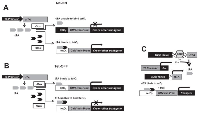
The Tet system can be used to conditionally activate gene expression in the mouse. A, In Tet-ON system Dox-binding to reverse tetracycline controlled transactivator (rtTA) leads to transcriptional activation of transgene. B, In the Tet-OFF system the tTA binds to tetO and induces transcription in the absence of Dox. In the presence of Dox the transgene expression is prevented. C, The boxed region indicates another application of the Tet-inducible transgenic mouse technology. In this example, tissue-specific (TS) Cre recom-binase can be used to globally activate rtTA (Tet-ON) in R26r-rtTA knock-in mice. When these 2 mice are crossed to a tetO transgenic mice, and when the triple transgenic mice are subjected to Dox-induction, the transgene of interest can be expressed in a Dox-inducible fashion. tTA indicates tetracycline-dependent transactivator; Dox, doxycycline (ligand); tetO7, Tet-responsive element.
The first demonstration of the Tet system in transgenic mice used the tTA approach in which E. coli’s Tn10 transposon fused to the VP16 transactivation domain from herpes simplex virus was used as a TetR for the Tet effector molecule.64 Gene induction became detectable in a few hours and reached its maximum level within a day.63 Several studies have indicated that the rtTA system is better suited for temporal control of rapid induction of gene expression.63
Recently, many investigators have combined the Tet-inducible system with the Tam-inducible Cre-loxP approach (Figure 5).63 This allows more flexibility producing more clinically relevant mouse models of human diseases. Several laboratories have integrated the Tet-inducible and Cre-loxP approaches and developed mice based on rtTA-dependent Dox-mediated gene induction following a Cre-mediated deletion to obtain a temporal and spatial or cell-, tissue-, and organ-specific gene targeting.65,66 Thus, gene targeting can be manipulated in specific cell types and lineages with a flexibility that is difficult to achieve with other methods. To make this system more sophisticated, a Tet-inducible CreERT2 has been targeted to the ROSA26 locus.67 By introducing Tet induction, this Tam-inducible Cre driver line permits highly selective dual Tet and Tam regulation of loxP recombination which could open new avenues for spatiotemporally controlled gene targeting in mice.
There are pitfalls inherent in Tet-inducible technology that is worth mentioning. The Tet system can be leaky.68 This can occur either through weak binding of rtTA to tetO even in the absence of Tet or Dox effectors, or through unwanted basal activity of the Tet-responsive promoter even in the absence of rtTA. This leakiness can result in unexpected phenotypes when Tet is absent.69 Although this leakiness can be tolerated in many experimental systems in which a phenotypic outcome are desired, it can be limiting when gene function is being investigated. Use of several Cre transgenic lines could obviate some of these difficulties. Refinements have also been made to rtTA (ie, rtTA2S-M2) to increase the sensitivity of the system and to reduce background expression.70 Additional control over background expression can also be achieved by using an inducible transcriptional repressor that can bind to the Tet-responsive promoter and thereby block background expression. For example, the TetR protein can be used to minimize leakiness in the system.71 In theory it is possible that the application of a tTA repressor with rtTA in the absence of Tet could prevent leaky expression through its binding to the TRE. This means that in the presence of Tet, Tet would bind to the tTA repressor and dissociate from the TRE, while at the same time the Tet-rtTA complex would bind to the TRE and induce transcription. Although initial efforts to test these possibilities have produced mixed results,72 refinements could be promising.
Another potential problem with the Tet system is that of tissue toxicity caused by overexpression of TetR proteins.68 This can also be avoided by producing multiple transgenic lines expressing varying levels of Tet repressor. Taken together, it is very important that proper control animals are used in Tet-inducible gene targeting experiments to avoid misleading results due to these pitfalls. Overall, the ability to regulate gene activity in spatiotemporal and reversible fashions has made the Tet-inducible approach a favorite technology of numerous mouse geneticists.
Targeting Single Nucleotide Polymorphisms Into the Mouse Genome
There are numerous examples of single nucleotide polymorphisms (SNPs) in both exons and introns, and also important functionally relevant regulatory noncoding sequences and enhancer sequences within intronic regions.73 Several epidemiological studies have suggested that SNPs may be involved in the susceptibility and pathogenesis of cardiac disease in humans,74 and it is known that functionally relevant SNPs can be in noncoding regulatory and intronic regions. For example, about one-third of mammalian coding transcripts are silenced by microRNAs (miRNAs), powerful regulators of gene expression that are involved in cardiac development and pathogenesis of cardiac disease.75,76 Although conventional or conditional gene targeting has been used in introducing SNPs, which were created by site-directed mutagenesis in the targeting vector, in most instances a residual loxP site is left behind, sometimes accompanied by a selectable marker gene such as neomycin.77 The concern is that insertion of a selectable marker and/or loxP or FRT site(s) in noncoding intronic regions could cause lethality, or a hypomorphic or other unwanted phenotype.78 – 80 This makes it difficult to separate the effect of neomycin or loxP from the effect of the SNP. At the vector design stage incorporating bioinformatic approaches such as TraFac,81 which provides information on the conserved cis sequences within a gene, or ORF and miR analysis to detect other embedded genes, can usually identify regions in the gene that should be avoided when placing loxPs, FRTs, and selectable cassettes. However, these approaches are not completely accurate, and there are undoubtedly other functionally relevant sequence motifs not yet identified.
Consequently, it may be worthwhile not to leave behind residual sequences that in and of themselves could complicate phenotype analysis. We have previously reported on the use of a Tag and Exchange gene targeting approach to introduce point mutations in the basic fibroblast growth factor gene to ablate the function of protein isoforms resulting from alternative, in frame translational start sites.82– 85 Alternative technologies have been used by others to introduce subtle mutations without the use of recombinase systems that necessarily leave residual sequences in intronic regions. The “Hit and Run” system was used to introduce a small mutation in the Hox2.6 gene without leaving residual sequences,86 and an “In and Out” approach used a sequence insertion and removal procedure to put a 4-bp insertion into the Hprt gene.87 Improvement of these approaches would be useful for future functional analysis on SNPs.
Nuclease-Mediated Gene Targeting
Zinc finger nucleases (ZFNs) are custom-engineered DNA-binding modules fused to the catalytic domain of Fok1 endonuclease that have been adopted for targeted editing of the mouse and rat genome (Figure 6).88,89 ZFNs are eukaryotic transcription factors that bind 3 bp of DNA per finger; binding site specificity depends on key residues within the ≈ 30aa ZFNs. The FokI cleavage domain can render a double-strand break at any user-specific sequence in the genome but must dimerize to be functional. Therefore, it is possible to cut within a unique genomic sequence of 18 to 24 nucleotides by designing a dimerizing pair of ZFNs, each containing a set of 3 to 4 strategically chosen ZFNs joined to a FokI catalytic domain (up to 6 fingers per monomer have been used). A donor DNA can be exogenously supplied to affect repair of the break by a homology-driven homologous recombination process, resulting in targeted gene replacement with a desired sequence. Alternatively, a double strand break stimulates the cellular process of nonhomologous end joining and results in the mis-repair of the DNA sequence. This error-prone process results in a KO genotype/phenotype.90
Figure 6. Nuclease-mediated gene targeting.

A, Diagrammatic representation of a pair of zinc finger nucleases (ZFNs) (top left) and transcription activator-like effector nucleases (TALENs) (top right) bound to DNA. Custom-made ZFNs and TALENs both have the FokI endonuclease fused to an array of zinc finger or transcription activator-like (TAL) effector (TALE) effector DNA-binding modules (gray triangles). Each ZFNs recognizes a 3-nucleotide sequence, whereas each of the DNA-binding modules of TALENs binds to a single nucleotide. ZFNs and TALENs arrays act as heterodimers, which create DNA cleavage at a user-specified genomic sequence. Zinc fingers (gray triangle) and TALENs (gray oval shapes) are shown with short vertical lines that indicate the main contacts of these nucleases with the DNA base pairs. FokI cleavage-encoding domains are shown as 2 large oval shapes (gray), with common cleavage sites, spaced by 4 bp, and indicated by vertical arrowheads. The spacer between the zinc finger or TALE binding sites is 6 bp, as indicated. In the presence of a homologous donor DNA (solid gray rectangular box, left), DNA repair can occur by homologous recombination using the donor as template. More recombination occurs near the original break, as indicated by the dark gray shading (right). The nonhomologous end joining results in mutations (eg, deletions, insertions, base substitutions) at the cleavage site (gray square, left). B, Scheme for producing gene-altered mice using nuclease-mediated gene targeting approach.
ZFNs can be introduced directly into fertilized single cell embryos in plasmid or mRNA form. The embryos are implanted into the foster mother and the newborn founder animals are identified by standard screening methods. KO mice and rats can be generated in about 4 months at remarkably high efficiencies (monoallelic 10%–15%, biallelic 1%) compared to the conventional ES cell based gene targeting approach in mice that usually takes about 9 months to a year. The ZFN break-induced events are somewhere between 1,000- and 1,000,000-fold more efficient than the conventional ES cell targeting and there is no need to use selection to find the targeted products. A major difficulty in reproducibly applying the ZFN technology is the fact that it is difficult to design the zinc finger set with the highest probability of successfully cutting the user-specified location in the genome. There is also a potential risk of off-target cleavage by ZFNs. This is also due to a lack of known fingers for some nucleotide triplets and context effects on the specificities of individual fingers in an array.
Another class of engineered nucleases, transcription activator-like effector (nucleases (TALENs) appears to overcome some of the challenges facing the ZFN technology.91 Similar to ZFNs, TALENs are made by fusing the catalytic domain of FokI to transcription activator-like effector repeat modules that are custom combined to target a unique stretch of genomic nucleotide sequence. Transcription activator-like effectors harbor ≈ 34 amino acid repeat domains that each binds 1 target base in the host genome with base specificity determined by 2 variable amino acids (Figure 6).92 TALENs have been successfully used in creating heritable changes in both zebrafish and rats.93,94 However, there are no gene-targeted mouse models generated using TALENs technology. Advantages of TALENs are that they are more designable than ZFNs because each module of TALENs binds 1 bp of DNA instead of 3 in ZFNs, and the TALEN specificity seems to be less influenced by neighboring DNA sequences. The TALENs also seem to be better than ZFNs at being restricted to their designed target. However, a limitation with TALENs is that the constructs for the TALENs are bigger than for ZFNs, largely because each module of 34 amino acids recognizes only 1 bp in the TALENs; whereas, each ZFN module of 30 amino acids recognizes 3. Although it is possible in principle to conditionally modify an allele for a tissue-specific disruption or overexpression using nuclease-mediated gene targeting techniques, a proof of principle experiment is still missing. Overall, nuclease-mediated gene targeting technology is at the cutting edge and needs to be perfected before it becomes ready to be applied for making conditional and inducible gene KOs and transgenic mice.
Mouse Models for miRNA Expression
miRNAs are 21 to 23 nucleotide long single-stranded RNAs. miRNAs are involved in numerous diverse cellular and developmental processes, and in the pathogenesis of several diseases, including cardiovascular disease.95 Sequence-specific base pairing of miRNA with target mRNAs reduces protein stability and translation. At least 30% of protein-coding genes appear to be conserved targets for miRNAs.95 The influence and importance of miRNAs as regulators of gene expression has led to their intensive study in the cardiovascular system.96 Only a limited number of individual miRNA KO mice have been described in the literature. Overall, several mouse models of miRNA expression have been successfully made by individual investigators using conditional mouse gene targeting approaches.97,98
The application of inducible technology in the field is growing rapidly.99 The analysis of miRNA KO alleles is complicated by the presence of several miRNA genes with related predicted specificities at multiple loci.100 Consequently, analysis of miRNA function will require the creation of tissue- or cell type-specific conditional and inducible as well as compound mutant mice of miRNA family members. The miRNA KO resource is a research toolbox of mutant alleles that can be used as reagents for studying miRNA function in a range of complementary approaches.101 The targeted alleles have been designed to be adaptable research tools that can be efficiently altered by recombinase-mediated cassette exchange to create reporter, conditional, and other allelic variants. Other examples of possible applications might include the variation of miRNA copy number and mutating individual miRNAs within clusters. This miRNA KO resource can be searched electronically and is available from ES cell repositories (http://www.knockoutmouse.org/martsearch).50 Finally, it is yet to be seen whether emerging technologies such as nuclease-mediated gene targeting can successfully manipulate miRNA in conditional or inducible fashion.
Manipulating Alternatively Splicing of Genes Through Gene Targeting
Many mammalian genes undergo alternative splicing.102 Alternative splicing results in the differential inclusion or exclusion of a specific exon in different cell types, retention of introns, exclusion of a portion of an exon, and mutually exclusive inclusion of exons.103 Importantly, any of these differential patterns have the potential to alter the open reading frame of the resultant mRNA or modify the cis-regulatory elements that control mRNA stability or translation. Thus, the proper regulation of alternative splicing is important for maintaining cellular protein repertoire, and changes in splicing can significantly alter cellular function. Gene targeting in mice has been used to create in vivo models to study the regulation and consequences of alternative splicing.104 –106 The evidence accumulated so far argues for an important role of alternative splicing in mammalian development and tissue function, and highlights the importance of various protein isoforms in vivo.
Various approaches have been used to generate mice with modified alternatively spliced forms (Figure 7). Briefly, conditional mouse genetics approaches can be used to manipulate alternative splicing of a gene in a tissue-specific fashion. Furthermore, a stop codon can be placed to eliminate expression of all alternatively spliced isoforms carrying an upstream exon, but allowing expression of downstream exons containing alternatively spliced variants. Another similar approach is to destroy the splice sites of the target exon. Furthermore, 1 exon can be replaced by another exon or an unrelated coding region to obtain the expression of an isoform in a tissue, where it is usually not expressed. This technique requires careful consideration of the targeting construct, as exonic regulatory sequences of the deleted exon might be important for tissue-specific splicing of the gene. Random retroviral insertion of marker genes and subsequent sequencing of the integration site has also generated many commercially available targeted ES cells. In this method, stop codons, loxPs, and other gene modification can be made along with the marker gene into the gene. A conventional gene KO is used to eliminate the whole or parts of the gene to disrupt specific alternatively spliced isoforms. In the case of a partial removal of a gene, the expression of the resultant truncated form of alternatively spliced variant mRNA and protein should be taken into consideration. Lastly, the utility of ZFNs and TALENs in conditional or inducible gene targeting to manipulate alternative splicing in mice remains to be tested.
Figure 7. Mouse genetic engineering to manipulate alternative splicing in mice.
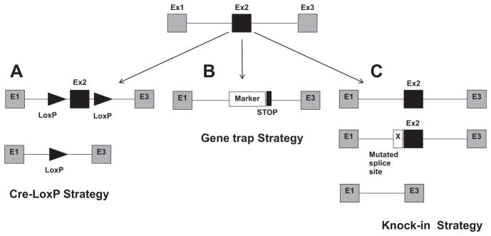
Three exons (1–3) in a gene are shown. Three examples are provided to splice out or delete Exon 2 (black box). A, Exon 2 is floxed and can be deleted in conditional/inducible fashion. B, One STOP codon can be introduced along with the marker gene into the gene using random retroviral insertion or gene trap method. C, Depletion of isoforms containing Exon 2 by causing the destruction of the splice site. See text for details.
Cardiac Cell Type-Specific Inducible Gene Targeting in Mice
Several important genes that are involved in human cardiovascular disease are expressed in multiple cardiac cell types (Figure 1). In addition, the foundation of many adult congenital cardiovascular diseases can be traced to abnormal cardiovascular development.3,6,107 Thus, careful mutational analysis of genes in multiple cardiac cell lineages during development and into the adult heart is required to delineate the complex function of many genes in the heart. Tam- and Tet-inducible strategies have been applied to achieve this end6,7,108 and have been successful in demonstrating gene function in cardiac structure and function, cardiac physiology and pathophysiology, cardiomechanics, cardiac calcium handling, and cardiac stress response.11 In clinical terms, the development of cardiac-inducible/reversible gene targeting approaches provide translational potential.9 Because of the success and feasibility of cardiac-specific inducible gene targeting, the number of inducible cardiac cell-type-specific Cre mice is increasing (see the tetmouse database: www.creline.org). Here, we briefly describe the importance and practical aspects of some of the major cardiac lineage-specific CreERT2 and rtTA driver lines (Table).
Myocardium
The human α-myosin heavy chain (αMHC)33,68,109,110 and rat troponin T (Tnnt2)111 promoters are prominently used in Tam- and/or Tet-inducible gene targeting strategies for the heart. Because both αMHC and Tnnt2 are significantly expressed in the heart during embryonic development, inducible strategies must be used to drive Cre or rtTA in the postnatal heart.112,113 Tamoxifen-regulated αMHC-CreERT2 mice are by far the most extensively used.33 Inducible myocardial gene targeting in adult hearts has also been achieved by RU486 (antiprogestin)-inducible αMHC-CrePR1 mice.109
There are some inherent pitfalls in using Tam or RU486 and an αMHC promoter to drive inducible cre expression in the postnatal heart. Tamoxifen is better tolerated than RU486 in both mouse embryos and adult mice, and therefore more investigators have preferred αMHC-CreERT2 over αMHC-CrePR1. Excess Cre levels in general can be toxic to cells, and in the heart there is evidence that high levels of cardiac cre driven by the αMHC promoter is cardiotoxic between 8 to 12 months of age.7,114 Another minor caveat with αMHC-CreERT2 mice is that Tam induction can temporarily cause cardiac dysfunction which, however, resolves after 1 to 2 weeks of treatment. In general, however, most of the problems associated with the use of CreERT2 mice can be avoided by using proper control mice.
Tet induction is another widely used approach to manipulate genes in myocardial-specific and inducible fashions in mouse embryos and adult mice. The rat αMhc promoter was initially used to drive tTA in transgenic mice.110 This strategy was inefficient for many investigators, probably due in part to promoter leakiness, thereby requiring several transgenic lines to get reliable data. Consequently, the mouse αMhc promoter was reengineered to drive low levels of tTA in transgenic mice.68 This resulted in more robust myocardial-specific transgene expression that can be controlled by Dox treatment. These investigators also found that modest to high levels of tTA can be cardiotoxic and can cause cardiac disease and lethality. Attempts to make αMHC-rtTA transgenic mice were not very successful.115
More recently, both rtTA and tetO binary components of the Tet-inducible system have been combined in transgenic mice (Tnnt2-rtTA/tetO mice) in which the rat cardiac Tnn2 promoter drives the expression of rtTA, and tetO drives the cre. This Tnnt2-rtTA effectively activates tetO cre expression in cardiomyocytes following Dox treatment. These investigators demonstrated that Tnnt2-rtTA/tetO mice can be used to drive Dox-dependent myocardial-specific gene deletion in embryos and adults. More importantly, there seems to be no effect on cardiac structure and function in the absence or presence of Dox in Tnnt2-rtTA/tetO mice alone. The obvious advantage of this system is that although it robustly drives cre expression in the entire myocardium, it also requires only 1 mouse line to cross with floxed mice to conditionally delete the gene. This system has already been adopted by other investigators116,117 and should attract more attention and scrutiny from the cardiac community.
One of the pitfalls in using Tet induction in Cre-loxP–based conditional KO experiments is that rtTA-driven cre expression acts as a trigger for Cre-mediated gene deletion. This leads to irreversible gene deletion in all of the descendent cells, even if Dox treatment is removed. This is in contrast to transgenic mice in which Dox allows for the induction of gene expression by addition or removal of Dox. Finally, there will always be concerns about differences in cardiac phenotypes that relate to characteristics of different myocardial promoters driving inducible cre. For example, the cardiac phenotypes of myocardial Smad4 conditional KO mice range from embryonic cardiac malformations (valves and myocardium) to cardiac hypertrophy in adults, depending on which myocardial promoters driving cre is used.118 Notably, the Smad4flox/flox mice used in all of these studies were of a common lineage,119 suggesting that the differences in the phenotypes related to the characteristics of the different myocardial promoters and not genetic background differences. Overall, however, it is possible to design carefully controlled experiments to harness the power of Tam- and Tet-inducible systems to regulate cre expression.
Cardiac Endothelium and Fibroblasts
There is evidence to suggest that defects in valve formation during embryonic development predispose patients to progressive postnatal valvular disease.31,107 The process of valve formation begins with an epithelial mesenchymal transition (EMT) (embryonic day [E] 9.5 to E10.5 in mice) that forms the endocardial cushions,120 which are the primordia of valves and septa, and which later become mature structures through remodeling (E10.5–E18.5) and valve elongation and maturation (E14.5–1 week after birth).121 During the maturation process the extracellular matrix of atrioventricular (AV) or outflow tract valves is organized into atrialis or ventricularis, spongiosa, and fibrosa layers.107 Cardiac cell lineage-specific analysis has found that the majority of valve mesenchyme is derived from the valve endocardium.122 Overall, there are 2 major cell types in adult cardiac valves that are represented by cardiac valve endocardium and valve interstitial cells or valvular fibroblasts (Figure 1).107
Tie2-Cre mice, which drive recombination in the entire endocardium and vascular endothelium, are the most extensively used Cre drivers in the cardiac valve community (reviewed 107). A Tam-inducible Tie2-CreERT2 has also been made and successfully applied in numerous systems.123 The obvious pitfall of this mouse line is that it is not specific to valve endocardium and that it also functions in the hematopoietic cell lineage.124 Recently, it has been reported that Tie2-Cre mice exhibit activity in the female germ line.125 Because this can result in the inheritance of a nontissue-specific KO allele in the second generation, careful breeding plans and appropriate controls are needed if Tie2-Cre promoters are used in inducible conditional gene targeting experiments. Some of the problems of Tie2-Cre mice are mitigated in VE-cadherin–CreERT2 mice,126 which also drive recombination in both the entire endocardium and vascular endothelium but which have minimal activity in the hematopoietic cell lineage. However, some investigators have reported less efficient cardiac endothelial-specific Cre-mediated gene deletion in VE-cadherin–CreERT2 mice as compared to Tie-2–Cre mice.127
As far as valvular fibroblasts are concerned, the only published information available is from the Tcf21-CreERT2 knockin mouse line, which has targeted insertion of the Tam-inducible Cre recombinase, MerCreMer, at the Tcf21 locus.128 Following Tam treatment at E10.5, E18.5 hearts were analyzed for reporter activity using the R26RYFP reporter.129 Reporter expression was detectable in the atrioventricular valves and absent from the outflow tract valves. Although reporter expression seems to be present in the valvular fibroblasts at E18.5, Tam induction at different time points in cardiac development and at postnatal stages is needed to capture the spatiotemporal nature and the cell type specificity of the reporter expression in valve development and homeostasis. In addition, Col2-CreERT2 mice have been made and need to be verified for inducible valve-specific conditional gene targeting in mice.130 A Col2a1-Cre driver mouse line provides some clues about the utility of the inducible Cre driver line.131 Col2a1-Cre–mediated recombination has been shown to occur in developing valves between E15.5 and E18.5 during heart development. However, on closer comparison of Col2a1-Cre mice to NFatc1-Cre mice that exclusively drive recombination in cushion endothelial cells, an intriguing similarity was found, suggesting that Col2a1-Cre recombination may in fact occur in cushion endocardium and not in cushion mesenchyme.131,132 Hence, this needs to be resolved. Also, there is no formal confirmation as to whether Col2a1-Cre mice can drive Cre expression in adult heart valves.
More inducible approaches need to be developed with promoters that only come on after cardiac stress or disease onset. For example, pressure-overload–induced cardiac hypertrophy is accompanied by cardiac fibrosis, which can be successfully marked with a noninducible Postn-Cre.133 Another example is that of myofibroblast activation, which predisposes valve disease. For these studies it is possible to manipulate genes in an inducible fashion using Postn and αSMA actin promoters. The existing αSMA-CreERT2 mice134 need to be characterized because they may provide an important tool to both the cardiac valve and cardiac hypertrophy and fibrosis research communities. In addition, because there is no inducible Cre-driver line for cardiac fibro-blasts, Postn-Cre-CreERT2 should be developed and characterized. This would provide the ability to turn genes on and off in cardiac fibroblasts which would open new avenues to uncouple and/or treat cardiac hypertrophy and fibrosis.
Finally, there are SM22a-(r)tTA and Tie2-(r)tTA mice that are available through several mouse repositories (see the tetmouse database). Recently, utility of the Tet inducible system has been shown in a valvulogenesis study.135 Collectively, there are limited resources and approaches available to the cardiac valve community for inducible conditional gene targeting in mouse models to study heart valve development and the pathogenesis of valvular heart diseases.
Cardiac Neural Crest Cells
Cardiac neural crest cells (NCCs) originate from the neural tube and migrate to the heart to form the aorticopulmonary septum of the heart. Specifically, NCC contributes to smooth muscle cells of the aortic wall, the ventricular and aorticopulmonary septa and both semilunar and AV valves. Disruption of cardiac NCCs commonly leads to heart deformations including persistent truncus ateriosus, ventricular septal defects, and aortic arch abnormalities (reviewed in 136). Studies in both mice and chick suggest they can also enter the heart from the venous inflow as well as the usual arterial outflow region, and may contribute to the adult semilunar and atrioventricular valves as well as part of the cardiac conduction system.137 There is still some debate whether NCC contributes to the AV valves in mice.122,137 For conditional gene targeting, the Wnt1-Cre is the most commonly used Cre-driver for the cardiac NCC during heart development.138 Because of the tremendous success of Wnt1-Cre mice in tracing NCC lineage, the Wnt1 promoter was also used to generate Wnt1-CreERT2 mice.139 Surprisingly, there has been no published literature so far about the use of Wnt1-CreERT2 mice in either cardiac NCC lineage mapping or NCC-specific Tam-dependent conditional deletion or overexpression of genes during heart development and in adults.
Epicardium and Epicardium-Derived Cells
Epicardium is the outermost mesothelial cellular layer of the heart, which plays essential roles in heart development and cardiac homeostasis, repair, and regeneration.140 –142 In heart development, epicardium gives rise to cardiac fibroblasts by epicardial EMT. It also contributes to cardiac endothelium and coronary smooth muscle cells. Under pathological conditions such as after myocardial infarction, epicardial cells also undergo EMT and produce myofibroblasts, fibroblasts, and smooth muscles.116,142 Inducible epicardial cell-specific conditional gene targeting is becoming a powerful tool to dissect epicardial cell function during heart development and adult heart under both physiological and pathophysiological conditions. The Wt1-CreERT2 and Tcf21-CreERT2 are the only well characterized inducible epicardial Cre mice currently available.128,143 Epicardial cell lineage cell tracking after Tam treatment showed that epicardial cells contribute to epicardium-derived cells and to cardiomyocytes.144 Lineage tracking also indicates that epicardium contributes to formation of the mammalian annulus fibrosis through EMT.141,143,145 Tam induction of Wt1-CreERT2 mice revealed that in uninjured postnatal hearts the epicardial cells were quiescent. Myocardial infarction stimulated formation of epicardium-derived cells, which remained in a thickened layer on the surface of the heart. The epicardium-derived cells did not adopt cardiomyocyte or coronary endothelial fates, but rather differentiated into mesenchymal cells expressing fibroblast and smooth muscle markers.116 Additional inducible epicardial Cre driver lines are needed to ensure and further validate these findings.
Tcf21-CreERT2 is another valuable Tam-inducible epicardial Cre driver line.128 Tam-inductions were performed at 2 embryonic time points (E10.5 and E14.5) to determine the ROSA26 reporter expression in epicardium and its derivatives. By contrast, hearts from E18.5 embryos that were induced at E14.5 had very little labeling of epicardial cells and showed reporter expression predominantly in cells scattered within the myocardium (perhaps epicardium-derived cells). The authors suggested that this pattern of expression was likely caused by migration of cells from the epicardium into the myocardium following epicardial EMT. Cre activity was also analyzed in 4-week-old hearts where it was detected in a vast majority of interstitial cells that were not endothelial or vascular smooth muscle cells. Tcf21 is heavily expressed in proepicardium at E9.5 during heart development.146 It remains to be seen whether Tcf21-CreERT2 mice can drive Cre-activity in the proepicardium during early stages of heart development. This will be very informative for the cardiac research community.
Cardiac Conduction System
Cardiac conduction system (CCS) function is essential for proper rhythmic contraction of the heart. The CCS is made of a network of cardiomyocytes, which generate and propagate electric impulses in the heart (Figure 1).147 Electric impulses are produced in the sinoatrial node, which propagate through fast-conducting atrial myocardium and later delayed at the AV node. From the AV node, the electric impulse is rapidly conducted through the ventricular conduction system, consisting of the AV (His) bundle and bundle branches and peripheral Purkinje network. Electric impulses are finally transmitted from Purkinje fibers to working myocardium. Several genes that are important for the CCS are also required for myocardial development and function.
Although there are several good inducible Cre mouse models available for specifically targeting genes in myocardium, there are fewer choices for the CCS. HCN4-CreERT2 knockin (at HCN4 locus) mice drive Tam-dependent Cre activity in the sinoatrial node and AV node, but not the AV bundle or bundle branches.148 Tamoxifen-inducible Cre activity in Cx40-CreERT2 knockin (at Cx40 locus) mice was seen in the AV bundle, bundle branches, and Purkinje fibers, but not the AV node.149 A major pitfall of Cx40-CreERT2 mice is that the Cre activity is also present in non-CCS (ie, atrial cardiomyocytes, coronary vessels). Recently, mink-CreERT2 BAC transgenic mice were made.150 Tam-induced Cre activity was found in the AV node, AV bundle, and bundle branches of adult transgenic mice. There are concerns that it will be difficult to interpret the findings based on the haploinsufficient HCN4-CreERT2 and Cx40-CreERT2 knockin mice because both HCN4 and Cx40 are important players in the CCS. This issue can be circumvented in the mink-CreERT2 transgenic mice, which are CCS-specific and have an intact endogenous mink locus.
Perspectives
Site-specific recombinases have proven to be remarkable tools for many aspects of mouse experimentation. However, these tools have limitations. Knowledge of their strengths and weaknesses is vital for optimizing their application. There are several good Cre driver lines to induce gene manipulations in myocardium. There is a need for more Cre driver lines for inducible gene targeting in epicardium. There is no satisfactory Cre driver line for inducible conditional gene targeting in cardiac valve endocardium and valve mesenchyme for both mouse embryos and adult mice. Serious efforts are needed to identify, develop, and validate these resources for the cardiac valve community.
Because conditional KO or transgenic mouse technology limits gene mutation or overexpression to a specific cell type/tissue/organ or to a specific life stage, generation of improved mouse models of adult cardiac diseases heavily depends on the progress and innovation in inducible conditional gene targeting technology. The difference between conventional conditional and inducible conditional gene targeting approaches has recently been exemplified by Braf gene inactivation in forebrain neurons where both common and distinct phenotypes were found.151 This also applies to cardiovascular disease studies. For example, although considerable progress has been made in the past 20 years in delineating mechanisms underlying cardiovascular disease by using conventional gene targeting strategies, future progress will come largely from inducible conditional gene targeting approaches. These can better model the human disease conditions and identify pathways involved in unintended side-effects of potential therapies.
Table.
Validated Mouse Models for Cardiac-Specific Inducible and/or Conditional Gene Targeting
| Cardiac Cell Type | Cre Mice | Cre Expression | Induction | Ref. |
|---|---|---|---|---|
| Myocardium | αMHC-CreERT2 | Myocardium | Tamoxifen | 33 |
| αMHC-CrePR1 | RU486 | 109 | ||
| αMHC-tTA | Doxycycline | 68 | ||
| Tnnt2-rtTA/tetO | Doxycycline | 111 | ||
| Endothelium | Tie2-CreERT2 | Cardiac and vascular endothelium | Tamoxifen | 123 |
| VE-cadherin–CreERT2 | Cardiac and vascular endothelium | Tamoxifen | 126 | |
| Tie2-(r)tTA | ND | Doxycycline | Tetmouse database | |
| Cardiac and valvular fibroblasts | Tcf21-CreERT2 | Epicardium, cardiac fibroblasts, valve interstitial cells | Tamoxifen | 128 |
| Col2-CreERT2 | ND | Tamoxifen | 130 | |
| αSMA-CreERT2 | ND | Tamoxifen | 134 | |
| SM22a-(r)tTA | ND | Doxycycline | Tetmouse database | |
| Cardiac neural crest cells | Wnt1-CreERT2 | ND | Tamoxifen | 139 |
| Epicardium | Wt1-CreERT2 | Epicardium, epicardium-drived cells | Tamoxifen | 143 |
| Tcf21-CreERT2 | Epicardium, cardiac fibroblasts, valve interstitial cells | Tamoxifen | 128 | |
| Cardiac conduction system | HCN4-CreERT2 | SA node, AV node | Tamoxifen | 148 |
| Cx40-CreERT2 | AV bundle, bundle branches, Purkinje fibers, atrial cardiomyocytes, coronary vessels | Tamoxifen | 149 | |
| mink-CreERT2 | AV node, AV bundle, bundle branches | Tamoxifen | 150 |
αMHC indicates α-myosin heavy chain; CreER, Cre with the mutated progesterone receptor ligand binding domain; Tie2, tyrosine-protein kinase receptor TIE-2; Tcf21, transcription factor 21; Col2, collagen 2; αSMA, alpha smooth muscle actin; SM22a, SM22alpha; Wnt1, wingless-related MMTV integration site 1; Wt1, Wilms tumor 1 homolog; HCN4, hyperpolarization-activated, cyclic nucleotide-gated K+4; Cx40, connexin 40; ND, not determined; mink, Kcne1l potassium voltage-gated channel; tTA, tetracycline-controlled transactivator; Tnnt2, rat troponin T; rtTA, reverse tetracycline controlled transactivator; tetO, tetracycline operator; AV, atrioventricular.
Acknowledgments
Sources of Funding
This study was supported by funding from Program for Developmental Biology and Neonatal Medicine at Indiana University College of Medicine (to M.A.), and the BIO5 Institute at University of Arizona (to T.D.).
Non-standard Abbreviations and Acronyms
- αMHC
α-myosin heavy chain
- AV
atrioventricular
- CCS
cardiac conduction system
- CMV
minimal cytomegalovirus promoter
- CreER
Cre with the mutant ligand-binding domain of human estrogen receptor
- Dox
doxycycline
- EMT
epithelial mesenchymal transition
- KO
knockout
- miRNA
MicroRNA
- NCC
neural crest cells
- rtTA
reverse tetracycline controlled transactivator
- (r)tTA
tTA or rtTA
- SNPs
single nucleotide polymorphisms
- TALENs
transcription activator-like effector nucleases
- Tam
tamoxifen
- Tet
tetracycline
- tetO
Tet operator
- TetR
Tet repressor
- Tnnt2
rat troponin T
- tTA
tetracycline-controlled transactivator
- ZFNs
zinc finger nucleases
Footnotes
Disclosures
None.
References
- 1.Jackson CF, Wenger NK. Cardiovascular disease in the elderly. Rev Esp Cardiol. 2011;64:697–712. doi: 10.1016/j.recesp.2011.05.001. [DOI] [PubMed] [Google Scholar]
- 2.Khairy P, Hosn JA, Broberg C, Cook S, Earing M, Gersony D, Kay J, Landzberg MJ, Nickolaus MJ, Opotowsky S, Valente AM, Warnes C, Webb G, Gurvitz MZ. Multicenter research in adult congenital heart disease. Int J Cardiol. 2008;129:155–159. doi: 10.1016/j.ijcard.2008.03.014. [DOI] [PubMed] [Google Scholar]
- 3.Doetschman T, Barnett JV, Runyan RB, Camenisch TD, Heimark RL, Granzier HL, Conway SJ, Azhar M. Transforming growth factor beta signaling in adult cardiovascular diseases and repair. Cell Tissue Res. 2012;347:203–223. doi: 10.1007/s00441-011-1241-3. [DOI] [PMC free article] [PubMed] [Google Scholar]
- 4.Gittenbergerde Groot AC, Bartelings MM, DeRuiter MC, Poelmann RE. Basics of cardiac development for the understanding of congenital heart malformations. Pediatr Res. 2005;57:169–176. doi: 10.1203/01.PDR.0000148710.69159.61. [DOI] [PubMed] [Google Scholar]
- 5.Snider P, Conway SJ. Probing human cardiovascular congenital disease using transgenic mouse models. Prog Mol Biol Transl Sci. 2011;100:83–110. doi: 10.1016/B978-0-12-384878-9.00003-0. [DOI] [PMC free article] [PubMed] [Google Scholar]
- 6.Yutzey KE, Robbins J. Principles of genetic murine models for cardiac disease. Circulation. 2007;115:792–799. doi: 10.1161/CIRCULATIONAHA.106.682534. [DOI] [PubMed] [Google Scholar]
- 7.Molkentin JD, Robbins J. With great power comes great responsibility: using mouse genetics to study cardiac hypertrophy and failure. J Mol Cell Cardiol. 2009;46:130–136. doi: 10.1016/j.yjmcc.2008.09.002. [DOI] [PMC free article] [PubMed] [Google Scholar]
- 8.Rentschler S, Jain R, Epstein JA. Tissue-tissue interactions during morphogenesis of the outflow tract. Pediatr Cardiol. 2010;31:408–413. doi: 10.1007/s00246-009-9611-2. [DOI] [PMC free article] [PubMed] [Google Scholar]
- 9.Robbins J. Twenty years of gene targeting: what we don’t know. Circ Res. 2011;109:722–723. doi: 10.1161/CIRCRESAHA.111.249912. [DOI] [PubMed] [Google Scholar]
- 10.Doetschman T. Interpretation of phenotype in genetically engineered mice. Lab Anim Sci. 1999;49:137–143. [PubMed] [Google Scholar]
- 11.Moga MA, Nakamura T, Robbins J. Genetic approaches for changing the heart and dissecting complex syndromes. J Mol Cell Cardiol. 2008;45:148–155. doi: 10.1016/j.yjmcc.2008.06.002. [DOI] [PMC free article] [PubMed] [Google Scholar]
- 12.Stevens LC, Little CC. Spontaneous testicular teratomas in an inbred strain of mice. Proc Natl Acad Sci U S A. 1954;40:1080–1087. doi: 10.1073/pnas.40.11.1080. [DOI] [PMC free article] [PubMed] [Google Scholar]
- 13.Stevens LC. Embryonic potency of embryoid bodies derived from a transplantable testicular teratoma of the mouse. Dev Biol. 1960;2:285–297. doi: 10.1016/0012-1606(60)90010-5. [DOI] [PubMed] [Google Scholar]
- 14.Evans MJ. The isolation and properties of a clonal tissue culture strain of pluripotent mouse teratoma cells. J Embryol Exp Morphol. 1972;28:163–176. [PubMed] [Google Scholar]
- 15.Brinster RL. The effect of cells transferred into the mouse blastocyst on subsequent development. Journal of Experimental Medicine. 1974;140:1049–1056. doi: 10.1084/jem.140.4.1049. [DOI] [PMC free article] [PubMed] [Google Scholar]
- 16.Papaioannou VE, McBurney MW, Gardner RL, Evans MJ. Fate of teratocarcinoma cells injected into early mouse embryos. Nature. 1975;258:70–73. doi: 10.1038/258070a0. [DOI] [PubMed] [Google Scholar]
- 17.Evans MJ, Kaufman MH. Establishment in culture of pluripotential cells from mouse embryos. Nature. 1981;292:154–156. doi: 10.1038/292154a0. [DOI] [PubMed] [Google Scholar]
- 18.Martin GR. Isolation of a pluripotent cell line from early mouse embryos cultured in medium conditioned by teratocarcinoma stem cells. Proc Natl Acad Sci U S A. 1981;78:7634–7638. doi: 10.1073/pnas.78.12.7634. [DOI] [PMC free article] [PubMed] [Google Scholar]
- 19.Bradley A, Evans M, Kaufman MH, Robertson E. Formation of germ-line chimaeras from embryo-derived teratocarcinoma cell lines. Nature. 1984;309:255–256. doi: 10.1038/309255a0. [DOI] [PubMed] [Google Scholar]
- 20.Robertson E, Bradley A, Kuehn M, Evans M. Germ-line transmission of genes introduced into cultured pluripotential cells by retroviral vector. Nature. 1986;323:445–448. doi: 10.1038/323445a0. [DOI] [PubMed] [Google Scholar]
- 21.Gossler A, Doetschman T, Korn R, Serfling E, Kemler R. Transgenesis by means of blastocyst-derived embryonic stem cell lines. Proc Natl Acad Sci U S A. 1986;83:9065–9069. doi: 10.1073/pnas.83.23.9065. [DOI] [PMC free article] [PubMed] [Google Scholar]
- 22.Doetschman T, Gregg RG, Maeda N, Hooper ML, Melton DW, Thompson S, Smithies O. Targetted correction of a mutant HPRT gene in mouse embryonic stem cells. Nature. 1987;330:576–578. doi: 10.1038/330576a0. [DOI] [PubMed] [Google Scholar]
- 23.Thomas KR, Capecchi MR. Site-directed mutagenesis by gene targeting in mouse embryo- derived stem cells. Cell. 1987;51:503–512. doi: 10.1016/0092-8674(87)90646-5. [DOI] [PubMed] [Google Scholar]
- 24.Koller BH, Hagemann LJ, Doetschman T, Hagaman JR, Huang S, Williams PJ, First NL, Maeda N, Smithies O. Germ-line transmission of a planned alteration made in a hypoxanthine phosphoribosyltransferase gene by homologous recombination in embryonic stem cells. Proc Natl Acad Sci U S A. 1989;86:8927–8931. doi: 10.1073/pnas.86.22.8927. [DOI] [PMC free article] [PubMed] [Google Scholar]
- 25.Thompson S, Clarke AR, Pow AM, Hooper ML, Melton DW. Germ line transmission and expression of a corrected HPRT gene produced by gene targeting in embryonic stem cells. Cell. 1989;56:313–321. doi: 10.1016/0092-8674(89)90905-7. [DOI] [PubMed] [Google Scholar]
- 26.Johnson RS, Sheng M, Greenberg ME, Kolodner RD, Papaioannou VE, Spiegelman BM. Targeting of nonexpressed genes in embryonic stem cells via homologous recombination. Science. 1989;245:1234–1236. doi: 10.1126/science.2506639. [DOI] [PubMed] [Google Scholar]
- 27.Mansour SL. Gene targeting in murine embryonic stem cells: introduction of specific alterations into the mammalian genome. Genet Anal Tech Appl. 1990;7:219–227. doi: 10.1016/0735-0651(90)90004-y. [DOI] [PubMed] [Google Scholar]
- 28.Koller BH, Smithies O. Altering genes in animals by gene targeting. [Review] Annual Review of Immunology. 1992;10:705–730. doi: 10.1146/annurev.iy.10.040192.003421. [DOI] [PubMed] [Google Scholar]
- 29.Collins FS, Rossant J, Wurst W. A mouse for all reasons. Cell. 2007;128:9–13. doi: 10.1016/j.cell.2006.12.018. [DOI] [PubMed] [Google Scholar]
- 30.Lindsay ME, Dietz HC. Lessons on the pathogenesis of aneurysm from heritable conditions. Nature. 2011;473:308–316. doi: 10.1038/nature10145. [DOI] [PMC free article] [PubMed] [Google Scholar]
- 31.Conway SJ, Doetschman T, Azhar M. The inter-relationship of periostin, TGF beta, and BMP in heart valve development and valvular heart diseases. Scientific World Journal. 2011;11:1509–1524. doi: 10.1100/tsw.2011.132. [DOI] [PMC free article] [PubMed] [Google Scholar]
- 32.Branda CS, Dymecki SM. Talking about a revolution: The impact of site-specific recombinases on genetic analyses in mice. Dev Cell. 2004;6:7–28. doi: 10.1016/s1534-5807(03)00399-x. [DOI] [PubMed] [Google Scholar]
- 33.Sohal DS, Nghiem M, Crackower MA, Witt SA, Kimball TR, Tymitz KM, Penninger JM, Molkentin JD. Temporally regulated and tissue-specific gene manipulations in the adult and embryonic heart using a tamoxifen-inducible Cre protein. Circ Res. 2001;89:20–25. doi: 10.1161/hh1301.092687. [DOI] [PubMed] [Google Scholar]
- 34.Lakso M, Sauer B, Mosinger B, Jr, Lee EJ, Manning RW, Yu SH, Mulder KL, Westphal H. Targeted oncogene activation by site-specific recombination in transgenic mice. Proc Natl Acad Sci U S A. 1992;89:6232–6236. doi: 10.1073/pnas.89.14.6232. [DOI] [PMC free article] [PubMed] [Google Scholar]
- 35.Gu H, Zou YR, Rajewsky K. Independent control of immunoglobulin switch recombination at individual switch regions evidenced through Cre-loxP-mediated gene targeting. Cell. 1993;73:1155–1164. doi: 10.1016/0092-8674(93)90644-6. [DOI] [PubMed] [Google Scholar]
- 36.Dymecki SM. Flp recombinase promotes site-specific DNA recombination in embryonic stem cells and transgenic mice. Proc Natl Acad Sci U S A. 1996;93:6191–6196. doi: 10.1073/pnas.93.12.6191. [DOI] [PMC free article] [PubMed] [Google Scholar]
- 37.Anastassiadis K, Fu J, Patsch C, Hu S, Weidlich S, Duerschke K, Buchholz F, Edenhofer F, Stewart AF. Dre recombinase, like Cre, is a highly efficient site-specific recombinase in E. coli, mammalian cells and mice. Dis Model Mech. 2009;2:508–515. doi: 10.1242/dmm.003087. [DOI] [PubMed] [Google Scholar]
- 38.Kuhn R, Schwenk F, Aguet M, Rajewsky K. Inducible gene targeting in mice. Science. 1995;269:1427–1429. doi: 10.1126/science.7660125. [DOI] [PubMed] [Google Scholar]
- 39.Utomo AR, Nikitin AY, Lee WH. Temporal, spatial, and cell type-specific control of Cre-mediated DNA recombination in transgenic mice. Nat Biotechnol. 1999;17:1091–1096. doi: 10.1038/15073. [DOI] [PubMed] [Google Scholar]
- 40.Saam JR, Gordon JI. Inducible gene knockouts in the small intestinal and colonic epithelium. J Biol Chem. 1999;274:38071–38082. doi: 10.1074/jbc.274.53.38071. [DOI] [PubMed] [Google Scholar]
- 41.Kaspar BK, Vissel B, Bengoechea T, Crone S, Randolph-Moore L, Muller R, Brandon EP, Schaffer D, Verma IM, Lee KF, Heinemann SF, Gage FH. Adeno-associated virus effectively mediates conditional gene modification in the brain. Proc Natl Acad Sci U S A. 2002;99:2320–2325. doi: 10.1073/pnas.042678699. [DOI] [PMC free article] [PubMed] [Google Scholar]
- 42.Brooks AI, Muhkerjee B, Panahian N, Cory-Slechta D, Federoff HJ. Nerve growth factor somatic mosaicism produced by herpes virus-directed expression of cre recombinase. Nat Biotechnol. 1997;15:57–62. doi: 10.1038/nbt0197-57. [DOI] [PubMed] [Google Scholar]
- 43.Schmidt EE, Taylor DS, Prigge JR, Barnett S, Capecchi MR. Illegitimate Cre-dependent chromosome rearrangements in transgenic mouse spermatids. Proc Natl Acad Sci U S A. 2000;97:13702–13707. doi: 10.1073/pnas.240471297. [DOI] [PMC free article] [PubMed] [Google Scholar]
- 44.Loonstra A, Vooijs M, Beverloo HB, Allak BA, van DE, Kanaar R, Berns A, Jonkers J. Growth inhibition and DNA damage induced by Cre recombinase in mammalian cells. Proc Natl Acad Sci U S A. 2001;98:9209–9214. doi: 10.1073/pnas.161269798. [DOI] [PMC free article] [PubMed] [Google Scholar]
- 45.Naiche LA, Papaioannou VE. Cre activity causes widespread apoptosis and lethal anemia during embryonic development. Genesis. 2007;45:768–775. doi: 10.1002/dvg.20353. [DOI] [PubMed] [Google Scholar]
- 46.Mahonen AJ, Airenne KJ, Lind MM, Lesch HP, Yla-Herttuala S. Optimized self-excising Cre-expression cassette for mammalian cells. Biochem Biophys Res Commun. 2004;320:366–371. doi: 10.1016/j.bbrc.2004.05.175. [DOI] [PubMed] [Google Scholar]
- 47.Azhar M, Yin M, Bommireddy R, Duffy JJ, Yang J, Pawlowski SA, Boivin GP, Engle SJ, Sanford LP, Grisham C, Singh RR, Babcock GF, Doetschman T. Generation of mice with a conditional allele for transforming growth factor beta 1 gene. Genesis. 2009;47:423–431. doi: 10.1002/dvg.20516. [DOI] [PMC free article] [PubMed] [Google Scholar]
- 48.Testa G, Schaft J, van der Hoeven F, Glaser S, Anastassiadis K, Zhang Y, Hermann T, Stremmel W, Stewart AF. A reliable lacZ expression reporter cassette for multipurpose, knockout-first alleles. Genesis. 2004;38:151–158. doi: 10.1002/gene.20012. [DOI] [PubMed] [Google Scholar]
- 49.Skarnes WC, Rosen B, West AP, et al. A conditional knockout resource for the genome-wide study of mouse gene function. Nature. 2011;474:337–342. doi: 10.1038/nature10163. [DOI] [PMC free article] [PubMed] [Google Scholar]
- 50.Ringwald M, Iyer V, Mason JC, et al. The IKMC web portal: a central point of entry to data and resources from the International Knockout Mouse Consortium. Nucleic Acids Res. 2011;39:D849–D855. doi: 10.1093/nar/gkq879. [DOI] [PMC free article] [PubMed] [Google Scholar]
- 51.Feil R, Brocard J, Mascrez B, LeMeur M, Metzger D, Chambon P. Ligand-activated site-specific recombination in mice. Proc Natl Acad Sci U S A. 1996;93:10887–10890. doi: 10.1073/pnas.93.20.10887. [DOI] [PMC free article] [PubMed] [Google Scholar]
- 52.Indra AK, Li M, Brocard J, Warot X, Bornert JM, Gerard C, Messaddeq N, Chambon P, Metzger D. Targeted somatic mutagenesis in mouse epidermis. Horm Res. 2000;54:296–300. doi: 10.1159/000053275. [DOI] [PubMed] [Google Scholar]
- 53.Levin ER. Integration of the extranuclear and nuclear actions of estrogen. Mol Endocrinol. 2005;19:1951–1959. doi: 10.1210/me.2004-0390. [DOI] [PMC free article] [PubMed] [Google Scholar]
- 54.Schwenk F, Kuhn R, Angrand PO, Rajewsky K, Stewart AF. Temporally and spatially regulated somatic mutagenesis in mice. Nucleic Acids Res. 1998;26:1427–1432. doi: 10.1093/nar/26.6.1427. [DOI] [PMC free article] [PubMed] [Google Scholar]
- 55.Hameyer D, Loonstra A, Eshkind L, Schmitt S, Antunes C, Groen A, Bindels E, Jonkers J, Krimpenfort P, Meuwissen R, Rijswijk L, Bex A, Berns A, Bockamp E. Toxicity of ligand-dependent Cre recombinases and generation of a conditional Cre deleter mouse allowing mosaic recombination in peripheral tissues. Physiol Genomics. 2007;31:32–41. doi: 10.1152/physiolgenomics.00019.2007. [DOI] [PubMed] [Google Scholar]
- 56.Zambrowicz BP, Imamoto A, Fiering S, Herzenberg LA, Kerr WG, Soriano P. Disruption of overlapping transcripts in the ROSA beta geo 26 gene trap strain leads to widespread expression of beta-galactosidase in mouse embryos and hematopoietic cells. TA Proc Natl Acad Sci U S A. 1997;94:3789–3794. doi: 10.1073/pnas.94.8.3789. [DOI] [PMC free article] [PubMed] [Google Scholar]
- 57.Soriano P. Generalized lacZ expression with the ROSA26 Cre reporter strain. Nat Genet. 1999;21:70–71. doi: 10.1038/5007. [DOI] [PubMed] [Google Scholar]
- 58.Glaser S, Lubitz S, Loveland KL, Ohbo K, Robb L, Schwenk F, Seibler J, Roellig D, Kranz A, Anastassiadis K, Stewart AF. The histone 3 lysine 4 methyltransferase, Mll2, is only required briefly in development and spermatogenesis. Epigenetics Chromatin. 2009;2:5. doi: 10.1186/1756-8935-2-5. [DOI] [PMC free article] [PubMed] [Google Scholar]
- 59.Danielian PS, Muccino D, Rowitch DH, Michael SK, McMahon AP. Modification of gene activity in mouse embryos in utero by a tamoxifen-inducible form of Cre recombinase. Curr Biol. 1998;8:1323–1326. doi: 10.1016/s0960-9822(07)00562-3. [DOI] [PubMed] [Google Scholar]
- 60.Vogt MA, Chourbaji S, Brandwein C, Dormann C, Sprengel R, Gass P. Suitability of tamoxifen-induced mutagenesis for behavioral phenotyping. Exp Neurol. 2008;211:25–33. doi: 10.1016/j.expneurol.2007.12.012. [DOI] [PubMed] [Google Scholar]
- 61.Bujard H. Controlling genes with tetracyclines. J Gene Med. 1999;1(5):372–374. doi: 10.1002/(SICI)1521-2254(199909/10)1:5<372::AID-JGM61>3.0.CO;2-T. [DOI] [PubMed] [Google Scholar]
- 62.Berens C, Hillen W. Gene regulation by tetracyclines. Constraints of resistance regulation in bacteria shape TetR for application in eukaryotes. Eur J Biochem. 2003;270:3109–3121. doi: 10.1046/j.1432-1033.2003.03694.x. [DOI] [PubMed] [Google Scholar]
- 63.Schonig K, Bujard H. Generating conditional mouse mutants via tetracycline-controlled gene expression. Methods Mol Biol. 2003;209:69–104. doi: 10.1385/1-59259-340-2:69. [DOI] [PubMed] [Google Scholar]
- 64.Furth PA, St Onge L, Boger H, Gruss P, Gossen M, Kistner A, Bujard H, Hennighausen L. Temporal control of gene expression in transgenic mice by a tetracycline-responsive promoter. Proc Natl Acad Sci U S A. 1994;91:9302–9306. doi: 10.1073/pnas.91.20.9302. [DOI] [PMC free article] [PubMed] [Google Scholar]
- 65.Belteki G, Haigh J, Kabacs N, Haigh K, Sison K, Costantini F, Whitsett J, Quaggin SE, Nagy A. Conditional and inducible transgene expression in mice through the combinatorial use of Cre-mediated recombination and tetracycline induction. Nucleic Acids Res. 2005;33(5):e51. doi: 10.1093/nar/gni051. [DOI] [PMC free article] [PubMed] [Google Scholar]
- 66.Yu HM, Liu B, Chiu SY, Costantini F, Hsu W. Development of a unique system for spatiotemporal and lineage-specific gene expression in mice. Proc Natl Acad Sci U S A. 2005;102:8615–8620. doi: 10.1073/pnas.0500124102. [DOI] [PMC free article] [PubMed] [Google Scholar]
- 67.Badea TC, Hua ZL, Smallwood PM, Williams J, Rotolo T, Ye X, Nathans J. New mouse lines for the analysis of neuronal morphology using CreER(T)/loxP-directed sparse labeling. PLoS One. 2009;4:e7859. doi: 10.1371/journal.pone.0007859. [DOI] [PMC free article] [PubMed] [Google Scholar]
- 68.Sanbe A, Gulick J, Hanks MC, Liang Q, Osinska H, Robbins J. Reen-gineering inducible cardiac-specific transgenesis with an attenuated myosin heavy chain promoter. Circ Res. 2003;92:609–616. doi: 10.1161/01.RES.0000065442.64694.9F. [DOI] [PubMed] [Google Scholar]
- 69.Lee P, Morley G, Huang Q, Fischer A, Seiler S, Horner JW, Factor S, Vaidya D, Jalife J, Fishman GI. Conditional lineage ablation to model human diseases. Proc Natl Acad Sci U S A. 1998;95:11371–11376. doi: 10.1073/pnas.95.19.11371. [DOI] [PMC free article] [PubMed] [Google Scholar]
- 70.Urlinger S, Baron U, Thellmann M, Hasan MT, Bujard H, Hillen W. Exploring the sequence space for tetracycline-dependent transcriptional activators: novel mutations yield expanded range and sensitivity. Proc Natl Acad Sci U S A. 2000;97:7963–7938. doi: 10.1073/pnas.130192197. [DOI] [PMC free article] [PubMed] [Google Scholar]
- 71.Backman CM, Zhang Y, Hoffer BJ, Tomac AC. Tetracycline-inducible expression systems for the generation of transgenic animals: a comparison of various inducible systems carried in a single vector. J Neurosci Methods. 2004;139:257–262. doi: 10.1016/j.jneumeth.2004.05.012. [DOI] [PubMed] [Google Scholar]
- 72.Dobrovolsky VN, Heflich RH. On the use of the T-REx tetracycline-inducible gene expression system in vivo. Biotechnol Bioeng. 2007;98:719–723. doi: 10.1002/bit.21454. [DOI] [PubMed] [Google Scholar]
- 73.Yalcin B, Wong K, Agam A, et al. Sequence-based characterization of structural variation in the mouse genome. Nature. 2011;477:326–329. doi: 10.1038/nature10432. [DOI] [PMC free article] [PubMed] [Google Scholar]
- 74.Ho E, Bhindi R, Ashley EA, Figtree GA. Genetic analysis in cardiovascular disease: a clinical perspective. Cardiol Rev. 2011;19:81–89. doi: 10.1097/CRD.0b013e318207ffac. [DOI] [PubMed] [Google Scholar]
- 75.Small EM, Frost RJ, Olson EN. MicroRNAs add a new dimension to cardiovascular disease. Circulation. 2010;121:1022–1032. doi: 10.1161/CIRCULATIONAHA.109.889048. [DOI] [PMC free article] [PubMed] [Google Scholar]
- 76.Cordes KR, Srivastava D. MicroRNA regulation of cardiovascular development. Circ Res. 2009;104:724–732. doi: 10.1161/CIRCRESAHA.108.192872. [DOI] [PMC free article] [PubMed] [Google Scholar]
- 77.van Berlo JH, Elrod JW, Aronow BJ, Pu WT, Molkentin JD. Serine 105 phosphorylation of transcription factor GATA4 is necessary for stress-induced cardiac hypertrophy in vivo. Proc Natl Acad Sci U S A. 2011;108:12331–12336. doi: 10.1073/pnas.1104499108. [DOI] [PMC free article] [PubMed] [Google Scholar]
- 78.Scacheri PC, Crabtree JS, Novotny EA, Garrett-Beal L, Chen A, Edgemon KA, Marx SJ, Spiegel AM, Chandrasekharappa SC, Collins FS. Bidirectional transcriptional activity of PGK-neomycin and unexpected embryonic lethality in heterozygote chimeric knockout mice. Genesis. 2001;30:259–263. doi: 10.1002/gene.1072. [DOI] [PubMed] [Google Scholar]
- 79.Ren SY, Angrand PO, Rijli FM. Targeted insertion results in a rhombomere 2-specific Hoxa2 knockdown and ectopic activation of Hoxa1 expression. Dev Dyn. 2002;225:305–315. doi: 10.1002/dvdy.10171. [DOI] [PubMed] [Google Scholar]
- 80.Pham CT, MacIvor DM, Hug BA, Heusel JW, Ley TJ. Long-range disruption of gene expression by a selectable marker cassette. Proc Natl Acad Sci U S A. 1996;93:13090–13095. doi: 10.1073/pnas.93.23.13090. [DOI] [PMC free article] [PubMed] [Google Scholar]
- 81.Jegga AG, Sherwood SP, Carman JW, Pinski AT, Phillips JL, Pestian JP, Aronow BJ. Detection and visualization of compositionally similar cis-regulatory element clusters in orthologous and coordinately controlled genes. Genome Res. 2002;12:1408–1417. doi: 10.1101/gr.255002. [DOI] [PMC free article] [PubMed] [Google Scholar]
- 82.Askew GR, Doetschman T, Lingrel JB. Site-directed point mutations in embryonic stem cells: a gene- targeting tag-and-exchange strategy. Molecular & Cellular Biology. 1993;13:4115–4124. doi: 10.1128/mcb.13.7.4115-4124.1993. [DOI] [PMC free article] [PubMed] [Google Scholar]
- 83.Zhou M, Sutliff RL, Paul RJ, Lorenz JN, Hoying JB, Haudenschild CC, Yin M, Coffin JD, Kong L, Kranias EG, Luo W, Boivin GP, Duffy JJ, Pawlowski SA, Doetschman T. Fibroblast growth factor 2 control of vascular tone. Nat Med. 1998;4:201–207. doi: 10.1038/nm0298-201. [DOI] [PMC free article] [PubMed] [Google Scholar]
- 84.Garmy-Susini B, Delmas E, Gourdy P, Zhou M, Bossard C, Bugler B, Bayard F, Krust A, Prats AC, Doetschman T, Prats H, Arnal JF. Role of fibroblast growth factor-2 isoforms in the effect of estradiol on endothelial cell migration and proliferation. Circ Res. 2004;94:1301–1309. doi: 10.1161/01.RES.0000127719.13255.81. [DOI] [PubMed] [Google Scholar]
- 85.Azhar M, Yin M, Zhou M, Li H, Mustafa M, Nusayr E, Keenan JB, Chen H, Pawlowski S, Gard C, Grisham C, Sanford LP, Doetschman T. Gene targeted ablation of high molecular weight fibroblast growth factor-2. Dev Dyn. 2009;238:351–357. doi: 10.1002/dvdy.21835. [DOI] [PMC free article] [PubMed] [Google Scholar]
- 86.Hasty P, Ramirez-Solis R, Krumlauf R, Bradley A. Introduction of a subtle mutation into the Hox-2.6 locus in embryonic stem cells. Nature. 1991;350:243–246. doi: 10.1038/350243a0. [DOI] [PubMed] [Google Scholar]
- 87.Valancius V, Smithies O. Testing an “in-out” targeting procedure for making subtle genomic modifications in mouse embryonic stem cells. Mol Cell Biol. 1991;11:1402–1408. doi: 10.1128/mcb.11.3.1402-1408.1991. [DOI] [PMC free article] [PubMed] [Google Scholar]
- 88.Geurts AM, Cost GJ, Freyvert Y, et al. Knockout rats via embryo microinjection of zinc-finger nucleases. Science. 2009;325:433. doi: 10.1126/science.1172447. [DOI] [PMC free article] [PubMed] [Google Scholar]
- 89.Carbery ID, Ji D, Harrington A, Brown V, Weinstein EJ, Liaw L, Cui X. Targeted genome modification in mice using zinc-finger nucleases. Genetics. 2010;186:451–459. doi: 10.1534/genetics.110.117002. [DOI] [PMC free article] [PubMed] [Google Scholar]
- 90.Carroll D. Genome engineering with zinc-finger nucleases. Genetics. 2011;188:773–782. doi: 10.1534/genetics.111.131433. [DOI] [PMC free article] [PubMed] [Google Scholar]
- 91.Bogdanove AJ, Voytas DF. TAL effectors: customizable proteins for DNA targeting. Science. 2011;333:1843–1846. doi: 10.1126/science.1204094. [DOI] [PubMed] [Google Scholar]
- 92.Miller JC, Tan S, Qiao G, et al. A TALE nuclease architecture for efficient genome editing. Nat Biotechnol. 2011;29:143–148. doi: 10.1038/nbt.1755. [DOI] [PubMed] [Google Scholar]
- 93.Huang P, Xiao A, Zhou M, Zhu Z, Lin S, Zhang B. Heritable gene targeting in zebrafish using customized TALENs. Nat Biotechnol. 2011;29:699–700. doi: 10.1038/nbt.1939. [DOI] [PubMed] [Google Scholar]
- 94.Tesson L, Usal C, Menoret S, Leung E, Niles BJ, Remy S, Santiago Y, Vincent AI, Meng X, Zhang L, Gregory PD, Anegon I, Cost GJ. Knockout rats generated by embryo microinjection of TALENs. Nat Biotechnol. 2011;29:695–696. doi: 10.1038/nbt.1940. [DOI] [PubMed] [Google Scholar]
- 95.Bartel DP. MicroRNAs: target recognition and regulatory functions. Cell. 2009;136:215–233. doi: 10.1016/j.cell.2009.01.002. [DOI] [PMC free article] [PubMed] [Google Scholar]
- 96.Dorn GW. MicroRNAs in cardiac disease. Transl Res. 2011;157:226–235. doi: 10.1016/j.trsl.2010.12.013. [DOI] [PMC free article] [PubMed] [Google Scholar]
- 97.Ventura A, Young AG, Winslow MM, Lintault L, Meissner A, Erkeland SJ, Newman J, Bronson RT, Crowley D, Stone JR, Jaenisch R, Sharp PA, Jacks T. Targeted deletion reveals essential and overlapping functions of the miR-17 through 92 family of miRNA clusters. Cell. 2008;132:875–886. doi: 10.1016/j.cell.2008.02.019. [DOI] [PMC free article] [PubMed] [Google Scholar]
- 98.Xiao C, Srinivasan L, Calado DP, Patterson HC, Zhang B, Wang J, Henderson JM, Kutok JL, Rajewsky K. Lymphoproliferative disease and autoimmunity in mice with increased miR-17–92 expression in lymphocytes. Nat Immunol. 2008;9:405–414. doi: 10.1038/ni1575. [DOI] [PMC free article] [PubMed] [Google Scholar]
- 99.Qiu L, Rivera-Perez JA, Xu Z. A non-specific effect associated with conditional transgene expression based on Cre-loxP strategy in mice. PLoS One. 2011;6:e18778. doi: 10.1371/journal.pone.0018778. [DOI] [PMC free article] [PubMed] [Google Scholar]
- 100.Griffiths-Jones S, Grocock RJ, van DS, Bateman A, Enright AJ. miR-Base: microRNA sequences, targets and gene nomenclature. Nucleic Acids Res. 2006;34:D140–D144. doi: 10.1093/nar/gkj112. [DOI] [PMC free article] [PubMed] [Google Scholar]
- 101.Prosser HM, Koike-Yusa H, Cooper JD, Law FC, Bradley A. A resource of vectors and ES cells for targeted deletion of microRNAs in mice. Nat Biotechnol. 2011;29:840–845. doi: 10.1038/nbt.1929. [DOI] [PMC free article] [PubMed] [Google Scholar]
- 102.Pan Q, Shai O, Lee LJ, Frey BJ, Blencowe BJ. Deep surveying of alternative splicing complexity in the human transcriptome by high-throughput sequencing. Nat Genet. 2008;40:1413–1415. doi: 10.1038/ng.259. [DOI] [PubMed] [Google Scholar]
- 103.Nilsen TW, Graveley BR. Expansion of the eukaryotic proteome by alternative splicing. Nature. 2010;463:457–463. doi: 10.1038/nature08909. [DOI] [PMC free article] [PubMed] [Google Scholar]
- 104.De ML, Spencer-Dene B, Revest JM, Hajihosseini M, Rosewell I, Dickson C. An important role for the IIIb isoform of fibroblast growth factor receptor 2 (FGFR2) in mesenchymal-epithelial signalling during mouse organogenesis. Development. 2000;127:483–492. doi: 10.1242/dev.127.3.483. [DOI] [PubMed] [Google Scholar]
- 105.Hajihosseini MK, Wilson S, De ML, Dickson C. A splicing switch and gain-of-function mutation in FgfR2-IIIc hemizygotes causes Apert/ Pfeiffer-syndrome-like phenotypes. Proc Natl Acad Sci U S A. 2001;98:3855–3860. doi: 10.1073/pnas.071586898. [DOI] [PMC free article] [PubMed] [Google Scholar]
- 106.Ver HM, Heymans S, Antoons G, Reed T, Periasamy M, Awede B, Lebacq J, Vangheluwe P, Dewerchin M, Collen D, Sipido K, Carmeliet P, Wuytack F. Replacement of the muscle-specific sarcoplasmic reticulum Ca(2+)-ATPase isoform SERCA2a by the nonmuscle SERCA2b homologue causes mild concentric hypertrophy and impairs contraction-relaxation of the heart. Circ Res. 2001;89:838–846. doi: 10.1161/hh2101.098466. [DOI] [PubMed] [Google Scholar]
- 107.Hinton RB, Yutzey KE. Heart valve structure and function in development and disease. Annu Rev Physiol. 2011;73:29–46. doi: 10.1146/annurev-physiol-012110-142145. [DOI] [PMC free article] [PubMed] [Google Scholar]
- 108.Conway SJ, Kruzynska-Frejtag A, Kneer PL, Machnicki M, Koushik SV. What cardiovascular defect does my prenatal mouse mutant have, and why? Genesis. 2003;35:1–21. doi: 10.1002/gene.10152. [DOI] [PubMed] [Google Scholar]
- 109.Minamino T, Gaussin V, DeMayo FJ, Schneider MD. Inducible gene targeting in postnatal myocardium by cardiac-specific expression of a hormone-activated Cre fusion protein. Circ Res. 2001;88:587–592. doi: 10.1161/01.res.88.6.587. [DOI] [PubMed] [Google Scholar]
- 110.Passman RS, Fishman GI. Regulated expression of foreign genes in vivo after germline transfer. J Clin Invest. 1994;94:2421–2425. doi: 10.1172/JCI117609. [DOI] [PMC free article] [PubMed] [Google Scholar]
- 111.Wu B, Zhou B, Wang Y, Cheng HL, Hang CT, Pu WT, Chang CP, Zhou B. Inducible cardiomyocyte-specific gene disruption directed by the rat Tnnt2 promoter in the mouse. Genesis. 2010;48:63–72. doi: 10.1002/dvg.20573. [DOI] [PMC free article] [PubMed] [Google Scholar]
- 112.Ng WA, Grupp IL, Subramaniam A, Robbins J. Cardiac myosin heavy chain mRNA expression and myocardial function in the mouse heart. Circ Res. 1991;68:1742–1750. doi: 10.1161/01.res.68.6.1742. [DOI] [PubMed] [Google Scholar]
- 113.Jiao K, Kulessa H, Tompkins K, Zhou Y, Batts L, Baldwin HS, Hogan BL. An essential role of Bmp4 in the atrioventricular septation of the mouse heart. Genes Dev. 2003;17:2362–2367. doi: 10.1101/gad.1124803. [DOI] [PMC free article] [PubMed] [Google Scholar]
- 114.Agah R, Frenkel PA, French BA, Michael LH, Overbeek PA, Schneider MD. Gene recombination in postmitotic cells. Targeted expression of Cre recombinase provokes cardiac-restricted, site-specific rearrangement in adult ventricular muscle in vivo. J Clin Invest. 1997;100:169–179. doi: 10.1172/JCI119509. [DOI] [PMC free article] [PubMed] [Google Scholar]
- 115.Valencik ML, McDonald JA. Codon optimization markedly improves doxycycline regulated gene expression in the mouse heart. Transgenic Res. 2001;10:269–275. doi: 10.1023/a:1016601928465. [DOI] [PubMed] [Google Scholar]
- 116.Zhou B, Honor LB, He H, et al. Adult mouse epicardium modulates myocardial injury by secreting paracrine factors. J Clin Invest. 2011;121:1894–1904. doi: 10.1172/JCI45529. [DOI] [PMC free article] [PubMed] [Google Scholar]
- 117.Hang CT, Yang J, Han P, Cheng HL, Shang C, Ashley E, Zhou B, Chang CP. Chromatin regulation by Brg1 underlies heart muscle development and disease. Nature. 2010;466:62–67. doi: 10.1038/nature09130. [DOI] [PMC free article] [PubMed] [Google Scholar]
- 118.Azhar M, Wang PY, Frugier T, Koishi K, Deng C, Noakes PG, McLennan IS. Myocardial deletion of Smad4 using a novel alpha skeletal muscle actin Cre recombinase transgenic mouse causes misalignment of the cardiac outflow tract. Int J Biol Sci. 2010;6:546–555. doi: 10.7150/ijbs.6.546. [DOI] [PMC free article] [PubMed] [Google Scholar]
- 119.Yang X, Li C, Herrera PL, Deng CX. Generation of Smad4/Dpc4 conditional knockout mice. Genesis. 2002;32:80–81. doi: 10.1002/gene.10029. [DOI] [PubMed] [Google Scholar]
- 120.Lencinas A, Tavares AL, Barnett JV, Runyan RB. Collagen gel analysis of epithelial-mesenchymal transition in the embryo heart: an in vitro model system for the analysis of tissue interaction, signal transduction, and environmental effects. Birth Defects Res C Embryo Today. 2011;93:298–311. doi: 10.1002/bdrc.20222. [DOI] [PubMed] [Google Scholar]
- 121.Markwald RR, Norris RA, Moreno-Rodriguez R, Levine RA. Developmental basis of adult cardiovascular diseases: valvular heart diseases. Ann N Y Acad Sci. 2010;1188:177–183. doi: 10.1111/j.1749-6632.2009.05098.x. [DOI] [PMC free article] [PubMed] [Google Scholar]
- 122.de Lange FJ, Moorman AF, Anderson RH, Manner J, Soufan AT, Gierde Vries C, Schneider MD, Webb S, van den Hoff MJ, Christoffels VM. Lineage and morphogenetic analysis of the cardiac valves. Circ Res. 2004;95:645–654. doi: 10.1161/01.RES.0000141429.13560.cb. [DOI] [PubMed] [Google Scholar]
- 123.Forde A, Constien R, Grone HJ, Hammerling G, Arnold B. Temporal Cre-mediated recombination exclusively in endothelial cells using Tie2 regulatory elements. Genesis. 2002;33:191–197. doi: 10.1002/gene.10117. [DOI] [PubMed] [Google Scholar]
- 124.Tang Y, Harrington A, Yang X, Friesel RE, Liaw L. The contribution of the Tie2+ lineage to primitive and definitive hematopoietic cells. Genesis. 2010;48:563–567. doi: 10.1002/dvg.20654. [DOI] [PMC free article] [PubMed] [Google Scholar]
- 125.de Lange WJ, Halabi CM, Beyer AM, Sigmund CD. Germ line activation of the Tie2 and SMMHC promoters causes noncell-specific deletion of floxed alleles. Physiol Genomics. 2008;35:1–4. doi: 10.1152/physiolgenomics.90284.2008. [DOI] [PMC free article] [PubMed] [Google Scholar]
- 126.Monvoisin A, Alva JA, Hofmann JJ, Zovein AC, Lane TF, Iruela-Arispe ML. VE-cadherin-CreERT2 transgenic mouse: a model for inducible recombination in the endothelium. Dev Dyn. 2006;235:3413–3422. doi: 10.1002/dvdy.20982. [DOI] [PubMed] [Google Scholar]
- 127.Zhang Y, Singh MK, Degenhardt KR, Lu MM, Bennett J, Yoshida Y, Epstein JA. Tie2Cre-mediated inactivation of plexinD1 results in congenital heart, vascular and skeletal defects. Dev Biol. 2009;325:82–93. doi: 10.1016/j.ydbio.2008.09.031. [DOI] [PMC free article] [PubMed] [Google Scholar]
- 128.Acharya A, Baek ST, Banfi S, Eskiocak B, Tallquist MD. Efficient inducible Cre-mediated recombination in Tcf21 cell lineages in the heart and kidney. Genesis. 2011;49:870–877. doi: 10.1002/dvg.20750. [DOI] [PMC free article] [PubMed] [Google Scholar]
- 129.Srinivas S, Watanabe T, Lin CS, William CM, Tanabe Y, Jessell TM, Costantini F. Cre reporter strains produced by targeted insertion of EYFP and ECFP into the ROSA26 locus. BMC Dev Biol. 2001;1:4. doi: 10.1186/1471-213X-1-4. [DOI] [PMC free article] [PubMed] [Google Scholar]
- 130.Nakamura E, Nguyen MT, Mackem S. Kinetics of tamoxifen-regulated Cre activity in mice using a cartilage-specific CreER(T) to assay temporal activity windows along the proximodistal limb skeleton. Dev Dyn. 2006;235:2603–2612. doi: 10.1002/dvdy.20892. [DOI] [PubMed] [Google Scholar]
- 131.Lincoln J, Kist R, Scherer G, Yutzey KE. Sox9 is required for precursor cell expansion and extracellular matrix organization during mouse heart valve development. Dev Biol. 2007;305:120–132. doi: 10.1016/j.ydbio.2007.02.002. [DOI] [PMC free article] [PubMed] [Google Scholar]
- 132.Wu B, Wang Y, Lui W, Langworthy M, Tompkins KL, Hatzopoulos AK, Baldwin HS, Zhou B. Nfatc1 coordinates valve endocardial cell lineage development required for heart valve formation. Circ Res. 2011;109:183–192. doi: 10.1161/CIRCRESAHA.111.245035. [DOI] [PMC free article] [PubMed] [Google Scholar]
- 133.Takeda N, Manabe I, Uchino Y, Eguchi K, Matsumoto S, Nishimura S, Shindo T, Sano M, Otsu K, Snider P, Conway SJ, Nagai R. Cardiac fibroblasts are essential for the adaptive response of the murine heart to pressure overload. J Clin Invest. 2010;120:254–265. doi: 10.1172/JCI40295. [DOI] [PMC free article] [PubMed] [Google Scholar]
- 134.Wendling O, Bornert JM, Chambon P, Metzger D. Efficient temporally-controlled targeted mutagenesis in smooth muscle cells of the adult mouse. Genesis. 2009;47:14–18. doi: 10.1002/dvg.20448. [DOI] [PubMed] [Google Scholar]
- 135.Tang S, Snider P, Firulli AB, Conway SJ. Trigenic neural crest-restricted Smad7 over-expression results in congenital craniofacial and cardiovascular defects. Dev Biol. 2010;344:233–247. doi: 10.1016/j.ydbio.2010.05.004. [DOI] [PMC free article] [PubMed] [Google Scholar]
- 136.Snider P, Olaopa M, Firulli AB, Conway SJ. Cardiovascular development and the colonizing cardiac neural crest lineage. ScientificWorld-Journal. 2007;7:1090–1113. doi: 10.1100/tsw.2007.189. [DOI] [PMC free article] [PubMed] [Google Scholar]
- 137.Nakamura T, Colbert MC, Robbins J. Neural crest cells retain multipotential characteristics in the developing valves and label the cardiac conduction system. Circ Res. 2006;98:1547–1554. doi: 10.1161/01.RES.0000227505.19472.69. [DOI] [PubMed] [Google Scholar]
- 138.Jiang X, Rowitch DH, Soriano P, McMahon AP, Sucov HM. Fate of the mammalian cardiac neural crest. Development. 2000;127:1607–1616. doi: 10.1242/dev.127.8.1607. [DOI] [PubMed] [Google Scholar]
- 139.Zervas M, Millet S, Ahn S, Joyner AL. Cell behaviors and genetic lineages of the mesencephalon and rhombomere 1. Neuron. 2004;43:345–357. doi: 10.1016/j.neuron.2004.07.010. [DOI] [PubMed] [Google Scholar]
- 140.Gittenbergerde Groot AC, Winter EM, Poelmann RE. Epicardium-derived cells (EPDCs) in development, cardiac disease and repair of ischemia. J Cell Mol Med. 2010;14:1056–1060. doi: 10.1111/j.1582-4934.2010.01077.x. [DOI] [PMC free article] [PubMed] [Google Scholar]
- 141.Zhou B, Pu WT. Epicardial epithelial-to-mesenchymal transition in injured heart. J Cell Mol Med. 2011;15:2781–2783. doi: 10.1111/j.1582-4934.2011.01450.x. [DOI] [PMC free article] [PubMed] [Google Scholar]
- 142.Vieira JM, Riley PR. Epicardium-derived cells: a new source of regenerative capacity. Heart. 2011;97:15–19. doi: 10.1136/hrt.2010.193292. [DOI] [PubMed] [Google Scholar]
- 143.Zhou B, Ma Q, Rajagopal S, Wu SM, Domian I, Rivera-Feliciano J, Jiang D, von GA, Ikeda S, Chien KR, Pu WT. Epicardial progenitors contribute to the cardiomyocyte lineage in the developing heart. Nature. 2008:454109–113. doi: 10.1038/nature07060. [DOI] [PMC free article] [PubMed] [Google Scholar]
- 144.Smart N, Bollini S, Dube KN, Vieira JM, Zhou B, Davidson S, Yellon D, Riegler J, Price AN, Lythgoe MF, Pu WT, Riley PR. De novo cardiomyocytes from within the activated adult heart after injury. Nature. 2011;474:640–644. doi: 10.1038/nature10188. [DOI] [PMC free article] [PubMed] [Google Scholar]
- 145.Zhou B, von GA, Ma Q, Hu YW, Pu WT. Genetic fate mapping demonstrates contribution of epicardium-derived cells to the annulus fibrosis of the mammalian heart. Dev Biol. 2010;338:251–261. doi: 10.1016/j.ydbio.2009.12.007. [DOI] [PMC free article] [PubMed] [Google Scholar]
- 146.Lu J, Chang P, Richardson JA, Gan L, Weiler H, Olson EN. The basic helix-loop-helix transcription factor capsulin controls spleen organo-genesis. Proc Natl Acad Sci U S A. 2000;97:9525–9530. doi: 10.1073/pnas.97.17.9525. [DOI] [PMC free article] [PubMed] [Google Scholar]
- 147.Arnolds DE, Chu A, McNally EM, Nobrega MA, Moskowitz IP. The emerging genetic landscape underlying cardiac conduction system function. Birth Defects Res A Clin Mol Teratol. 2011;91:578–585. doi: 10.1002/bdra.20800. [DOI] [PMC free article] [PubMed] [Google Scholar]
- 148.Hoesl E, Stieber J, Herrmann S, Feil S, Tybl E, Hofmann F, Feil R, Ludwig A. Tamoxifen-inducible gene deletion in the cardiac conduction system. J Mol Cell Cardiol. 2008;45:62–69. doi: 10.1016/j.yjmcc.2008.04.008. [DOI] [PubMed] [Google Scholar]
- 149.Beyer S, Kelly RG, Miquerol L. Inducible Cx40-Cre expression in the cardiac conduction system and arterial endothelial cells. Genesis. 2011;49:83–91. doi: 10.1002/dvg.20687. [DOI] [PubMed] [Google Scholar]
- 150.Arnolds DE, Moskowitz IP. Inducible recombination in the cardiac conduction system of minK: CreERT(2) BAC transgenic mice. Genesis. 2011;49:878–884. doi: 10.1002/dvg.20759. [DOI] [PubMed] [Google Scholar]
- 151.Friedel RH, Wurst W, Wefers B, Kuhn R. Generating conditional knockout mice. Methods Mol Biol. 2011;693:205–231. doi: 10.1007/978-1-60761-974-1_12. [DOI] [PubMed] [Google Scholar]