


FIGURE 5.
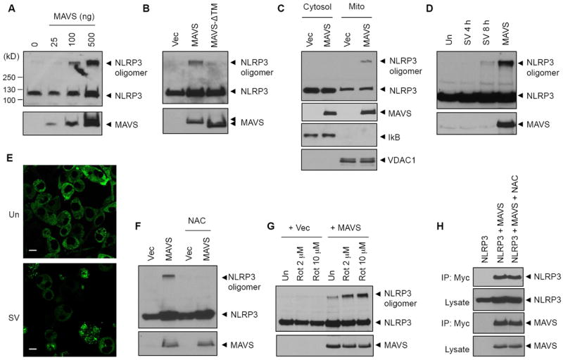
NLRP3 oligomerization by MAVS. (A, B) 293T cells were transfected with a construct for NLRP3 (0.3 μg) and increasing amount of MAVS construct (A), or NLRP3 and MAVS or MAVS-ΔTM constructs (0.3 μg, B) as indicated. Cell lysates were immunoblotted with anti-Flag (NLRP3) or anti-Myc (MAVS) antibodies. (C) 293T cells were transfected with a NLRP3 construct together with an empty vector (Vec) or MAVS construct. Cell lysates were fractionated into cytosolic or mitochondrial fraction, and then immunoblotted with anti-Flag (NLRP3), anti-Myc (MAVS), anti-IκB (cytosol) or anti-VDAC1 (mitochondria) antibodies. (D) 293T cells were transfected with NLRP3 (0.3 μg) followed by infection with Sendai virus (SV, 10 HA/ml) for the indicated times, or transfected with NLRP3 together with MAVS (0.3 μg) as indicated. Cell lysates were immunoblotted for NLRP3 and MAVS. (E) Confocal images of NLRP-GFP-expressing BMDMs left untreated (Un) or infected with Sendai virus (SV, 10 HA/ml, 8 h) as indicated. Bar: 10 μm. (F-G) 293T cells were transfected with a NLRP3 construct (0.3 μg) together with an empty vector or MAVS construct (0.3 μg), in the presence or absence of NAC (15 mM, 18 h) (F) or followed by treatment with rotenone for 6 h as indicated. Cell lysates were immunoblotted for NLRP3 or MAVS as described above. (H) 293T cells were transfected with a Flag-NLRP3 (0.3 μg) plasmid alone, or together with a Myc-MAVS (0.3 μg) plasmid in the presence or absence of NAC (15 mM, 18 h) as indicated. Cell lysates were immunoprecipitated using anti-Myc antibody, and the immunoprecipitates were then immunoblotted with anti-Flag (NLRP3) or anti-Myc (MAVS) antibody, respectively.