


Figure 3.
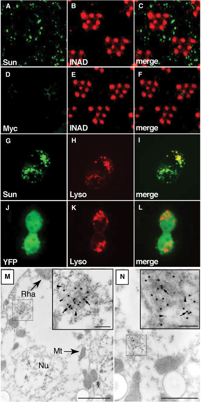
Immunolocalization of Sun. (A–F) Fly heads were embedded in LR White and 0.5 μm cross-sections of compound eyes were stained with rabbit anti-Myc and a rhabdomeral marker, rat anti-INAD. (A) Section from myc-sun transgenic fly, P[w+;c-sun];cn bw, stained with rabbit anti-Myc polyclonal antibodies. (B) Same section as in (A) stained with rat anti-INAD antibodies. (C) Merged images from (A, B). (D) Section from a white-eyed (cn bw) nontransgenic fly stained with anti-Myc antibodies. (E) Same section as in (D) stained with anti-INAD. (F) Merged images from (D, E). (G–I) 293 cells transfected with a plasmid encoding a YFP-Sun fusion protein. (G) Localization of YFP. (H) Same cells as in (J) stained with LysoTracker. (I) Merged images from (G, H). (J–L) 293 cells transfected with a plasmid encoding YFP. (J) Localization of YFP. (K) Same cells as (G) stained with LysoTracker. (L) Merged images from (J, K). (M, N) Immuno-EM localization of Myc-Sun and Rh1 in the cell bodies of P[c-sun];cn bw photoreceptor cells. Sections were stained with anti-Myc rabbit polyclonal antibodies (15 nm gold particles; several are indicated by arrows) and anti-Rh1 mouse monoclonal antibodies (5 nm gold particles, several are indicated by arrowheads). The dashed and solid boxes show lower and higher magnification views of Myc-Sun and Rh1 positive lysosomal-like vesicles (M) and MVB-like vesicles (N). The small (inset) and large-scale bars represent 0.1 and 1.0 μm, respectively. Mt, mitochondrion; Nu, nucleus; Rha, rhabdomere.