


Fig 5.
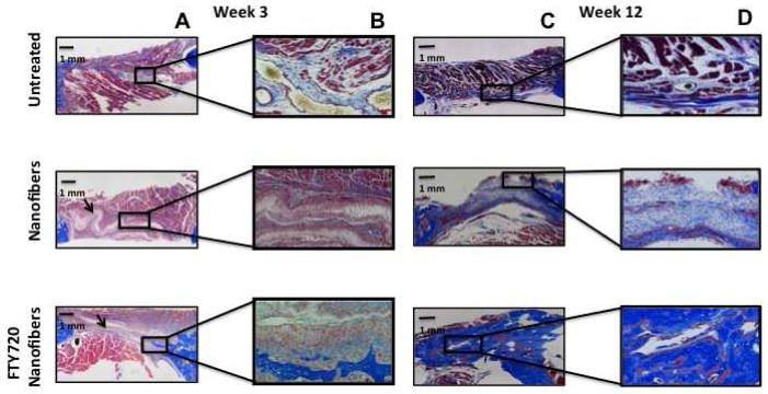
Assessment of new bone formation in rat critical size mandibular defects by Masson’s trichrome staining. Defects were left untreated or treated with unloaded nanofibers or FTY720-loaded nanofibers. (A) Representative images of mandibular defect 3 weeks post-surgery (original magnification x10). (B) High magnification image of a region with new bone formation 3 weeks post surgery (original magnification x40). (C) Representative images of mandibular defect 12 weeks post-surgery (original magnification x10). (D) High magnification image of a region with new bone formation 12 weeks post surgery (original magnification x40).