



Abstract
The pathogenesis of Systemic Sclerosis (SSc) is extremely complex, and despite extensive studies, the exact mechanisms involved are not well understood. Numerous recent studies of early events in SSc pathogenesis have suggested that unknown etiologic factors in a genetically receptive host trigger structural and functional microvascular endothelial cell abnormalities. These alterations result in the attraction, transmigration, and accumulation of immune and inflammatory cells in the perivascular tissues, which in turn induce the phenotypic conversion of endothelial cells and quiescent fibroblasts into activated myofibroblasts, a process known as endothelial to mesenchymal transition or EndoMT. The activated myofibroblasts are the effector cells responsible for the severe and frequently progressive fibrotic process and the fibroproliferative vasculopathy that are the hallmarks of SSc. Thus, according to this hypothesis the endothelial and vascular alterations, which include the phenotypic conversion of endothelial cells into activated myofibroblasts, play a crucial role in the development of the progressive fibrotic process affecting skin and multiple internal organs. The role of endothelial cell and vascular alterations, the potential contribution of endothelial to mesenchymal cell transition in the pathogenesis of the tissue fibrosis, and fibroproliferative vasculopathy in SSc will be reviewed here.
1. Introduction
Scleroderma or Systemic Sclerosis (SSc) is an autoimmune disease of unknown etiology characterized by progressive fibrosis of skin and multiple internal organs and prominent and often severe alterations in the microvasculature [1]. Although SSc is the third most common systemic inflammatory autoimmune disease and has the highest case-specific mortality among this group of idiopathic disorders, there is currently no effective disease-modifying therapy for SSc. Therefore, there is an urgent unmet need for the development of effective disease-modifying therapies to improve the devastating health consequences and high mortality caused by the disease. The cells responsible for the severe fibroproliferative process in SSc are activated myofibroblasts, a unique population of mesenchymal cells displaying unique biological functions including increased production of fibrillar type l and type lll collagens, initiation of expression of α-smooth muscle actin (α-SMA), a molecular marker of activated myofibroblasts, and reduction in the expression of genes encoding extracellular matrix (ECM)-degradative enzymes. The accumulation of myofibroblasts in affected tissues and the persistence of their elevated biosynthetic functions are crucial determinants of the extent and rate of progression of the fibrotic process in SSc, and of its clinical course, response to therapy, prognosis, and overall mortality. The origins of the myofibroblasts responsible for the exaggerated and uncontrolled production of collagen and other ECM proteins in SSc have not been completely elucidated. Extensive research studies have shown that these cells originate from several sources, including expansion of resident tissue fibroblasts and migration and tissue accumulation of bone marrow-derived circulating fibrocytes, or from epithelial cells which have undergone epithelial to mesenchymal transition (EMT). More recent studies, however, have demonstrated that endothelial cells are also capable of undergoing endothelial to mesenchymal transition (EndoMT) and that this transition might be an important source of the mesenchymal cells participating in the fibroproliferative vasculopathy and the fibrotic process in SSc. Thus, this novel mechanism of generation of activated myofibroblasts may represent an important and currently unexplored target for the development of disease-modifying therapeutic interventions for this currently incurable disease.
2. SSc Pathogenesis: Overview
The pathogenesis of SSc is extremely complex, and despite numerous studies that examined several aspects of its intricate picture, the exact mechanisms involved are not well understood. However, it is apparent that the clinical and pathologic manifestations of the disease are the result of three distinct processes: (1) fibroproliferative vascular lesions of small arteries and arterioles, (2) excessive and often progressive deposition of collagen and other ECM macromolecules in skin and various internal organs, and (3) alterations of humoral and cellular immunity characterized by innate immunity alterations, involvement of macrophages and T- and B-lymphocytes, and the production of numerous disease-specific autoantibodies [2–4]. It has not been established which of these processes is of primary importance or how they are temporally related during the development and progression of the disease. However, numerous recent studies have clarified some of the early events in SSc pathogenesis [5–11]. A current hypothesis for SSc pathogenesis posits that there is a sequence of pathogenetic events initiated by unknown etiologic factors in a genetically receptive host which trigger microvascular injury characterized by structural and functional endothelial cell abnormalities. The endothelial cell abnormalities result in the increased production and release of numerous and potent mediators including cytokines, chemokines, polypeptide growth factors, and various other substances such as nitric oxide, prostaglandins, reactive oxygen species (ROS), and thrombogenic and pro-coagulant activities or in the reduction of important compounds such as prostacyclin. The endothelial cell dysfunction triggers the chemokine and cytokine-mediated attraction of specific inflammatory cellular elements from the bloodstream and bone marrow and their transmigration into the surrounding tissues. These events lead to the establishment of a chronic inflammatory process with participation of macrophages and T- and B-lymphocytes, with further production and secretion of cytokines and growth factors that induce the tissue accumulation of activated myofibroblasts, the effector cells responsible for the fibrotic process [12–14]. This sequence of events, diagrammatically illustrated in Figure 1, results in the development of a severe and often progressive fibroproliferative vasculopathy and in the exaggerated and widespread accumulation of fibrotic tissue in the skin and multiple internal organs, which are the most salient characteristics of the disease.
Figure 1.
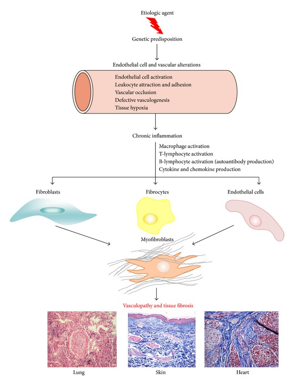
Overall scheme illustrating a current understanding of SSc pathogenesis. Hypothetical sequence of events involved in tissue fibrosis and fibroproliferative vasculopathy in SSc. An unknown causative agent induces activation of immune and inflammatory cells in genetically predisposed hosts resulting in chronic inflammation. Activated inflammatory and immune cells secrete cytokines, chemokines, and growth factors which cause fibroblast activation, differentiation of endothelial and epithelial cells into myofibroblasts, and recruitment of fibrocytes from the bone marrow and the peripheral blood circulation. The activated myofibroblasts produce exaggerated amounts of ECM resulting in tissue fibrosis.
The purpose of this review is to discuss recent studies that have substantially advanced the current understanding of SSc pathogenesis regarding the endothelial cell and vascular abnormalities and the role of endothelial to mesenchymal transition (EndoMT) in the pathogenesis of this currently incurable disease. However, the genetic, innate, and acquired immunological abnormalities and the cytokine, chemokine, and growth factor abnormalities, all of which play an extremely important role in SSc pathogenesis, will not be reviewed here owing to the availability of numerous outstanding reviews about these topics that have been recently published [15–22].
3. Vascular Abnormalities in SSc
Vascular dysfunction is one of the earliest and most noticeable manifestations of SSc as indicated by the occurrence of Raynaud phenomenon, nailfold capillary microvascular alterations, and digital ulcers almost universally in SSc patients often preceding the appearance of clinical evidence of tissue fibrosis [23, 24]. Furthermore, there is a remarkable microvascular fibroproliferative vasculopathy present in essentially all SSc affected organs that is responsible for the most important symptoms and clinical manifestations of SSc and often leads to serious and even fatal complications. Although the effects of vascular abnormalities and dysfunction in patients with SSc are most dramatic when they involve the pulmonary and renal arterioles, causing renal crisis [25, 26] and pulmonary artery hypertension [27–29], respectively, there are numerous other important clinical manifestations of the disease that are caused or mediated by the prominent fibroproliferative vasculopathy. These include capillary rarefaction and capillary loop dilation in the nailfold capillaries [30, 31], cutaneous and mucosal telangiectasias [32–34], erectile dysfunction resulting from alterations in penile blood flow [35–37], and cardiac dysfunction including nonartherosclerotic myocardial infarcts [38, 39], gastric antral vascular ectasia [40–42], central retinal artery occlusion [43, 44], and involvement of larger vessels [45, 46]. Histopathologically, the affected vessels display marked narrowing or even complete occlusion of the vessel lumen with remarkable accumulation of mesenchymal cells and fibrous tissue in the subendothelial compartment and associated endothelial cell abnormalities, which include swelling and apoptotic changes, as well as thickening of the basement membrane. Occasionally, endothelial cell detachment and intravascular platelet thrombi are found. On transmission, electron microscopy universal morphological changes of endothelial cells and basement membrane duplication and lamellation are characteristic alterations. The histopathological changes in the microvasculature of several affected organs are illustrated in Figure 2.
Figure 2.
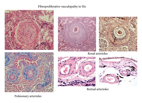
Histopathology of fibroproliferative vasculopathy in small vessels of various affected organs. Histopathology of microvascular arterioles from SSc lung, kidney, and retinal vessels displaying prominent endothelial fibroproliferative alterations causing severe narrowing of vessel lumen and thickening of vessel walls.
4. Mechanisms of Vascular and Endothelial Cell Injury in SSc
The initial events responsible for the vascular and endothelial cell injury and their subsequent activation are not known although numerous putative etiologic factors have been suggested. Some of these include exogenous chemical substances, vasculotropic viral pathogens, antiendothelial cell antibodies, cellular products from inflammatory cells, tissue hypoxia, or ROS generated during episodes of ischemia/reperfusion [8–10, 47–52]. The injured/activated endothelial cells may undergo apoptosis or may detach from the vascular endothelium, leaving a denuded vascular lumen which triggers the release of endothelial cell precursors from the bone marrow in attempts to repair the endothelial lining defects. Supporting this notion are the observations of increased numbers of circulating endothelial cells and endothelial cell precursors in SSc patients [53–55]. The activation of endothelial cells also induces the expression of cell adhesion molecules such as ICAM, VCAM-1, and E-selectin [56, 57]. The induced expression of cell adhesion molecules by the endothelial cells leads to recruitment and activation of chronic inflammatory cells, including T- and B-lymphocytes and profibrotic macrophage populations and their accumulation in the perivascular tissue and in the interstitium of parenchymal organs. The activated chronic inflammatory cells are responsible for the increased production of transforming growth factor-β (TGF-β), connective tissue growth factor (CTGF), and other profibrotic polypeptide growth factors which together with the mediators released by the endothelial cells, such as endothelin-1, induce subsequent pathogenetic events leading to the severe tissue fibrosis and fibroproliferative vasculopathy characteristic of the disease [1–11]. Besides the endothelial cell abnormalities, other vascular alterations include increased proliferation of smooth muscle cells in the medial layer of affected vessels, marked accumulation of fibrotic tissue in the subendothelial compartment, and initiation of platelet aggregation and intravascular thrombosis, eventually causing microvascular occlusion [58, 59]. These multiple events result in tissue hypoxia which can cause activation of hypoxia-dependent profibrogenic processes, including further increases in production of TGF-β and interstitial collagens as well as other ECM macromolecules [60, 61]. In addition to the structural vascular changes described above, there are also functional vascular alterations which include a reduction in endothelium dependent vasodilator molecules and dysfunction of the neurovascular and neuroendothelial control of vasodilation [62–65], as well as a relative deficiency of vasodilator molecules such as prostacyclin and nitric oxide.
The injured or cytokine/growth factor-activated endothelial cells also produce increased amounts of the potent profibrotic and vasoconstrictor polypeptide, endothelin-1 [66, 67], and numerous other vasoactive and prothrombogenic compounds that are capable of directly stimulating various target cells such as vascular smooth muscle cells and fibroblasts [8–10, 66, 67]. The important role of endothelin-1 in the development of SSc-associated tissue fibrosis and fibroproliferative vasculopathy has received increasing attention recently. Indeed, elevated levels of endothelin-1 have been found in plasma and bronchoalveolar lavage of SSc patients [68–70] and correlate with clinical parameters and subsets of the disease [71, 72]. Numerous studies have demonstrated that endothelin-1 is a potent inducer of proliferation and ECM production by fibroblastic cells [73–76]. The exaggerated vasoconstrictor response to the increased endothelin levels causes vascular hypoxia and further endothelial injury, thus establishing and maintaining a vicious cycle of endothelial injury and fibrosis. The chronic inflammatory cells accumulated in the perivascular environment also participate in the maintenance of a powerful profibrotic cycle since the numerous cytokines, chemokines, and growth factors they produce can in turn induce further activation of the endothelial cells and their production of profibrotic mediators [67]. The mutual interaction between inflammatory and endothelial cells has been validated by a recent study describing the upregulation of endothelin-1 and TGF-β in human microvascular endothelial cells induced by interferon-γ, one of the potent cytokines released by the infiltrating inflammatory cells [77]. Additional alterations which contribute to the severe vascular dysfunction and rarefaction in SSc are the result of disordered angiogenesis [78–84] and impaired differentiation of bone marrow stem cells into endothelial cells [85].
5. Endothelial to Mesenchymal Transition (EndoMT) in the Pathogenesis of SSc
One of the most characteristic histopathologic alterations in SSc is a severe fibroproliferative vasculopathy affecting the microvasculature as well as some larger vessels [86]. The proliferative vasculopathy of SSc has two distinct components. The first one is a marked proliferation of smooth muscle cells in the media of medium size and small size arterioles, a process which plays a crucial role in SSc-associated pulmonary hypertension. The second component is most prominent in the small arterioles of parenchymal organs, such as the lungs and kidneys, and is characterized by the subendothelial accumulation of activated fibroblasts or myofibroblasts and the production of abundant fibrotic tissue. The origin of mesenchymal cells responsible for the fibrotic microvascular occlusion in SSc is not known, but recent studies have suggested that at least some of these cells may result from EndoMT, that is, the transdifferentiation of endothelial cells into subintimal fibroblasts induced by locally-secreted cytokines and growth factors. During EndoMT, endothelial cells lose their specific endothelial cell markers, such as vascular endothelial cadherin (VE cadherin) and von Willebrand factor, and acquire a mesenchymal or myofibroblastic phenotype initiating expression of α-SMA, vimentin, and type I collagen. In addition, these cells become motile and are capable of migrating into surrounding tissues. EndoMT has been described as an important process during cardiac valve and pulmonary artery embryonic development [87–89]. More recently, EndoMT has emerged as a possible mechanism in the pathogenesis of tissue fibrosis in various diseases, including diabetic nephropathy, cardiac fibrosis, intestinal fibrosis, portal hypertension, and pulmonary hypertension [90–100]. Although there is some experimental evidence supporting the participation of EndoMT in SSc, further studies will be required to conclusively demonstrate that EndoMT plays a role in the pathogenesis of SSc-associated fibroproliferative vasculopathy and progressive tissue fibrosis. A firm demonstration of the occurrence of EndoMT in SSc and a further understanding of the molecular mechanisms involved may lead to the pharmacologic modulation or abrogation of this pathway in SSc.
6. Molecular Mechanisms of EndoMT
The molecular mechanisms involved in the EndoMT process have not been fully elucidated, and despite the remarkable importance of this process to normal development and to various pathologic conditions including SSc, only a few studies have examined the molecular changes and the regulatory events occurring in endothelial cells during their transdifferentiation into mesenchymal cells or myofibroblasts. However, substantial recent evidence has accumulated demonstrating the crucial role of TGF-β signaling [101–104] in the initiation of EndoMT during normal development as well as in various diseases.
6.1. Role of TGF-β in EndoMT
TGF-β is a pleiotropic growth factor involved in numerous physiologic and pathologic processes including embryogenesis, cellular development and differentiation, immunologic system development, inflammatory response functions, and wound repair [105–107]. TGF-β plays a key role in the pathogenesis of fibrotic diseases by stimulating the production of various collagens and other ECM components by mesenchymal cells and by inhibiting the expression of various relevant metalloproteinases [103, 104, 108–114]. Although the precise mechanisms mediating the potent profibrotic effects of TGF-β have not been completely elucidated, it appears that TGF-β may cause the establishment of an autocrine signaling cascade capable of continuous activation of profibrotic gene expression in the target cells [115]. However, extensive studies have shown that besides causing a potent stimulation of the expression of genes participating in the exaggerated production and accumulation of ECM, TGF-β is also involved in the generation of myofibroblasts through EndoMT [101–104, 116–121]. Indeed, studies in experimentally induced cardiac hypertrophy showed that TGF-β was a crucial mediator causing endothelial cells to undergo EndoMT [96]. Although the detailed molecular events and the intracellular cascades activated by TGF-β that result in the remarkable phenotypic change of endothelial cells to mesenchymal cells have not been entirely elucidated, recent studies in cultured human cutaneous microvascular endothelial cells [103], primary cultures of murine pulmonary endothelial cells [116], and cultured pancreatic microvascular endothelial cells [121] demonstrated that both Smad-dependent and Smad-independent pathways are involved. The intracellular signaling pathways that are likely to be involved in EndoMT induction by TGF-β are illustrated in Figure 3.
Figure 3.
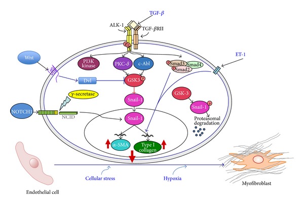
Signaling pathways involved in EndoMT. The diagram shows the numerous putative pathways that may participate in the EndoMT process and may be involved in SSc pathogenesis. One central pathway initiated following ligand-binding activation of the Smad-independent TGF-β pathway causes phosphorylation of GSK-3β mediated by PKC-δ and the c-Abl nonreceptor kinase. Phosphorylation of GSK-3β at serine 9 (ser9) causes its inhibition which then allows Snail-1 to enter the nucleus. Nuclear accumulation of Snail-1 results in marked stimulation of Snail-1 expression which then leads to acquisition of the myofibroblast phenotype with stimulation of α-SMA. The inhibition of GSK-3β ser9 phosphorylation by specific inhibition of PKC-δ or c-Abl activity allows GSK-3β to phosphorylate Snail-1 targeting it for proteosomal degradation and thus effectively abolishes the acquisition of the myofibroblastic phenotype and the fibrotic response. Other pathways such as those involving ET-1, Wnt, NOTCH, hypoxia, and cellular stress responses may also participate although the molecular events have not been fully elucidated. Modified from Piera-Velazquez and Jimenez [101].
Given the crucial role of TGF-β in the development of tissue fibrosis and its participation in the pathogenesis of numerous fibrotic diseases, we recently examined the mechanisms involved in the induction of EndoMT by this pleotropic growth factor and studied the intracellular transduction pathways involved in this process employing primary pulmonary endothelial cells in culture [116]. In our study, we examined the transdifferentiation of murine pulmonary endothelial cells into mesenchymal cells in vitro and the signaling pathways involved in this process and made the following observations: (1) primary murine pulmonary endothelial cells undergo EndoMT in response to TGF-β with initiation of expression of α-SMA, assembly of typical intracellular α-SMA stress fibers, and loss of VE-cadherin in vitro; (2) TGF-β induction of EndoMT was associated with a strong upregulation in the expression of the transcriptional repressor Snail-1 indicating that Snail-1 is directly involved in TGF-β-induced α-SMA expression; and (3) induction of α-SMA expression in pulmonary endothelial cells was mediated by the c-Abl kinase and by protein kinase c-δ (PKC-δ), as specific inhibition of their kinase activity with imatinib mesylate and rottlerin, respectively, or by knockdown of their corresponding transcripts with specific siRNA abrogated the marked increase in TGF-β induced α-SMA and Snail-1 expression and protein levels. These studies collectively showed that these effects are mediated by the transcriptional repressor Snail-1 [103, 116]. Snail-1 is a zinc-finger transcription factor that forms a complex with Smad3/Smad4. The active Smad3/Smad4/Snail-1 complex causes potent inhibition of the expression of E-cadherin by directly binding to specific sequences within the gene promoter and blocking its transcription. Besides inhibition of E-cadherin, Snail-1 induces numerous transcriptional events that lead to the expression of a mesenchymal-cell-specific phenotype. Snail-1 levels are regulated by complex phosphorylation events mediated by intracellular kinases including c-Abl kinase, PKC-δ, PI3K, p38 MAP kinase and glycogen synthase kinase 3β (GSK-3β). The role of PKC-δ and c-Abl kinases has been demonstrated employing specific kinase inhibitors and/or specific knockdown with small interfering RNAs [116], whereas the role of PI3K, p38 MAPK and GSK-3β was demonstrated employing specific inhibitors of the corresponding pathways [103]. Numerous studies have shown a crucial role of GSK-3β in the regulation of Snail-1 effects. GSK-3β phosphorylation results in its inactivation which in turn induces the nuclear accumulation of Snail-1 followed by a profound increase in the expression of its corresponding gene. In contrast, in the absence of GSK-3β phosphorylation, the GSK-3β kinase is active and induces the proteosomal degradation of Snail-1, thus abrogating the endothelial to mesenchymal cellular phenotypic conversion. The role of GSK-3β in the regulation of Snail-1 stability, and therefore, in the expression of its potent transcriptional effects is illustrated in Figure 3.
6.2. Regulation of EndoMT by the Wnt and NOTCH Signaling Pathways
Although not extensively studied in EndoMT, it has recently become apparent that several important regulatory pathways including the canonical Wnt pathway and the NOTCH pathway may also participate in the regulation of EndoMT as illustrated in Figure 3.
6.2.1. Wnt Signaling
The Wnt proteins comprise a large family of secreted glycoproteins with complex canonical and noncanonical intracellular signaling pathways that play crucial roles during embryonic development and organogenesis [122–124]. Wnt proteins and pathways have been recently implicated in the pathogenesis of numerous diseases, including SSc and other fibrotic diseases [125–129]. TGF-β appears to be the major factor activating the canonical Wnt pathway [130, 131]. This process is probably mediated by a decrease of Dickkopf-related protein 1 (Dkk-1), a potent Wnt pathway inhibitor, as indicated by the observations that the addition of recombinant Dkk-1 blocked the stimulatory effects of TGF-β on the canonical Wnt pathway in fibroblasts [132, 133].
Although there is extensive published literature regarding the role of Wnt pathway activation in the phenotypic conversion of epithelial cells into mesenchymal cells, also known as EMT [134–137], the possibility that Wnt may participate in EndoMT is just beginning to be explored. Indeed, a recent study examined the role of Wnt7 and the Wnt7 antagonist Dkk-1 on EndoMT in primary aortic endothelial cells in culture and in transgenic mice with an endothelial-specific Wnt-7b deletion [138]. The results showed that Dkk-1 inhibition of the Wnt pathway enhanced EndoMT, whereas Wnt-7b expression preserved the endothelial cell phenotype.
6.2.2. NOTCH Signaling
The NOTCH proteins are members of the group of proteins collectively known as morphogens owing to their crucial roles in cell fate decisions during morphogenesis and embryonic development, particularly in relation to cardiovascular development and to regulation of central nervous system polarity and vertebrate segmentation [139, 140]. However, involvement of NOTCH proteins in a broad spectrum of disorders is just becoming apparent [141–147]. The role of NOTCH signaling in EndoMT was first described by Noseda et al. [148], and it was suggested that the NOTCH pathway may be crucial for heart valve and cardiac cushion development and/or vascular smooth muscle differentiation. Numerous subsequent studies have confirmed and extended these observations and have examined the molecular mechanisms involved and the important interactions with the TGF-β pathways [149–154]. Studies to examine the participation of NOTCH proteins in the EndoMT process in SSc have not been described, although the demonstration of activation of NOTCH signaling in affected SSc skin suggests that the NOTCH proteins may play a role in SSc pathogenesis and thus may represent a potential target for SSc disease modifying therapeutic approaches [155, 156].
6.3. Caveolin-1 Regulation of EndoMT
Another recently identified mechanism of regulation and fine tuning of TGF-β activity involves Caveolin-1 (Cav-1), the most important member of a family of proteins found in lipid rafts. Cav-1 plays an important role in TGF-β signaling regulation owing to its participation in TGF-β receptor (TβR) internalization [157–159]. TβRs are internalized both by Cav-1-associated lipid rafts and by early endosome antigen 1 (EEA-1) nonlipid raft pathways. Non-lipid raft associated internalization increases TGF-β signaling, whereas caveolin-associated internalization increases TβR degradation, thereby effectively decreasing or abolishing TGF-β signaling [157]. The localization of the TβRs in the EEA-1 positive compartment is responsible for downstream Smad activation, whereas their localization in Cav-1 containing lipid rafts has been shown to cause subsequent receptor ubiquitination and rapid degradation and turnover [158, 159]. Despite the important interactions between Cav-1 and TGF-β and the numerous studies that supported the role of Cav-1 in the pathogenesis of SSc [160–164], the possibility that Cav-1 may participate in the regulation of EndoMT has not been explored in detail, although a recent study examined the contribution of Cav-1 to EndoMT employing Cav-1 knockout mice [165]. The results indicated that Cav-1 may be a crucial regulator of EndoMT in murine pulmonary endothelial cells. In these studies, it was shown that pulmonary endothelial cells isolated from Cav-1 knockout mice displayed spontaneous EndoMT and that Cav-1 deficiency potentiated the EndoMT effect induced by TGF-β [165].
6.4. Role of Other Growth Factors in EndoMT
The most severe clinical and pathologic manifestations of SSc are the result of a fibrotic process characterized by the excessive and often progressive deposition of collagen and other connective tissue macromolecules in skin and numerous internal organs. Numerous studies have shown that tissue fibrosis in SSc is the result of an upregulated expression of genes encoding collagen and other extracellular matrix proteins in affected organs. The exact mechanisms responsible for the establishment of the fibrotic process in SSc have not been precisely determined [2–6], although it has become very clear that several growth factors play a crucial role [166].
Besides TGF-β, the most important growth factor, involved in SSc tissue fibrosis and in EndoMT, other growth factors and profibrogenic molecules, including platelet derived growth factors [167], vascular endothelial growth factor [168], and insulin-derived growth factor [169], may also participate in EndoMT although their role in this process has not been studied to our knowledge. However, some studies that examined the role of other profibrotic growth factors have been described. One of the profibrotic polypeptides shown to participate in EndoMT is endothelin-1. One study showed that endothelial cell-derived endothelin-1 promotes cardiac fibrosis and heart failure in diabetic hearts through stimulation of EndoMT as these effects did not occur in hearts from transgenic mice with endothelial cell specific endothelin-1 deletion [170].
Connective Tissue Growth Factor (CTGF), also known as CCN2, is another pleotropic growth factor that has emerged as an important mediator of normal and pathological tissue fibrotic responses [171–174] and has been suggested to play a crucial role in SSc tissue fibrosis. TGF-β causes potent stimulation of CTGF synthesis in fibroblasts, vascular smooth muscle cells, and endothelial cells, and numerous studies have shown that CTGF represents a downstream mediator of TGF-β fibrogenic effects [175–177]. Despite the important role of CTGF in the pathogenesis of tissue fibrosis and its potential participation in SSc owing to the well-recognized functional interactions with TGF-β, its possible participation in the EndoMT process has not been investigated, although a very recent study showed that elevated levels of CTGF in SSc microvascular endothelial cells were capable of stimulating fibroblast activation and increased motility and invasion in in vitro studies. Further investigation indicated that these effects were mediated by CTGF-induced increased expression of TGF-β in the target fibroblasts [178]. Another very recent study demonstrated that CTGF is one of the target genes of Snail-1 and showed a remarkable increase in CTGF expression in endothelial cells following experimentally-induced overexpression of Snail-1 [179].
6.5. Role of MicroRNAs in the Regulation of EndoMT
MicroRNAs (miRNAs) are small (~22 nucleotides), evolutionarily conserved noncoding RNAs which play important roles in the regulation of the expression of a large number of protein coding genes at the posttranscriptional level [180–183]. The mechanisms involved in posttranscriptional regulation of gene expression by miRNAs are complex and require the sequence-specific complementary binding to the 3′ untranslated region (UTR) of target mRNAs suppressing their expression by either inhibiting mRNA translation or facilitating their degradation [184–186]. Recent interest has been devoted to elucidating their participation in tissue fibrosis and fibrotic diseases [187–190]. Indeed, several miRNAs have been shown to be involved in SSc tissue fibrosis [191–197], displaying either profibrotic or antifibrotic effects. Furthermore, it has been shown that numerous miRNAs display strong modulation of their expression by TGF-β [198], although the implications of these TGF-β-miRNA interactions have not been fully elucidated. Moreover, several studies have described potential modulatory effects of miRNA on EMT [199], although their participation in EndoMT has not been examined in detail. However, recent reports described results indicating that miRNA21 partially mediated the TGF-β-induced EndoMT in human umbilical vein endothelial cells [200] and that several miRNAs were either increased or decreased during TGF-β2-induced EndoMT in murine cardiac endothelial cells [120].
7. Conclusions and Future Directions
Scleroderma or Systemic Sclerosis (SSc) is a systemic autoimmune disease of unknown etiology characterized by progressive fibrosis of skin and multiple internal organs and severe alterations in the microvasculature [1]. SSc is the third most common systemic inflammatory autoimmune disease and has the highest case-specific mortality among this group of idiopathic disorders. Whereas remarkable therapeutic advances have recently been accomplished for Rheumatoid Arthritis and Systemic Lupus Erythematosus, there is currently no effective disease-modifying therapy for SSc. Therefore, there is an urgent unmet need for the development of effective disease-modifying therapies to improve the devastating health consequences and high mortality caused by the disease. The effector cells ultimately responsible for the severe fibroproliferative process in SSc are activated myofibroblasts. These cells display very active protein synthesis producing increased amounts of ECM proteins and acquiring a motile and contractile phenotype expressing a high level of α-SMA [12–14]. Although it is widely recognized that there are numerous inflammatory and immunological events in the pathogenesis of SSc, myofibroblasts have been recognized as the crucial determinant of the fibrotic process in SSc and other fibrotic disorders [6, 11–13, 201–203]. Furthermore, their accumulation in affected tissues and the persistence of their elevated biosynthetic functions are the primary determinants of the extent and severity of the clinical manifestations in SSc, and of its clinical course, response to therapy, prognosis, and overall mortality. Thus, activated myofibroblasts have become an important target for SSc disease-modifying therapeutic approaches [204–206].
Extensive research studies have shown that these cells originate from several sources [94, 207], including expansion and phenotypic activation of resident tissue fibroblasts and migration and tissue accumulation of bone marrow-derived circulating fibrocytes, or from epithelial cells which have undergone EMT. More recent studies, however, have demonstrated that endothelial cells are also capable of undergoing a phenotypic change to activated mesenchymal cells in a complex process known as EndoMT. Although there are very few studies that have examined the possible participation of EndoMT in the initiation and progression of the fibrotic and fibroproliferative processes in SSc, it is expected that given its potential importance in the pathogenesis of this currently incurable disease this area of investigation may attract further scientific attention.
Despite the relatively recent research interest in the role of EndoMT in the SSc pathogenesis, important components of the complex pathway of TGF-β-induced EndoMT and the molecular mechanisms involved in the generation of activated tissue myofibroblasts have already been identified. These observations suggest that targeting components of these pathways may be a feasible therapeutic goal to modify crucial steps in the development of SSc fibroproliferative vasculopathy [208]. Furthermore, the important role that miRNAs have been shown to play in the regulation of gene expression has clearly opened the possibility of developing a novel therapeutic approach for SSc by targeting these extremely versatile noncoding RNA species. Obviously, subsequent preclinical studies employing suitable animal models will be required to further support the potential therapeutic role of EndoMT and/or miRNA modulation for the fibrosis and proliferative vasculopathy of SSc.
Acknowledgments
This work is supported by the National Institute of Arthritis and Musculoskeletal and Skin Diseases, part of the National Institutes of Health, under Award no. AM19606. The content is solely the responsibility of the author and does not necessarily represent the official views of the National Institutes of Health. The expert assistance of Kenneth Brown is duly acknowledged. The author thanks Drs. Joel Rosenbloom and Sonsoles Piera-Velazquez for the valuable comments and for critical reading of this paper.
References
- 1.Gabrielli A, Avvedimento EV, Krieg T. Scleroderma. The New England Journal of Medicine. 2009;360(19):1989–2003. doi: 10.1056/NEJMra0806188. [DOI] [PubMed] [Google Scholar]
- 2.Jimenez SA, Derk CT. Following the molecular pathways toward an understanding of the pathogenesis of systemic sclerosis. Annals of Internal Medicine. 2004;140(1):37–50. [PubMed] [Google Scholar]
- 3.Varga J, Abraham D. Systemic sclerosis: a prototypic multisystem fibrotic disorder. Journal of Clinical Investigation. 2007;117(3):557–567. doi: 10.1172/JCI31139. [DOI] [PMC free article] [PubMed] [Google Scholar]
- 4.Katsumoto TR, Whitfield ML, Connolly MK. The pathogenesis of systemic sclerosis. Annual Review of Pathology: Mechanisms of Disease. 2011;6:509–537. doi: 10.1146/annurev-pathol-011110-130312. [DOI] [PubMed] [Google Scholar]
- 5.Denton CP, Black CM, Abraham DJ. Mechanisms and consequences of fibrosis in systemic sclerosis. Nature Clinical Practice Rheumatology. 2006;2:134–144. doi: 10.1038/ncprheum0115. [DOI] [PubMed] [Google Scholar]
- 6.Wynn TA. Common and unique mechanisms regulate fibrosis in various fibroproliferative diseases. Journal of Clinical Investigation. 2007;117(3):524–529. doi: 10.1172/JCI31487. [DOI] [PMC free article] [PubMed] [Google Scholar]
- 7.Varga JA, Trojanowska M. Fibrosis in systemic sclerosis. Rheumatic Disease Clinics of North America. 2008;34(1):115–143. doi: 10.1016/j.rdc.2007.11.002. [DOI] [PMC free article] [PubMed] [Google Scholar]
- 8.Matucci-Cerinic M, Kahaleh B, Wigley FM. Systemic sclerosis, (scleroderma, SSc) is a vascular disease. Arthritis and Rheumatism. 2013;65(8):1953–1962. doi: 10.1002/art.37988. [DOI] [PubMed] [Google Scholar]
- 9.Kahaleh B. Vascular disease in scleroderma: mechanisms of vascular injury. Rheumatic Disease Clinics of North America. 2008;34(1):57–71. doi: 10.1016/j.rdc.2007.12.004. [DOI] [PubMed] [Google Scholar]
- 10.Trojanowska M. Cellular and molecular aspects of vascular dysfunction in systemic sclerosis. Nature Reviews Rheumatology. 2010;6(8):453–460. doi: 10.1038/nrrheum.2010.102. [DOI] [PMC free article] [PubMed] [Google Scholar]
- 11.Wynn TA, Ramalingam TR. Mechanisms of fibrosis: therapeutic translation for fibrotic disease. Nature Medicine. 2012;18:1028–1040. doi: 10.1038/nm.2807. [DOI] [PMC free article] [PubMed] [Google Scholar]
- 12.Krieg T, Abraham D, Lafyatis R. Fibrosis in connective tissue disease: the role of the myofibroblast and fibroblast-epithelial cell interactions. Arthritis Research and Therapy. 2007;9(supplement 2, article S4) doi: 10.1186/ar2188. [DOI] [PMC free article] [PubMed] [Google Scholar]
- 13.Abraham DJ, Eckes B, Rajkumar V, Krieg T. New developments in fibroblast and myofibroblast biology: implications for fibrosis and scleroderma. Current Rheumatology Reports. 2007;9(2):136–143. doi: 10.1007/s11926-007-0008-z. [DOI] [PubMed] [Google Scholar]
- 14.Kirk TZ, Mark ME, Chua CC, Chua BH, Mayes MD. Myofibroblasts from scleroderma skin synthesize elevated levels of collagen and tissue inhibitor of metalloproteinase (TIMP-1) with two forms of TIMP-1. The Journal of Biological Chemistry. 1995;270(7):3423–3428. doi: 10.1074/jbc.270.7.3423. [DOI] [PubMed] [Google Scholar]
- 15.Romano E, Manetti M, Guiducci S, Ceccarelli C, Allanore Y, Matucci-Cerinic M. The genetics of systemic sclerosis: an update. Clinical and Experimental Rheumatology. 2011;29(2):S75–S86. [PubMed] [Google Scholar]
- 16.Mayes MD. The genetics of scleroderma: looking into the postgenomic era. Current Opinion in Rheumatology. 2012;24(6):677–684. doi: 10.1097/BOR.0b013e328358575b. [DOI] [PMC free article] [PubMed] [Google Scholar]
- 17.Broen JCA, Coenen MJH, Radstake TRDJ. Genetics of systemic sclerosis: an update. Current Rheumatology Reports. 2012;14(1):11–21. doi: 10.1007/s11926-011-0221-7. [DOI] [PMC free article] [PubMed] [Google Scholar]
- 18.Martín JE, Bossini-Castillo L, Martín J. Unraveling the genetic component of systemic sclerosis. Human Genetics. 2012;131(7):1023–1037. doi: 10.1007/s00439-011-1137-z. [DOI] [PubMed] [Google Scholar]
- 19.Chizzolini C, Brembilla NC, Montanari E, Truchetet M. Fibrosis and immune dysregulation in systemic sclerosis. Autoimmunity Reviews. 2011;10(5):276–281. doi: 10.1016/j.autrev.2010.09.016. [DOI] [PubMed] [Google Scholar]
- 20.Gu YS, Kong J, Cheema GS, Keen CL, Wick G, Gershwin ME. The immunobiology of systemic sclerosis. Seminars in Arthritis and Rheumatism. 2008;38(2):132–160. doi: 10.1016/j.semarthrit.2007.10.010. [DOI] [PubMed] [Google Scholar]
- 21.York MR. Novel insights on the role of the innate immune system in systemic sclerosis. Expert Review of Clinical Immunology. 2011;7(4):481–489. doi: 10.1586/eci.11.40. [DOI] [PubMed] [Google Scholar]
- 22.Wynn TA. Cellular and molecular mechanisms of fibrosis. Journal of Pathology. 2008;214(2):199–210. doi: 10.1002/path.2277. [DOI] [PMC free article] [PubMed] [Google Scholar]
- 23.Steen V, Denton CP, Pope JE, Matucci-Cerinic M. Digital ulcers: overt vascular disease in systemic sclerosis. Rheumatology. 2009;48(supplement 3):iii19–iii24. doi: 10.1093/rheumatology/kep105. [DOI] [PubMed] [Google Scholar]
- 24.Herrick AL. The pathogenesis, diagnosis and treatment of Raynaud phenomenon. Nature Reviews Rheumatology. 2012;8:469–479. doi: 10.1038/nrrheum.2012.96. [DOI] [PubMed] [Google Scholar]
- 25.Steen VD. Scleroderma renal crisis. Rheumatic Disease Clinics of North America. 2003;29(2):315–333. doi: 10.1016/s0889-857x(03)00016-4. [DOI] [PubMed] [Google Scholar]
- 26.Mouthon L, Bérezné A, Bussone G, Noël L, Villiger PM, Guillevin L. Scleroderma renal crisis: a rare but severe complication of systemic sclerosis. Clinical Reviews in Allergy and Immunology. 2011;40(2):84–91. doi: 10.1007/s12016-009-8191-5. [DOI] [PubMed] [Google Scholar]
- 27.Denton CP, Black CM. Pulmonary hypertension in systemic sclerosis. Rheumatic Disease Clinics of North America. 2003;29(2):335–349. doi: 10.1016/s0889-857x(03)00024-3. [DOI] [PubMed] [Google Scholar]
- 28.Ramirez A, Varga J. Pulmonary arterial hypertension in systematic sclerosis: clinical manifestations, pathophysiology, evaluation, and management. Treatments in Respiratory Medicine. 2004;3(6):339–352. doi: 10.2165/00151829-200403060-00002. [DOI] [PubMed] [Google Scholar]
- 29.Chatterjee S. Pulmonary hypertension in systemic sclerosis. Seminars in Arthritis and Rheumatism. 2011;41(1):19–37. doi: 10.1016/j.semarthrit.2010.08.004. [DOI] [PubMed] [Google Scholar]
- 30.Cutolo M, Sulli A, Smith V. Assessing microvascular changes in systemic sclerosis diagnosis and management. Nature Reviews Rheumatology. 2010;6(10):578–587. doi: 10.1038/nrrheum.2010.104. [DOI] [PubMed] [Google Scholar]
- 31.Grassi W, Medico PD, Izzo F, Cervini C. Microvascular involvement in systemic sclerosis: capillaroscopic findings. Seminars in Arthritis and Rheumatism. 2001;30(6):397–402. doi: 10.1053/sarh.2001.20269. [DOI] [PubMed] [Google Scholar]
- 32.Shah AA, Wigley FM, Hummers LK. Telangiectases in scleroderma: a potential clinical marker of pulmonary arterial hypertension. Journal of Rheumatology. 2010;37(1):98–104. doi: 10.3899/jrheum.090697. [DOI] [PMC free article] [PubMed] [Google Scholar]
- 33.El-Omar MM, Jenkins AP, Hollowood K, Banerjee AK, Thompson RPH. Gastric telangiectasis: a rare cause of severe blood loss in CREST syndrome. Postgraduate Medical Journal. 1994;70(822):302–304. doi: 10.1136/pgmj.70.822.302. [DOI] [PMC free article] [PubMed] [Google Scholar]
- 34.Jharap B, Koudstaal LG, Neefjes-Borst EA, van Weyenberg SJB. Colonic telangiectasias in progressive systemic sclerosis. Endoscopy. 2012;44(supplement 2):E42–E43. doi: 10.1055/s-0031-1291521. [DOI] [PubMed] [Google Scholar]
- 35.Lally EV, Jimenez SA. Impotence in progressive systemic sclerosis. Annals of Internal Medicine. 1981;95(2):150–153. doi: 10.7326/0003-4819-95-2-150. [DOI] [PubMed] [Google Scholar]
- 36.Sukenik S, Horowitz J, Buskila D, Abarbanel JM, Lismer L, Avinoach I. Impotence in systemic sclerosis. Annals of Internal Medicine. 1987;106(6):910–911. doi: 10.7326/0003-4819-106-6-910_3. [DOI] [PubMed] [Google Scholar]
- 37.Foocharoen C, Tyndall A, Hachulla E, et al. Erectile dysfunction is frequent in systemic sclerosis and associated with severe disease: a study of the EULAR scleroderma trial and research group. Arthritis Research and Therapy. 2012;14(1, article R37) doi: 10.1186/ar3748. [DOI] [PMC free article] [PubMed] [Google Scholar]
- 38.Derk CT, Jimenez SA. Acute myocardial infarction in systemic sclerosis patients: a case series. Clinical Rheumatology. 2007;26(6):965–968. doi: 10.1007/s10067-006-0211-8. [DOI] [PubMed] [Google Scholar]
- 39.Allanore Y, Meune C. Primary myocardial involvement in systemic sclerosis: evidence for a microvascular origin. Clinical and Experimental Rheumatology. 2010;28(5):S48–S53. [PubMed] [Google Scholar]
- 40.Watson M, Hally RJ, McCue PA, Varga J, Jiménez SA. Gastric antral vascular ectasia (watermelon stomach) in patients with systemic sclerosis. Arthritis and Rheumatism. 1996;39(2):341–346. doi: 10.1002/art.1780390226. [DOI] [PubMed] [Google Scholar]
- 41.Ingraham KM, O’Brien MS, Shenin MAX, Derk CT, Steen VD. Gastric antral vascular ectasia in systemic sclerosis: demographics and disease predictors. Journal of Rheumatology. 2010;37(3):603–607. doi: 10.3899/jrheum.090600. [DOI] [PubMed] [Google Scholar]
- 42.Hung EW, Mayes MD, Sharif R, Assassi S, Machicao VI, Hosing C. Gastric antral vascular ectasia and its clinical correlates in patients with early diffuse systemic sclerosis in the SCOT trial. Journal of Rheumatology. 2013;40(4):455–460. doi: 10.3899/jrheum.121087. [DOI] [PMC free article] [PubMed] [Google Scholar]
- 43.Busquets J, Lee Y, Santamarina L, et al. Acute retinal artery occlusion in systemic sclerosis: a rare manifestation of systemic sclerosis fibroproliferative vasculopathy. Seminars in Arthritis and Rheumatism. 2013 doi: 10.1016/j.semarthrit.2012.12.025. [DOI] [PMC free article] [PubMed] [Google Scholar]
- 44.Minasian M, Stanford M, Graham E, Denton CP, Black C. Bilateral ischaemic retinal vasculopathy in scleroderma. The British Journal of Ophthalmology. 2005;89(8):1064–1065. doi: 10.1136/bjo.2004.061499. [DOI] [PMC free article] [PubMed] [Google Scholar]
- 45.Taylor MH, McFadden JA, Bolster MB, Silver RM. Ulnar artery involvement in systemic sclerosis (scleroderma) Journal of Rheumatology. 2002;29(1):102–106. [PubMed] [Google Scholar]
- 46.Youssef P, Englert H, Bertouch J. Large vessel occlusive disease associated with CREST syndrome and scleroderma. Annals of the Rheumatic Diseases. 1993;52(6):464–466. doi: 10.1136/ard.52.6.464. [DOI] [PMC free article] [PubMed] [Google Scholar]
- 47.Müller-Ladner U, Distler O, Ibba-Manneschi L, Neumann E, Gay S. Mechanisms of vascular damage in systemic sclerosis. Autoimmunity. 2009;42(7):587–595. doi: 10.1080/08916930903002487. [DOI] [PubMed] [Google Scholar]
- 48.Abraham D, Distler O. How does endothelial cell injury start? The role of endothelin in systemic sclerosis. Arthritis Research and Therapy. 2007;9(supplement 2, article S2) doi: 10.1186/ar2186. [DOI] [PMC free article] [PubMed] [Google Scholar]
- 49.Pandey JP, LeRoy EC. Human cytomegalovirus and the vasculopathies of autoimmune diseases (especially scleroderma), allograft rejection, and coronary restenosis. Arthritis and Rheumatism. 1998;41(1):10–15. doi: 10.1002/1529-0131(199801)41:1<10::AID-ART2>3.0.CO;2-P. [DOI] [PubMed] [Google Scholar]
- 50.Neidhart M, Kuchen S, Distler O, et al. Increased serum levels of antibodies against human cytomegalovirus and prevalence of autoantibodies in systemic sclerosis. Arthritis and Rheumatism. 1999;42:389–392. doi: 10.1002/1529-0131(199902)42:2<389::AID-ANR23>3.0.CO;2-P. [DOI] [PubMed] [Google Scholar]
- 51.Renaudineau Y, Revelen R, Levy Y, et al. Anti-endothelial cell antibodies in systemic sclerosis. Clinical and Diagnostic Laboratory Immunology. 1999;6(2):156–160. doi: 10.1128/cdli.6.2.156-160.1999. [DOI] [PMC free article] [PubMed] [Google Scholar]
- 52.Drenk F, Deicher HRG. Pathophysiological effects of endothelial cytotoxic activity derived from sera of patients with progressive systemic sclerosis. Journal of Rheumatology. 1988;15(3):468–474. [PubMed] [Google Scholar]
- 53.Del Papa N, Colombo G, Fracchiolla N, et al. Circulating endothelial cells as a marker of ongoing vascular disease in systemic sclerosis. Arthritis and Rheumatism. 2004;50(4):1296–1304. doi: 10.1002/art.20116. [DOI] [PubMed] [Google Scholar]
- 54.Avouac J, Juin F, Wipff J, et al. Circulating endothelial progenitor cells in systemic sclerosis: association with disease severity. Annals of the Rheumatic Diseases. 2008;67(10):1455–1460. doi: 10.1136/ard.2007.082131. [DOI] [PubMed] [Google Scholar]
- 55.Allanore Y, Batteux F, Avouac J, Assous N, Weill B, Kahan A. Levels of circulating endothelial progenitor cells in systemic sclerosis. Clinical and Experimental Rheumatology. 2007;25(1):60–66. [PubMed] [Google Scholar]
- 56.Kuryliszyn-Moskal A, Klimiuk PA, Sierakowski S. Soluble adhesion molecules (sVCAM-1, sE-selectin), vascular endothelial growth factor (VEGF) and endothelin-1 in patients with systemic sclerosis: relationship to organ systemic involvement. Clinical Rheumatology. 2005;24(2):111–116. doi: 10.1007/s10067-004-0987-3. [DOI] [PubMed] [Google Scholar]
- 57.Andersen GN, Caidahl K, Kazzam E, et al. Correlation between increased nitric oxide production and markers of endothelial activation in systemic sclerosis: findings with the soluble adhesion molecules E-selectin, intercellular adhesion molecule 1, and vascular cell adhesion molecule 1. Arthritis and Rheumatism. 2000;43(5):1085–1093. doi: 10.1002/1529-0131(200005)43:5<1085::AID-ANR19>3.0.CO;2-7. [DOI] [PubMed] [Google Scholar]
- 58.Mulligan-Kehoe MJ, Simons M. Vascular disease in scleroderma: angiogenesis and vascular repair. Rheumatic Disease Clinics of North America. 2008;34(1):73–79. doi: 10.1016/j.rdc.2007.12.006. [DOI] [PubMed] [Google Scholar]
- 59.Cerinic MM, Valentini G, Sorano GG, et al. Blood coagulation, fibrinolysis, and markers of endothelial dysfunction in systemic sclerosis. Seminars in Arthritis and Rheumatism. 2003;32(5):285–295. doi: 10.1053/sarh.2002.50011. [DOI] [PubMed] [Google Scholar]
- 60.Stenmark KR, Fagan KA, Frid MG. Hypoxia-induced pulmonary vascular remodeling: cellular and molecular mechanisms. Circulation Research. 2006;99(7):675–691. doi: 10.1161/01.RES.0000243584.45145.3f. [DOI] [PubMed] [Google Scholar]
- 61.Beyer C, Schett G, Gay S, Distler O, Distler JHW. Hypoxia. Hypoxia in the pathogenesis of systemic sclerosis. Arthritis Research & Therapy. 2009;11(2):p. 220. doi: 10.1186/ar2598. [DOI] [PMC free article] [PubMed] [Google Scholar]
- 62.Anderson ME, Moore TL, Hollis S, Clark S, Jayson MIV, Herrick AL. Endothelial-dependent vasodilatation is impaired in patients with systemic sclerosis, as assessed by low dose iontophoresis. Clinical and Experimental Rheumatology. 2003;21(3, article 403) [PubMed] [Google Scholar]
- 63.Livi R, Teghini L, Generini S, Matucci-Cerinic M. The loss of endothelium-dependent vascular tone control in systemic sclerosis. Chest. 2001;119(2):672–673. doi: 10.1378/chest.119.2.672. [DOI] [PubMed] [Google Scholar]
- 64.Kahaleh B, Matucci-Cerinic M. Raynaud’s phenomenon and scleroderma: dysregulated neuroendothelial control of vascular tone. Arthritis and Rheumatism. 1995;38(1):1–4. doi: 10.1002/art.1780380102. [DOI] [PubMed] [Google Scholar]
- 65.Cailes J, Winter S, du Bois RM, Evans TW. Defective endothelially mediated pulmonary vasodilation in systemic sclerosis. Chest. 1998;114(1):178–184. doi: 10.1378/chest.114.1.178. [DOI] [PubMed] [Google Scholar]
- 66.Leask A. The role of endothelin-1 signaling in the fibrosis observed in systemic sclerosis. Pharmacological Research. 2011;63(6):502–503. doi: 10.1016/j.phrs.2011.01.011. [DOI] [PubMed] [Google Scholar]
- 67.Giordano N, Papakostas P, Pecetti G, Nuti R. Cytokine modulation by endothelin-1 and possible therapeutic implications in systemic sclerosis. Journal of Biological Regulators and Homeostatic Agents. 2011;25(4):487–492. [PubMed] [Google Scholar]
- 68.Yamane K, Kashiwagi H, Suzuki N, et al. Elevated plasma levels of endothelin-1 in systemic sclerosis. Arthritis and Rheumatism. 1991;34(2):243–244. doi: 10.1002/art.1780340220. [DOI] [PubMed] [Google Scholar]
- 69.Yamane K, Miyauchi T, Suzuki N, et al. Significance of plasma endothelin-1 levels in patients with systemic sclerosis. Journal of Rheumatology. 1992;19(10):1566–1571. [PubMed] [Google Scholar]
- 70.Cambrey AD, Harrison NK, Dawes KE, et al. Increased levels of endothelin-1 in bronchoalveolar lavage fluid from patients with systemic sclerosis contribute to fibroblast mitogenic activity in vitro. The American Journal of Respiratory Cell and Molecular Biology. 1994;11(4):439–445. doi: 10.1165/ajrcmb.11.4.7917311. [DOI] [PubMed] [Google Scholar]
- 71.Vancheeswaran R, Magoulas T, Efrat G, et al. Circulating endothelin-1 levels in systemic sclerosis subsets—a marker of fibrosis or vasular dysfunction? Journal of Rheumatology. 1994;21(10):1838–1844. [PubMed] [Google Scholar]
- 72.Morelli S, Ferri C, Polettini E, et al. Plasma endothelin-1 levels, pulmonary hypertension, and lung fibrosis in patients with systemic sclerosis. The American Journal of Medicine. 1995;99(3):255–260. doi: 10.1016/s0002-9343(99)80157-0. [DOI] [PubMed] [Google Scholar]
- 73.Xu S, Denton CP, Holmes A, Dashwood MR, Abraham DJ, Black CM. Endothelins: effect on matrix biosynthesis and proliferation in normal and scleroderma fibroblasts. Journal of Cardiovascular Pharmacology. 1998;31(1):S360–S363. doi: 10.1097/00005344-199800001-00101. [DOI] [PubMed] [Google Scholar]
- 74.Shi-Wen X, Denton CP, Dashwood MR, et al. Fibroblast matrix gene expression and connective tissue remodeling: role of endothelin-1. Journal of Investigative Dermatology. 2001;116(3):417–425. doi: 10.1046/j.1523-1747.2001.01256.x. [DOI] [PubMed] [Google Scholar]
- 75.Shi-Wen X, Renzoni EA, Kennedy L, et al. Endogenous endothelin-1 signaling contributes to type I collagen and CCN2 overexpression in fibrotic fibroblasts. Matrix Biology. 2007;26(8):625–632. doi: 10.1016/j.matbio.2007.06.003. [DOI] [PubMed] [Google Scholar]
- 76.Shi-wen X, Kennedy L, Renzoni EA, et al. Endothelin is a downstream mediator of profibrotic responses to transforming growth factor β in human lung fibroblasts. Arthritis and Rheumatism. 2007;56(12):4189–4194. doi: 10.1002/art.23134. [DOI] [PubMed] [Google Scholar]
- 77.Chrobak I, Lenna S, Stawski L, Trojanowska M. Interferon-γ promotes vascular remodeling in human microvascular endothelial cells by upregulating endothelin (ET)-1 and transforming growth factor (TGF) β2. Journal of Cellular Physiology. 2013;228(8):1774–1783. doi: 10.1002/jcp.24337. [DOI] [PMC free article] [PubMed] [Google Scholar]
- 78.Koch AE, Distler O. Vasculopathy and disordered angiogenesis in selected rheumatic diseases: rheumatoid arthritis and systemic sclerosis. Arthritis Research and Therapy. 2007;9(supplement 2, article S3) doi: 10.1186/ar2187. [DOI] [PMC free article] [PubMed] [Google Scholar]
- 79.Distler JHW, Gay S, Distler O. Angiogenesis and vasculogenesis in systemic sclerosis. Rheumatology. 2006;45(3):iii26–iii27. doi: 10.1093/rheumatology/kel295. [DOI] [PubMed] [Google Scholar]
- 80.Wipff J, Avouac J, Borderie D, et al. Disturbed angiogenesis in systemic sclerosis: high levels of soluble endoglin. Rheumatology. 2008;47(7):972–975. doi: 10.1093/rheumatology/ken100. [DOI] [PubMed] [Google Scholar]
- 81.Del Papa N, Quirici N, Soligo D, et al. Bone marrow endothelial progenitors are defective in systemic sclerosis. Arthritis and Rheumatism. 2006;54(8):2605–2615. doi: 10.1002/art.22035. [DOI] [PubMed] [Google Scholar]
- 82.Kuwana M, Okazaki Y, Yasuoka H, Kawakami Y, Ikeda Y. Defective vasculogenesis in systemic sclerosis. The Lancet. 2004;364(9434):603–610. doi: 10.1016/S0140-6736(04)16853-0. [DOI] [PubMed] [Google Scholar]
- 83.Distler O, Distler JHW, Scheid A, et al. Uncontrolled expression of vascular endothelial growth factor and its receptors leads to insufficient skin angiogenesis in patients with systemic sclerosis. Circulation Research. 2004;95(1):109–116. doi: 10.1161/01.RES.0000134644.89917.96. [DOI] [PubMed] [Google Scholar]
- 84.Nevskaya T, Bykovskaia S, Lyssuk E, et al. Circulating endothelial progenitor cells in systemic sclerosis: relation to impaired angiogenesis and cardiovascular manifestations. Clinical and Experimental Rheumatology. 2008;26(3):421–429. [PubMed] [Google Scholar]
- 85.Cipriani P, Guiducci S, Miniati I, et al. Impairment of endothelial cell differentiation from bone marrow-derived mesenchymal stem cells: new insight into the pathogenesis of systemic sclerosis. Arthritis and Rheumatism. 2007;56(6):1994–2004. doi: 10.1002/art.22698. [DOI] [PubMed] [Google Scholar]
- 86.Fleming JN, Schwartz SM. The pathology of scleroderma vascular disease. Rheumatic Disease Clinics of North America. 2008;34(1):41–55. doi: 10.1016/j.rdc.2008.01.001. [DOI] [PubMed] [Google Scholar]
- 87.de Lange FJ, Moorman AFM, Anderson RH, et al. Lineage and morphogenetic analysis of the cardiac valves. Circulation Research. 2004;95(6):645–654. doi: 10.1161/01.RES.0000141429.13560.cb. [DOI] [PubMed] [Google Scholar]
- 88.Arciniegas E, Neves CY, Carrillo LM, Zambrano EA, Ramírez R. Endothelial-mesenchymal transition occurs during embryonic pulmonary artery development. Endothelium. 2005;12(4):193–200. doi: 10.1080/10623320500227283. [DOI] [PubMed] [Google Scholar]
- 89.de Vlaming A, Sauls K, Hajdu Z, et al. Atrioventricular valve development: new perspectives on an old theme. Differentiation. 2012;84(1):103–116. doi: 10.1016/j.diff.2012.04.001. [DOI] [PMC free article] [PubMed] [Google Scholar]
- 90.Piera-Velazquez S, Li Z, Jimenez SA. Role of endothelial-mesenchymal transition (EndoMT) in the pathogenesis of fibrotic disorders. The American Journal of Pathology. 2011;179(3):1074–1080. doi: 10.1016/j.ajpath.2011.06.001. [DOI] [PMC free article] [PubMed] [Google Scholar]
- 91.He J, Xu Y, Kanasaki K. Role of endothelial-to-mesenchymal transition in renal fibrosis of chronic kidney disease. Clinical and Experimental Nephrology. 2013;17(4):488–497. doi: 10.1007/s10157-013-0781-0. [DOI] [PubMed] [Google Scholar]
- 92.Li J, Bertram JF. Endothelial-myofibroblast transition, a new player in diabetic renal fibrosis. Nephrology. 2010;15(5):507–512. doi: 10.1111/j.1440-1797.2010.01319.x. [DOI] [PubMed] [Google Scholar]
- 93.Rieder F, Kessler SP, West GA, et al. Inflammation-induced endothelial-to-mesenchymal transition: a novel mechanism of intestinal fibrosis. The American Journal of Pathology. 2011;179(5):2660–2673. doi: 10.1016/j.ajpath.2011.07.042. [DOI] [PMC free article] [PubMed] [Google Scholar]
- 94.Lebleu VS, Taduri G, O'Connell J, et al. Origin and function of myofibroblasts in kidney fibrosis. Nature Medicine. 2013;19(8):1047–1053. doi: 10.1038/nm.3218. [DOI] [PMC free article] [PubMed] [Google Scholar]
- 95.Zeisberg EM, Potenta SE, Sugimoto H, Zeisberg M, Kalluri R. Fibroblasts in kidney fibrosis emerge via endothelial-to-mesenchymal transition. Journal of the American Society of Nephrology. 2008;19(12):2282–2287. doi: 10.1681/ASN.2008050513. [DOI] [PMC free article] [PubMed] [Google Scholar]
- 96.Zeisberg EM, Tarnavski O, Zeisberg M, et al. Endothelial-to-mesenchymal transition contributes to cardiac fibrosis. Nature Medicine. 2007;13(8):952–961. doi: 10.1038/nm1613. [DOI] [PubMed] [Google Scholar]
- 97.Kizu A, Medici D, Kalluri R. Endothelial-mesenchymal transition as a novel mechanism for generating myofibroblasts during diabetic nephropathy. The American Journal of Pathology. 2009;175(4):1371–1373. doi: 10.2353/ajpath.2009.090698. [DOI] [PMC free article] [PubMed] [Google Scholar]
- 98.Speca S, Giusti I, Rieder F, Latella G. Cellular and molecular mechanisms of intestinal fibrosis. World Journal of Gastroenterology. 2012;18(28):3635–3661. doi: 10.3748/wjg.v18.i28.3635. [DOI] [PMC free article] [PubMed] [Google Scholar]
- 99.Sato Y, Nakanuma Y. Role of endothelial-mesenchymal transition in idiopathic portal hypertension. Histology and Histopathology. 2013;28:145–154. doi: 10.14670/HH-28.145. [DOI] [PubMed] [Google Scholar]
- 100.Arciniegas E, Frid MG, Douglas IS, Stenmark KR. Perspectives on endothelial-to-mesenchymal transition: potential contribution to vascular remodeling in chronic pulmonary hypertension. The American Journal of Physiology—Lung Cellular and Molecular Physiology. 2007;293(1):L1–L8. doi: 10.1152/ajplung.00378.2006. [DOI] [PubMed] [Google Scholar]
- 101.Piera-Velazquez S, Jimenez SA. Molecular mechanisms of endothelial to mesenchymal cell transition (EndoMT) in experimentally induced fibrotic diseases. Fibrogenesis and Tissue Repair. 2012;5(supplement 1, article S7) doi: 10.1186/1755-1536-5-S1-S7. [DOI] [PMC free article] [PubMed] [Google Scholar]
- 102.Goumans M, Liu Z, Dijke PT. TGF-β signaling in vascular biology and dysfunction. Cell Research. 2009;19(1):116–127. doi: 10.1038/cr.2008.326. [DOI] [PubMed] [Google Scholar]
- 103.Medici D, Potenta S, Kalluri R. Transforming growth factor-β2 promotes Snail-mediated endothelial—mesenchymal transition through convergence of Smad-dependent and Smad-independent signalling. Biochemical Journal. 2011;437(3):515–520. doi: 10.1042/BJ20101500. [DOI] [PMC free article] [PubMed] [Google Scholar]
- 104.van Meeteren LA, Dijke PT. Regulation of endothelial cell plasticity by TGF-β . Cell and Tissue Research. 2012;347(1):177–186. doi: 10.1007/s00441-011-1222-6. [DOI] [PMC free article] [PubMed] [Google Scholar]
- 105.Sporn MB, Roberts AB, Wakefield LM, Assoian RK. Transforming growth factor factor-β: biological function and chemical structure. Science. 1986;233(4763):532–534. doi: 10.1126/science.3487831. [DOI] [PubMed] [Google Scholar]
- 106.Roberts AB, Flanders KC, Heine UI, et al. Transforming growth factor-beta: multifunctional regulator of differentiation and development. Philosophical transactions of the Royal Society of London B. 1990;327(1239):145–154. doi: 10.1098/rstb.1990.0050. [DOI] [PubMed] [Google Scholar]
- 107.Blobe GC, Schiemann WP, Lodish HF. Role of transforming growth factor β in human disease. The New England Journal of Medicine. 2000;342(18):1350–1358. doi: 10.1056/NEJM200005043421807. [DOI] [PubMed] [Google Scholar]
- 108.Border WA, Noble NA. Transforming growth factor β in tissue fibrosis. The New England Journal of Medicine. 1994;331(19):1286–1292. doi: 10.1056/NEJM199411103311907. [DOI] [PubMed] [Google Scholar]
- 109.Ignotz RA, Massagué J. Transforming growth factor-β stimulates the expression of fibronectin and collagen and their incorporation into the extracellular matrix. The Journal of Biological Chemistry. 1986;261(9):4337–4345. [PubMed] [Google Scholar]
- 110.Varga J, Jimenez SA. Stimulation of normal human fibroblast collagen production and processing by transforming growth factor. Biochemical and Biophysical Research Communications. 1986;138(2):974–980. doi: 10.1016/s0006-291x(86)80591-5. [DOI] [PubMed] [Google Scholar]
- 111.Varga J, Rosenbloom J, Jimenez SA. Transforming growth factor β (TGFβ) causes a persistent increase in steady-state amounts of type I and type III collagen and fibronectin mRNAs in normal human dermal fibroblasts. Biochemical Journal. 1987;247(3):597–604. doi: 10.1042/bj2470597. [DOI] [PMC free article] [PubMed] [Google Scholar]
- 112.Roberts AB, Heine UI, Flanders KC, Sporn MB. Transforming growth factor-β. Major role in regulation of extracellular matrix. Annals of the New York Academy of Sciences. 1990;580:225–232. doi: 10.1111/j.1749-6632.1990.tb17931.x. [DOI] [PubMed] [Google Scholar]
- 113.McAnulty RJ, Campa JS, Cambrey AD, Laurent GJ. The effect of transforming growth factor β on rates of procollagen synthesis and degradation in vitro. Biochimica et Biophysica Acta. 1991;1091(2):231–235. doi: 10.1016/0167-4889(91)90066-7. [DOI] [PubMed] [Google Scholar]
- 114.Edwards DR, Murphy G, Reynolds JJ, et al. Transforming growth factor beta modulates the expression of collagenase and metalloproteinase inhibitor. EMBO Journal. 1987;6(7):1899–1904. doi: 10.1002/j.1460-2075.1987.tb02449.x. [DOI] [PMC free article] [PubMed] [Google Scholar]
- 115.Ihn H. Autocrine TGF-β signaling in the pathogenesis of systemic sclerosis. Journal of Dermatological Science. 2008;49(2):103–113. doi: 10.1016/j.jdermsci.2007.05.014. [DOI] [PubMed] [Google Scholar]
- 116.Li Z, Jimenez SA. Protein kinase Cδ and c-Abl kinase are required for transforming growth factor β induction of endothelial-mesenchymal transition in vitro. Arthritis and Rheumatism. 2011;63(8):2473–2483. doi: 10.1002/art.30317. [DOI] [PMC free article] [PubMed] [Google Scholar]
- 117.Yoshimatsu Y, Watabe T. Roles of TGF-β signals in endothelial-mesenchymal transition during cardiac fibrosis. International Journal of Inflammation. 2011;2011:8 pages. doi: 10.4061/2011/724080.724080 [DOI] [PMC free article] [PubMed] [Google Scholar]
- 118.Lin F, Wang N, Zhang TC. The role of endothelial-mesenchymal transition in development and pathological process. IUBMB Life. 2012;64(9):717–723. doi: 10.1002/iub.1059. [DOI] [PubMed] [Google Scholar]
- 119.Garcia J, Sandi MJ, Cordelier P, et al. Tie1 deficiency induces endothelial-mesenchymal transition. EMBO Reports. 2012;13(5):431–439. doi: 10.1038/embor.2012.29. [DOI] [PMC free article] [PubMed] [Google Scholar]
- 120.Ghosh AK, Nagpal V, Covington JW, Michaels MA, Vaughan DE. Molecular basis of cardiac endothelial-to-mesenchymal transition (EndMT): differential expression of microRNAs during EndMT. Cellular Signalling. 2012;24(5):1031–1036. doi: 10.1016/j.cellsig.2011.12.024. [DOI] [PMC free article] [PubMed] [Google Scholar]
- 121.Mihira H, Suzuki HI, Akatsu Y, et al. TGF-β-induced mesenchymal transition of MS-1 endothelial cells requires Smad-dependent cooperative activation of Rho signals and MRTF-A. Journal of Biochemistry. 2012;151(2):145–156. doi: 10.1093/jb/mvr121. [DOI] [PubMed] [Google Scholar]
- 122.Reis M, Liebner S. Wnt signaling in the vasculature. Experimental Cell Research. 2013;319(9):1317–1323. doi: 10.1016/j.yexcr.2012.12.023. [DOI] [PubMed] [Google Scholar]
- 123.Niehrs C. The complex world of WNT receptor signalling. Nature Reviews Molecular Cell Biology. 2012;13:767–779. doi: 10.1038/nrm3470. [DOI] [PubMed] [Google Scholar]
- 124.Clevers H, Nusse R. Wnt/β-catenin signaling and disease. Cell. 2012;149(6):1192–1205. doi: 10.1016/j.cell.2012.05.012. [DOI] [PubMed] [Google Scholar]
- 125.Herr P, Hausmann G, Basler K. WNT secretion and signalling in human disease. Trends in Molecular Medicine. 2012;18(8):483–493. doi: 10.1016/j.molmed.2012.06.008. [DOI] [PubMed] [Google Scholar]
- 126.Lafyatis R. Connective tissue disease: SSc-fibrosis takes flight with Wingless inhibition. Nature Reviews Rheumatology. 2012;8:441–442. doi: 10.1038/nrrheum.2012.99. [DOI] [PMC free article] [PubMed] [Google Scholar]
- 127.Beyer C, Schramm A, Akhmetshina A, et al. β-catenin is a central mediator of pro-fibrotic Wnt signaling in systemic sclerosis. Annals of the Rheumatic Diseases. 2012;71(5):761–767. doi: 10.1136/annrheumdis-2011-200568. [DOI] [PMC free article] [PubMed] [Google Scholar]
- 128.Wei J, Fang F, Lam AP, et al. Wnt/β-catenin signaling is hyperactivated in systemic sclerosis and induces Smad-dependent fibrotic responses in mesenchymal cells. Arthritis and Rheumatism. 2012;64(8):2734–2745. doi: 10.1002/art.34424. [DOI] [PMC free article] [PubMed] [Google Scholar]
- 129.Lam AP, Flozak AS, Russell S, et al. Nuclear β-catenin is increased in systemic sclerosis pulmonary fibrosis and promotes lung fibroblast migration and proliferation. The American Journal of Respiratory Cell and Molecular Biology. 2011;45(5):915–922. doi: 10.1165/rcmb.2010-0113OC. [DOI] [PMC free article] [PubMed] [Google Scholar]
- 130.Attisano L, Labbé E. TGFβ and Wnt pathway cross-talk. Cancer and Metastasis Reviews. 2004;23(1-2):53–61. doi: 10.1023/a:1025811012690. [DOI] [PubMed] [Google Scholar]
- 131.Minoo P, Li C. Cross-talk between transforming growth factor-β and Wingless/Int pathways in lung development and disease. International Journal of Biochemistry and Cell Biology. 2010;42(6):809–812. doi: 10.1016/j.biocel.2010.02.011. [DOI] [PMC free article] [PubMed] [Google Scholar]
- 132.Zhang P, Cai Y, Soofi A, Dressler GR. Activation of Wnt11 by transforming growth factor-β drives mesenchymal gene expression through non-canonical Wnt protein signaling in renal epithelial cells. The Journal of Biological Chemistry. 2012;287(25):21290–21302. doi: 10.1074/jbc.M112.357202. [DOI] [PMC free article] [PubMed] [Google Scholar]
- 133.Akhmetshina A, Palumbo K, Dees C, et al. Activation of canonical Wnt signalling is required for TGF-β-mediated fibrosis. Nature Communications. 2012;3, article 734 doi: 10.1038/ncomms1734. [DOI] [PMC free article] [PubMed] [Google Scholar]
- 134.Kalluri R, Neilson EG. Epithelial-mesenchymal transition and its implications for fibrosis. Journal of Clinical Investigation. 2003;112(12):1776–1784. doi: 10.1172/JCI20530. [DOI] [PMC free article] [PubMed] [Google Scholar]
- 135.Nieto MA. The ins and outs of the epithelial to mesenchymal transition in health and disease. Annual Review of Cell and Developmental Biology. 2011;27:347–376. doi: 10.1146/annurev-cellbio-092910-154036. [DOI] [PubMed] [Google Scholar]
- 136.Thiery JP, Acloque H, Huang RYJ, Nieto MA. Epithelial-mesenchymal transitions in development and disease. Cell. 2009;139(5):871–890. doi: 10.1016/j.cell.2009.11.007. [DOI] [PubMed] [Google Scholar]
- 137.Lee K, Nelson CM. New insights into the regulation of epithelial-mesenchymal transition and tissue fibrosis. International Review of Cell and Molecular Biology. 2012;294:171–221. doi: 10.1016/B978-0-12-394305-7.00004-5. [DOI] [PubMed] [Google Scholar]
- 138.Cheng SL, Shao JS, Behrmann A, Krchma K, Towler DA. Dkk1 and MSX2-wnt7b signaling reciprocally regulate the endothelial-mesenchymal transition in aortic endothelial cells. Arteriosclerosis, Thrombosis, and Vascular Biology. 2013;33:1679–1689. doi: 10.1161/ATVBAHA.113.300647. [DOI] [PMC free article] [PubMed] [Google Scholar]
- 139.Bray SJ. Notch signalling: a simple pathway becomes complex. Nature Reviews Molecular Cell Biology. 2006;7(9):678–689. doi: 10.1038/nrm2009. [DOI] [PubMed] [Google Scholar]
- 140.Kopan R, Ilagan MXG. The canonical notch signaling pathway: unfolding the activation mechanism. Cell. 2009;137(2):216–233. doi: 10.1016/j.cell.2009.03.045. [DOI] [PMC free article] [PubMed] [Google Scholar]
- 141.Louvi A, Artavanis-Tsakonas S. Notch and disease: a growing field. Seminars in Cell and Developmental Biology. 2012;23(4):473–480. doi: 10.1016/j.semcdb.2012.02.005. [DOI] [PMC free article] [PubMed] [Google Scholar]
- 142.Penton AL, Leonard LD, Spinner NB. Notch signaling in human development and disease. Seminars in Cell and Developmental Biology. 2012;23(4):450–457. doi: 10.1016/j.semcdb.2012.01.010. [DOI] [PMC free article] [PubMed] [Google Scholar]
- 143.Benson RA, Lowrey JA, Lamb JR, Howie SEM. The Notch and Sonic hedgehog signalling pathways in immunity. Molecular Immunology. 2004;41(6-7):715–725. doi: 10.1016/j.molimm.2004.04.017. [DOI] [PubMed] [Google Scholar]
- 144.Radtke F, Wilson A, Mancini SJC, MacDonald HR. Notch regulation of lymphocyte development and function. Nature Immunology. 2004;5(3):247–253. doi: 10.1038/ni1045. [DOI] [PubMed] [Google Scholar]
- 145.Laitman LE, Dahan S. Taking inflammatory bowel disease up a Notch. Immunologic Research. 2012;54(1–3):69–74. doi: 10.1007/s12026-012-8314-6. [DOI] [PubMed] [Google Scholar]
- 146.Bonegio R, Susztak K. Notch signaling in diabetic nephropathy. Experimental Cell Research. 2012;318(9):986–992. doi: 10.1016/j.yexcr.2012.02.036. [DOI] [PMC free article] [PubMed] [Google Scholar]
- 147.Sethi N, Kang Y. Notch signalling in cancer progression and bone metastasis. The British Journal of Cancer. 2011;105(12):1805–1810. doi: 10.1038/bjc.2011.497. [DOI] [PMC free article] [PubMed] [Google Scholar]
- 148.Noseda M, McLean G, Niessen K, et al. Notch activation results in phenotypic and functional changes consistent with endothelial-to-mesenchymal transformation. Circulation Research. 2004;94(7):910–917. doi: 10.1161/01.RES.0000124300.76171.C9. [DOI] [PubMed] [Google Scholar]
- 149.Noseda M, Fu Y, Niessen K, et al. Smooth muscle α-actin is a direct target of Notch/CSL. Circulation Research. 2006;98(12):1468–1470. doi: 10.1161/01.RES.0000229683.81357.26. [DOI] [PubMed] [Google Scholar]
- 150.Niessen K, Fu Y, Chang L, Hoodless PA, McFadden D, Karsan A. Slug is a direct Notch target required for initiation of cardiac cushion cellularization. Journal of Cell Biology. 2008;182(2):315–325. doi: 10.1083/jcb.200710067. [DOI] [PMC free article] [PubMed] [Google Scholar]
- 151.Fu YX, Chang A, Chang L, et al. Differential regulation of transforming growth factor β signaling pathways by Notch in human endothelial cells. The Journal of Biological Chemistry. 2009;284(29):19452–19462. doi: 10.1074/jbc.M109.011833. [DOI] [PMC free article] [PubMed] [Google Scholar]
- 152.Fu Y, Chang ACY, Fournier M, Chang L, Niessen K, Karsan A. RUNX3 maintains the mesenchymal phenotype after termination of the notch signal. The Journal of Biological Chemistry. 2011;286(13):11803–11813. doi: 10.1074/jbc.M111.222331. [DOI] [PMC free article] [PubMed] [Google Scholar]
- 153.Chang ACY, Fu Y, Garside VC, et al. Notch initiates the endothelial-to-mesenchymal transition in the atrioventricular canal through autocrine activation of soluble guanylyl cyclase. Developmental Cell. 2011;21(2):288–300. doi: 10.1016/j.devcel.2011.06.022. [DOI] [PubMed] [Google Scholar]
- 154.Garside VC, Chang AC, Karsan A, Hoodless PA. Co-ordinating Notch, BMP, and TGF-β signaling during heart valve development. Cellular and Molecular Life Sciences. 2013;70(16):2899–2917. doi: 10.1007/s00018-012-1197-9. [DOI] [PMC free article] [PubMed] [Google Scholar]
- 155.Beyer C, Distler JH. Morphogen pathways in systemic sclerosis. Current Rheumatology Reports. 2013;15(1, article 299) doi: 10.1007/s11926-012-0299-6. [DOI] [PubMed] [Google Scholar]
- 156.Beyer C, Dees C, Distler JH. Morphogen pathways as molecular targets for the treatment of fibrosis in systemic sclerosis. Archives of Dermatological Research. 2013;305(1):1–8. doi: 10.1007/s00403-012-1304-7. [DOI] [PubMed] [Google Scholar]
- 157.Di Guglielmo GM, Le Roy C, Goodfellow AF, Wrana JL. Distinct endocytic pathways regulate TGF-β receptor signalling and turnover. Nature Cell Biology. 2003;5(5):410–421. doi: 10.1038/ncb975. [DOI] [PubMed] [Google Scholar]
- 158.Hayes S, Chawla A, Corvera S. TGFβ receptor internalization into EEA1-enriched early endosomes: role in signaling to Smad2. Journal of Cell Biology. 2002;158(7):1239–1249. doi: 10.1083/jcb.200204088. [DOI] [PMC free article] [PubMed] [Google Scholar]
- 159.Runyan CE, Schnaper HW, Poncelet A. The role of internalization in transforming growth factor β1-induced Smad2 association with Smad anchor for receptor activation (SARA) and Smad2-dependent signaling in human mesangial cells. The Journal of Biological Chemistry. 2005;280(9):8300–8308. doi: 10.1074/jbc.M407939200. [DOI] [PubMed] [Google Scholar]
- 160.Del Galdo F, Lisanti MR, Jimenez SA. Caveolin-1, transforming growth factor-β receptor internalization, and the pathogenesis of systemic sclerosis. Current Opinion in Rheumatology. 2008;20(6):713–719. doi: 10.1097/bor.0b013e3283103d27. [DOI] [PMC free article] [PubMed] [Google Scholar]
- 161.Del Galdo F, Sotgia F, De Almeida CJ, et al. Decreased expression of caveolin 1 in patients with systemic sclerosis: crucial role in the pathogenesis of tissue fibrosis. Arthritis and Rheumatism. 2008;58(9):2854–2865. doi: 10.1002/art.23791. [DOI] [PMC free article] [PubMed] [Google Scholar]
- 162.Tourkina E, Richard M, Gööz P, et al. Antifibrotic properties of caveolin-1 scaffolding domain in vitro and in vivo. The American Journal of Physiology—Lung Cellular and Molecular Physiology. 2008;294(5):L843–L861. doi: 10.1152/ajplung.00295.2007. [DOI] [PubMed] [Google Scholar]
- 163.Le Saux O, Teeters K, Miyasato S, et al. The role of caveolin-1 in pulmonary matrix remodeling and mechanical properties. The American Journal of Physiology—Lung Cellular and Molecular Physiology. 2008;295(6):L1007–L1017. doi: 10.1152/ajplung.90207.2008. [DOI] [PMC free article] [PubMed] [Google Scholar]
- 164.Tourkina E, Bonner M, Oates J, et al. Altered monocyte and fibrocyte phenotype and function in scleroderma interstitial lung disease: reversal by caveolin-1 scaffolding domain peptide. Fibrogenesis and Tissue Repair. 2011;4(1, article 15) doi: 10.1186/1755-1536-4-15. [DOI] [PMC free article] [PubMed] [Google Scholar]
- 165.Li Z, Wermuth PJ, Benn BS, Lisanti MP, Jimenez SA. Caveolin-1 deficiency induces spontaneous endothelial-to-mesenchymal transition in murine pulmonary endothelial cells in vitro. The American Journal of Pathology. 2013;182(2):325–331. doi: 10.1016/j.ajpath.2012.10.022. [DOI] [PMC free article] [PubMed] [Google Scholar]
- 166.Jiménez SA, Castro SV, Piera-Velázquez S. Role of growth factors in the pathogenesis of tissue fibrosis in systemic sclerosis. Current Rheumatology Reviews. 2010;6(4):283–294. doi: 10.2174/157339710793205611. [DOI] [PubMed] [Google Scholar]
- 167.Trojanowska M. Role of PDGF in fibrotic diseases and systemic sclerosis. Rheumatology. 2008;47, supplement:v2–v4. doi: 10.1093/rheumatology/ken265. [DOI] [PubMed] [Google Scholar]
- 168.Bielecki M, Kowal K, Lapinska A, Chwiesko-Minarowska S, Chyczewski L, Kowal-Bielecka O. Peripheral blood mononuclear cells from patients with systemic sclerosis spontaneously secrete increased amounts of vascular endothelial growth factor (VEGF) already in the early stage of the disease. Advances in Medical Sciences. 2011;56(2):255–263. doi: 10.2478/v10039-011-0025-z. [DOI] [PubMed] [Google Scholar]
- 169.Krein PM, Winston BW. Roles for insulin-like growth factor I and transforming growth factor-β in fibrotic lung disease. Chest. 2002;122(6, supplement):289S–293S. doi: 10.1378/chest.122.6_suppl.289s. [DOI] [PubMed] [Google Scholar]
- 170.Widyantoro B, Emoto N, Nakayama K, et al. Endothelial cell-derived endothelin-1 promotes cardiac fibrosis in diabetic hearts through stimulation of endothelial-to-mesenchymal transition. Circulation. 2010;121(22):2407–2418. doi: 10.1161/CIRCULATIONAHA.110.938217. [DOI] [PubMed] [Google Scholar]
- 171.Grotendorst GR. Connective tissue growth factor: a mediator of TGf-β action on fibroblasts. Cytokine and Growth Factor Reviews. 1997;8(3):171–179. doi: 10.1016/s1359-6101(97)00010-5. [DOI] [PubMed] [Google Scholar]
- 172.Leask A, Abraham DJ. The role of connective tissue growth factor, a multifunctional matricellular protein, in fibroblast biology. Biochemistry and Cell Biology. 2003;81(6):355–363. doi: 10.1139/o03-069. [DOI] [PubMed] [Google Scholar]
- 173.Leask A, Abraham DJ. All in the CCN family: essential matricellular signaling modulators emerge from the bunker. Journal of Cell Science. 2006;119(23):4803–4810. doi: 10.1242/jcs.03270. [DOI] [PubMed] [Google Scholar]
- 174.Shi-Wen X, Leask A, Abraham D. Regulation and function of connective tissue growth factor/CCN2 in tissue repair, scarring and fibrosis. Cytokine and Growth Factor Reviews. 2008;19(2):133–144. doi: 10.1016/j.cytogfr.2008.01.002. [DOI] [PubMed] [Google Scholar]
- 175.Leask A, Denton CP, Abraham DJ. Insights into the molecular mechanism of chronic fibrosis: the role of connective tissue growth factor in scleroderma. Journal of Investigative Dermatology. 2004;122(1):1–6. doi: 10.1046/j.0022-202X.2003.22133.x. [DOI] [PubMed] [Google Scholar]
- 176.Denton CP, Abraham DJ. Transforming growth factor-β and connective tissue growth factor: key cytokines in scleroderma pathogenesis. Current Opinion in Rheumatology. 2001;13(6):505–511. doi: 10.1097/00002281-200111000-00010. [DOI] [PubMed] [Google Scholar]
- 177.Igarashi A, Nashiro K, Kikuchi K, et al. Significant correlation between connective tissue growth factor gene expression and skin sclerosis in tissue sections from patients with systemic sclerosis. Journal of Investigative Dermatology. 1995;105(2):280–284. doi: 10.1111/1523-1747.ep12318465. [DOI] [PubMed] [Google Scholar]
- 178.Serratì S, Chillà A, Laurenzana A, et al. Systemic sclerosis endothelial cells recruit and activate dermal fibroblasts by induction of a connective tissue growth factor (CCN2)/transforming growth factor β-dependent mesenchymal-to-mesenchymal transition. Arthritis and Rheumatism. 2013;65(1):258–269. doi: 10.1002/art.37705. [DOI] [PubMed] [Google Scholar]
- 179.Lee SW, Won JY, Kim WJ, et al. Snail as a potential target molecule in cardiac fibrosis: paracrine action of endothelial cells on fibroblasts through Snail and CTGF axis. Molecular Therapy. 2013 doi: 10.1038/mt.2013.146. [DOI] [PMC free article] [PubMed] [Google Scholar]
- 180.Bartel DP. MicroRNAs: genomics, biogenesis, mechanism, and function. Cell. 2004;116(2):281–297. doi: 10.1016/s0092-8674(04)00045-5. [DOI] [PubMed] [Google Scholar]
- 181.Liu X, Fortin K, Mourelatos Z. MicroRNAs: biogenesis and molecular functions. Brain Pathology. 2008;18(1):113–121. doi: 10.1111/j.1750-3639.2007.00121.x. [DOI] [PMC free article] [PubMed] [Google Scholar]
- 182.Treiber T, Treiber N, Meister G. Regulation of microRNA biogenesis and function. Thrombosis and Haemostasis. 2012;107(4):605–610. doi: 10.1160/TH11-12-0836. [DOI] [PubMed] [Google Scholar]
- 183.Cannell IG, Kong YW, Bushell M. How do microRNAs regulate gene expression? Biochemical Society Transactions. 2008;36(6):1224–1231. doi: 10.1042/BST0361224. [DOI] [PubMed] [Google Scholar]
- 184.Eulalio A, Huntzinger E, Izaurralde E. Getting to the root of miRNA-mediated gene silencing. Cell. 2008;132(1):9–14. doi: 10.1016/j.cell.2007.12.024. [DOI] [PubMed] [Google Scholar]
- 185.Fabian MR, Sonenberg N, Filipowicz W. Regulation of mRNA translation and stability by microRNAs. Annual Review of Biochemistry. 2010;79:351–379. doi: 10.1146/annurev-biochem-060308-103103. [DOI] [PubMed] [Google Scholar]
- 186.Iorio MV, Croce CM. MicroRNA dysregulation in cancer: diagnostics, monitoring and therapeutics. A comprehensive review. EMBO Molecular Medicine. 2012;4(3):143–159. doi: 10.1002/emmm.201100209. [DOI] [PMC free article] [PubMed] [Google Scholar]
- 187.Jiang X, Tsitsiou E, Herrick SE, Lindsay MA. MicroRNAs and the regulation of fibrosis. FEBS Journal. 2010;277(9):2015–2021. doi: 10.1111/j.1742-4658.2010.07632.x. [DOI] [PMC free article] [PubMed] [Google Scholar]
- 188.Vettori S, Gay S, Distler O. Role of MicroRNAs in fibrosis. Open Rheumatology Journal. 2012;6:130–139. doi: 10.2174/1874312901206010130. [DOI] [PMC free article] [PubMed] [Google Scholar]
- 189.Patel V, Noureddine L. MicroRNAs and fibrosis. Current Opinion in Nephrology and Hypertension. 2012;21(4):410–416. doi: 10.1097/MNH.0b013e328354e559. [DOI] [PMC free article] [PubMed] [Google Scholar]
- 190.Bowen T, Jenkins RH, Fraser DJ. MicroRNAs, transforming growth factor β-1, and tissue fibrosis. Journal of Pathology. 2013;229(2):274–285. doi: 10.1002/path.4119. [DOI] [PubMed] [Google Scholar]
- 191.Maurer B, Stanczyk J, Jüngel A, et al. MicroRNA-29, a key regulator of collagen expression in systemic sclerosis. Arthritis and Rheumatism. 2010;62(6):1733–1743. doi: 10.1002/art.27443. [DOI] [PubMed] [Google Scholar]
- 192.Zhu H, Li Y, Qu S, et al. MicroRNA expression abnormalities in limited cutaneous scleroderma and diffuse cutaneous scleroderma. Journal of Clinical Immunology. 2012;32(3):514–522. doi: 10.1007/s10875-011-9647-y. [DOI] [PubMed] [Google Scholar]
- 193.Honda N, Jinnin M, Kajihara I, et al. TGF-β-mediated downregulation of microRNA-196a contributes to the constitutive upregulated type I collagen expression in scleroderma dermal fibroblasts. Journal of Immunology. 2012;188(7):3323–3331. doi: 10.4049/jimmunol.1100876. [DOI] [PubMed] [Google Scholar]
- 194.Sing T, Jinnin M, Yamane K, et al. microRNA-92a expression in the sera and dermal fibroblasts increases in patients with scleroderma. Rheumatology. 2012;51(9):1550–1556. doi: 10.1093/rheumatology/kes120. [DOI] [PubMed] [Google Scholar]
- 195.Peng WJ, Tao JH, Mei B, et al. MicroRNA-29: a potential therapeutic target for systemic sclerosis. Expert Opinion on Therapeutic Targets. 2012;16(9):875–879. doi: 10.1517/14728222.2012.708339. [DOI] [PubMed] [Google Scholar]
- 196.Honda N, Jinnin M, Kira-Etoh T, et al. miR-150 down-regulation contributes to the constitutive type I collagen overexpression in scleroderma dermal fibroblasts via the induction of integrin β3. The American Journal of Pathology. 2013;182(1):206–216. doi: 10.1016/j.ajpath.2012.09.023. [DOI] [PubMed] [Google Scholar]
- 197.Makino K, Jinnin M, Aoi J, et al. Discoidin domain receptor 2-microRNA 196a-mediated negative feedback against excess type I collagen expression is impaired in scleroderma dermal fibroblasts. Journal of Investigative Dermatology. 2013;133:110–119. doi: 10.1038/jid.2012.252. [DOI] [PubMed] [Google Scholar]
- 198.Martin J, Jenkins RH, Bennagi R, et al. Post-transcriptional regulation of transforming growth factor β-1 by microRNA-744. PLoS ONE. 2011;6(10) doi: 10.1371/journal.pone.0025044.e25044 [DOI] [PMC free article] [PubMed] [Google Scholar]
- 199.Hong JP, Li XM, Li MX, Zheng FL. VEGF suppresses epithelial-mesenchymal transition by inhibiting the expression of Smad3 and miR-192, a Smad3-dependent microRNA. International Journal of Molecular Medicine. 2013;31(6):1436–1442. doi: 10.3892/ijmm.2013.1337. [DOI] [PubMed] [Google Scholar]
- 200.Kumarswamy R, Volkmann I, Jazbutyte V, Dangwal S, Park D, Thum T. Transforming growth factor-β-induced endothelial-to-mesenchymal transition is partly mediated by MicroRNA-21. Arteriosclerosis, Thrombosis, and Vascular Biology. 2012;32(2):361–369. doi: 10.1161/ATVBAHA.111.234286. [DOI] [PubMed] [Google Scholar]
- 201.Hu B, Phan SH. Myofibroblasts. Current Opinion in Rheumatology. 2013;25(1):71–77. doi: 10.1097/BOR.0b013e32835b1352. [DOI] [PMC free article] [PubMed] [Google Scholar]
- 202.Hinz B, Phan SH, Thannickal VJ, et al. Recent developments in myofibroblast biology: paradigms for connective tissue remodeling. The American Journal of Pathology. 2012;180(4):1340–1355. doi: 10.1016/j.ajpath.2012.02.004. [DOI] [PMC free article] [PubMed] [Google Scholar]
- 203.Watsky MA, Weber KT, Sun Y, Postlethwaite A. New insights into the mechanism of fibroblast to myofibroblast transformation and associated pathologies. International Review of Cell and Molecular Biology. 2010;282:165–192. doi: 10.1016/S1937-6448(10)82004-0. [DOI] [PubMed] [Google Scholar]
- 204.Gilbane AJ, Denton CP, Holmes AM. Scleroderma pathogenesis: a pivotal role for fibroblasts as effector cells. Arthritis Research and Therapy. 2013;15(3, article 215) doi: 10.1186/ar4230. [DOI] [PMC free article] [PubMed] [Google Scholar]
- 205.Wei J, Bhattacharyya S, Tourtellotte WG, Varga J. Fibrosis in systemic sclerosis: emerging concepts and implications for targeted therapy. Autoimmunity Reviews. 2011;10(5):267–275. doi: 10.1016/j.autrev.2010.09.015. [DOI] [PMC free article] [PubMed] [Google Scholar]
- 206.Leask A. Towards an anti-fibrotic therapy for scleroderma: targeting myofibroblast differentiation and recruitment. Fibrogenesis and Tissue Repair. 2010;3(1, article 8) doi: 10.1186/1755-1536-3-8. [DOI] [PMC free article] [PubMed] [Google Scholar]
- 207.Postlethwaite AE, Shigemitsu H, Kanangat S. Cellular origins of fibroblasts: possible implications for organ fibrosis in systemic sclerosis. Current Opinion in Rheumatology. 2004;16(6):733–738. doi: 10.1097/01.bor.0000139310.77347.9c. [DOI] [PubMed] [Google Scholar]
- 208.Manetti M, Guiducci S, Matucci-Cerinic M. The origin of the myofibroblast in fibroproliferative vasculopathy: does the endothelial cell steer the pathophysiology of systemic sclerosis? Arthritis and Rheumatism. 2011;63(8):2164–2167. doi: 10.1002/art.30316. [DOI] [PubMed] [Google Scholar]