


FIGURE 3.
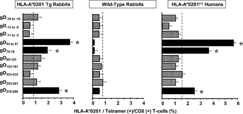
HLA-A*0201 Tg rabbit recognizes human HLA-A*0201–restricted CD8+ T cell epitopes from gD. Five HLA Tg rabbits and five wild type rabbits were infected ocularly with 2 × 10⁵ PFU per eye of HSV-1. Twelve days p.i., spleens were harvested and the percentage HLA-A*0201/tetramer (+) CD8+ T cells specific to each of 10 HSV-1 gD epitopes was determined by FACS. In the numbering system used for gD peptides in this study, the first amino acid in the mature gD is indicated as AA1. Thus, amino acids in the 25 aa leader sequence are indicated as negative numbers relative to AA1. Similar tetramer analysis was performed in PBMCs derived from five HSV-seropositive HLA-A*0201(+) human blood samples. The results are representative of two independent experiments. Asterisk indicates a significant T cell response (p < 0.0005) compared with an irrelevant tetramer. The backgrounds were determined for all ten tetramers using PBMCs from each of the five different HLA-A2(+) HSV-uninfected individuals, five uninfected HLA Tg rabbits, and five uninfected wild type rabbits (controls). The dashed vertical lines in each panel designate maximum background cutoff levels of epitope-specific CD8+ T cells in uninfected HLA-Tg rabbits, uninfected WT rabbits, and seronegative HLA-A2+ individuals.