



Abstract
Although NO donors have been shown to confer late preconditioning (PC) against myocardial ischemia/reperfusion injury in healthy rabbits, it is unknown whether concurrent systemic disorders affect NO donor-induced cardioprotection. Since many patients with coronary artery disease have hypercholesterolemia (HC), we examined the effect of this condition on late PC induced by the NO donor diethylenetriamine/nitric oxide (DETA/NO). Chronically instrumented rabbits were fed a normal diet (normocholesterolemia, NC) or a diet enriched with 1% cholesterol (HC) for 4 weeks. Plasma cholesterol levels were significantly elevated and the arterial pressure response to the endothelium-dependent vasodilator bradykinin was blunted in cholesterol diet-fed rabbits. Conscious rabbits underwent a 30-minute coronary occlusion followed by 3 days of reperfusion. When NC rabbits were pretreated with DETA/NO (0.1 mg/kg, i.v. × 4, group II, n = 7) 24 hours before the 30-minute occlusion, infarct size was reduced by 52% (29.7 ± 3.4% versus 62.4 ± 4.0% of the region at risk in NC controls [group I, n = 5], P < 0.05), indicating that DETA/NO induced a late PC effect against myocardial infarction. In contrast, when HC rabbits were pretreated with the same dose of DETA/NO (group IV, n = 6), infarct size was not significantly reduced (61.0 ± 5.7% versus 68.1 ± 4.5% of the region at risk in HC [group III, n = 5], P = NS), suggesting that DETA/NO failed to induce a delayed cardioprotective effect. These data demonstrate, for the first time, that HC blunts NO donor-induced late PC against myocardial infarction, implying that the inhibitory effects of HC on ischemia-induced and NO donor-induced late PC are caused by disruption of biochemical pathways distal to the generation of NO that triggers these adaptations.
Keywords: Myocardial ischemia, myocardial reperfusion, diethylenetriamine/nitric oxide, NO donor
Introduction
The late phase of ischemic preconditioning (PC) is a cardioprotective phenotypic shift whereby exposure to a brief ischemic stress increases the tolerance of the heart to stunning and infarction 24 h to 72 h later [2–4, 6–9, 23, 28, 30, 31, 46, 54]. In addition to ischemia, a delayed cardioprotective effect can be elicited by pretreatment with nitric oxide (NO) donors [1, 22, 34, 44, 45, 49]. In our previous studies in conscious rabbits, administration of the NO donor diethylenetriamine/nitric oxide (DETA/NO) induced a powerful late PC effect against myocardial stunning [34, 44, 45] and infarction [34, 44]. However, our previous observations regarding DETA/NO-induced late PC against ischemia/reperfusion injury [44, 45] were made in healthy rabbits. Concurrent clinical abnormalities, such as hypercholesterolemia (HC), may interfere with the biochemical pathways that underlie the PC phenomenon [14, 16]. Indeed, Szilvassy et al. [39] first reported that in conscious rabbits, the early PC protection induced by rapid pacing was lost in diet-induced HC. Komamura et al. [26] and Ueda et al. [50] found that the infarct-limiting effect of the early phase of ischemic PC was blunted by HC in rabbits fed a cholesterol diet, and Kocic et al. [25] reported that the protective effect of the early phase of ischemic PC was prevented by hyperlipidemia in isolated stunned rat papillary muscles. In addition, a recent study [29] in patients found that hyperlipidemia prevents the expected decrease in ST-segment elevation after repeated balloon inflations during angioplasty, suggesting that HC blocks early PC in humans as well.
In contrast to early PC, the effect of HC on late PC has not yet been extensively investigated. To our knowledge, only two reports [37, 38] are available, both of which examined the hemodynamic and electrophysiological changes caused by rapid ventricular overpacing as the end-points in cholesterol-fed rabbits. However, except the study we reported, no study has dealt with the effect of HC on late PC against injury induced by coronary occlusion/reperfusion. Although we have previously found that HC abrogates the infarct-limiting effect of the late phase of ischemic PC in conscious rabbits [48], it is unknown whether HC affects NO donor-induced late PC as well. iNOS-derived NO is a mediator of late PC [8]. Since it has been shown that HC decreases cardiac NO content [15, 17, 36], we reasoned that administration of exogenous NO would be useful to discern whether HC impairs the role of NO as a mediator of late PC: if it does, exogenous NO should not induce a late PC state. Therefore, the aim of this study was to determine whether administration of the NO donor DETA/NO induces a delayed cardioprotective effect in HC rabbits.
Methods
This study was performed in accordance with the guidelines of the Animal Care and Use Committee of the University of Louisville (KY) School of Medicine and with the Guide for the Care and Use of Laboratory Animals (Department of Health and Human Services, Publication No. [NIH] 86-23)
Experimental preparation
The experimental preparation is similar to that described in detail previously [7, 9, 35, 44] except that rabbits were rendered hypercholesterolemic. Briefly, New Zealand White male rabbits (weight, 2.0~2.5 kg; age, 3–4 months) were instrumented under sterile conditions with a balloon occluder around a major branch of the left coronary artery, a 10-MHz pulsed Doppler ultrasonic crystal in the center of the region to be rendered ischemic, and bipolar ECG leads on the chest wall. The rabbits were fed a standard rabbit chow until the cholesterol-enriched diet began. Beginning on day 3 after surgery, the animals were assigned to two groups: an age-matched normocholesterolemic group (NC), which was continuously fed a normal diet, and a hypercholesterolemic group (HC), which was fed a diet enriched with 1% cholesterol (Purina Test Diets, Richmond, IN) for 4 weeks. All rabbits were restricted to 100 g of chow per day and had free access to drinking water at all times. Two blood samples (2 ml each) for measurement of the plasma lipid profile were collected from each rabbit, one during open chest surgery and the other during sacrifice. The response of arterial pressure to endothelium-dependent (bradykinin) and endothelium-independent (nitroglycerin) vasodilators was tested in some cholesterol-fed rabbits at the beginning of the cholesterol diet and at the completion of the experiment (just prior to sacrifice). Arterial pressure was measured by cannulating the ear dorsal artery with a 22-gauge angiocatheter as previously described [7, 9, 35, 44]. After sacrifice of the HC rabbits, a segment of descending thoracic aorta was collected and stained with oil red O stain [13] to verify the absence of atherosclerotic lesions.
Experimental protocol
At the end of the 4-week diet feeding, each diet group of rabbits was divided into two sub-groups, generating a total of four groups, i.e., NC-control (group I), NC-DETA/NO (group II), HC-control (group III), and HC-DETA/NO (group IV). All experiments were performed while the rabbits were conscious. Groups I and III received vehicle (PBS), whereas groups II and IV received 4 consecutive intravenous boluses of DETA/NO (0.1 mg/kg each) every 25 minutes (total dose, 0.4 mg/kg). This dose was selected because in our previous studies [34, 44, 45, 51] it induced a robust late PC protection and triggered the signaling cascade of late PC [34, 51]. DETA/NO (Alexis Corp) was dissolved in PBS (total volume infused, 4 ml). To remove oxygen from the solution, the PBS was bubbled with nitrogen for at least 30 minutes before dissolving DETA/NO. Twenty-four hours later, rabbits were subjected to a 30-min coronary artery occlusion followed by 3 days of reperfusion. Diazepam was administered 20 min before the onset of ischemia (4 mg/kg i.p.) to relieve the stress caused by the coronary occlusion. Successful coronary occlusion was verified by observing the development of ST-segment elevation and the changes in the QRS complex on the ECG and the appearance of paradoxical systolic wall thinning on the ultrasonic crystal recordings.
Measurement of region at risk and infarct size
At the conclusion of the study, the rabbits were given heparin (1,000 U i.v.), after which they were anesthetized with sodium pentobarbital (50 mg/kg i.v.) and euthanized with KCl. The heart was excised and perfused with Krebs-Henseleit solution through an aortic cannula using a Langendorff apparatus. To delineate infarcted from viable myocardium, the heart was then perfused with a 1% solution of 2,3,5-triphenyltetrazolium chloride in phosphate buffer (pH 7.4, 37 °C) at a pressure of 60 mmHg (~30 ml over 5 min). To delineate the occluded-reperfused coronary vascular bed, the coronary artery was then tied at the site of the previous occlusion and the aortic root was perfused with a 5% solution of phthalo blue dye (Heucotech, Fairless Hill, PA) in normal saline (~10 ml over 3 min). As a result of this procedure, the portion of the left ventricle (LV) supplied by the previously occluded coronary artery (region at risk) was identified by the absence of blue dye, whereas the rest of the LV was stained dark blue. The heart was then cut into 6-7 transverse slices, and all atrial and right ventricular tissues were excised. The slices were weighed, fixed in a 10% neutral buffered formaldehyde solution, and photographed (Nikon D100 Digital Camera with a Nikkor AF28-105 mm lens plus Promaster Spectrum 7 close-up lenses). Color pictures of heart slices were projected onto a paper screen at a ten-fold magnification and the borders of the infarcted, ischemic-reperfused, and non-ischemic regions were traced. The tracings were then scanned and the corresponding areas measured by computerized planimetry (Adobe Photoshop, version 6.0). From these measurements infarct size was calculated as a percentage of the region at risk using methods analogous to those employed in previous studies [35, 40, 44].
Statistical analysis
Data are reported as means ± SEM. For intragroup comparisons, arterial pressure response was analyzed by a two-way repeated-measures ANOVA (time and dose) followed by Student’s t-tests for paired data with the Bonferroni correction. For intergroup comparisons, data were analyzed by a one-way ANOVA followed by unpaired Student’s t-tests with the Bonferroni correction. The relationship between infarct size and risk region size was compared among groups with an ANCOVA using the size of the risk region as the covariate [35, 40, 44]. The correlation between infarct size and risk region size was assessed by linear regression analysis using the least-squares method. ANOVA was performed using the SigmaStat for Windows version 2.0 and ANCOVA using SPSS for Windows 10.
Results
Exclusions and heart rate
A total of 28 rabbits were instrumented for this study (14 for each of the two diets). Seven rabbits were assigned to each of the four groups. Five out of the 28 rabbits were excluded from the study: 4 died of ventricular fibrillation during coronary occlusion (2 in group I, 1 in group III, and 1 in group IV), and 1 (in group III) was excluded because of malfunction of the balloon occluder. Therefore, a total of 5 rabbits completed the experimental protocol in groups I and III, and 6 in group IV. All 7 rabbits in group II were included into the analysis.
As seen in Table 1, there were no appreciable differences in heart rate among groups, either during administration of DETA/NO (groups II and IV) on day 1 or during coronary occlusion and reperfusion on day 2.
Table 1.
Heart rate (bpm)
| During administration of DETA/NO on day 1
| |||
|---|---|---|---|
| Group | Baseline | End of 4 injections | 1 h after the fourth injection |
| II (NC DETA/NO) | 244 ± 8 | 239 ± 7 | 246 ± 10 |
| IV (HC DETA/NO) | 251 ± 11 | 246 ± 11 | 248 ± 9 |
| During coronary occlusion and reperfusion on day 2
| ||||||
|---|---|---|---|---|---|---|
| Group | Baseline | Pre-occlusion (after diazepam) | Occlusion
|
Reperfusion
|
||
| 15 min | 30 min | 15 min | 1 h | |||
| I (NC control) | 250 ± 9 | 242 ± 6 | 262 ± 10 | 255 ± 8 | 248 ± 11 | 252 ± 8 |
| II (NC DETA/NO) | 246 ± 7 | 238 ± 8 | 270 ± 11 | 266 ± 8 | 252 ± 10 | 242 ± 9 |
| III (HC control) | 239 ± 9 | 232 ± 10 | 254 ± 10 | 258 ± 9 | 250 ± 8 | 253 ± 8 |
| IV (HC DETA/NO) | 247 ± 8 | 243 ± 10 | 255 ± 9 | 265 ± 11 | 256 ± 6 | 255 ± 7 |
Groups II(n = 7) and IV (n = 6) received 4 bolus i.v. injections of 0.1 mg/kg DETA/NO at 25-minute intervals on day 1. Heart rate was measured at baseline (before the first bolus), end of 4 injections (75 minutes after the first bolus), and 1 h after the fourth injection. On day 2, all groups (n = 5, 7, 5, and 6, respectively, in groups I, II, III, and IV) underwent a 30-minute coronary occlusion followed by 3 days of reperfusion. Heart rate was measured at baseline (before diazepam), pre-occlusion (immediately before coronary occlusion; i.e., 20 minutes after diazepam), at 15 and 30 minutes into coronary occlusion, and at selected times after reperfusion (for the sake of brevity, only the 15-minute and 1-hour measurements are reported). Values are mean ± SEM
Plasma cholesterol levels and atherosclerotic lesions
As seen in Table 2, the plasma lipid profile was similar at baseline between groups and did not change in rabbits fed a normal diet for four weeks. In contrast, a marked increase in plasma levels of total cholesterol, % LDL-cholesterol and the LDL/HDL ratio and a decrease in % HDL-cholesterol were noted in rabbits fed a 1% cholesterol-enriched diet for four weeks, although triglycerides were not significantly altered. Despite the marked changes in lipid profile, postmortem oil red O staining did not reveal any macroscopic atherosclerotic lesion in the descending thoracic aorta of any HC rabbit (data not shown).
Table 2.
Plasma lipid profile in rabbits
| Normal diet (n = 12) | 1% cholesterol diet (n = 11) | |||
|---|---|---|---|---|
| Baseline | 4 weeks later | Baseline | 4 weeks later | |
| Total cholesterol (mg/dL) | 54.1 ± 3.8 | 61.5 ± 4.3 | 59.7 ± 4.2 | 1130.5 ± 91.7* |
| % LDL-cholesterol | 50.1 ± 4.7 | 52.1 ± 3.4 | 46.8 ± 4.9 | 85.1 ± 0.7* |
| % HDL-cholesterol | 47.4 ± 4.7 | 43.8 ± 3.1 | 50.1 ± 4.7 | 13.1 ± 0.8* |
| LDL/HDL ratio | 1.3 ± 0.3 | 1.3 ± 0.2 | 1.2 ± 0.2 | 6.8 ± 0.5* |
| Triglycerides (mg/dL) | 80.7 ± 7.2 | 85.6 ± 14.0 | 78.2 ± 10.7 | 89.6 ± 14.6 |
Values are mean ± SEM.
P < 0.05 vs. baseline value
Vasodilator response
Studies were conducted in six cholesterol diet-fed rabbits to confirm that HC impairs NO activity. The response of arterial blood pressure to vasodilators was tested at baseline (before starting the cholesterol diet) and after 4 weeks on the cholesterol diet (just prior to sacrifice of the animal). As shown in Fig. 1, endothelium-dependent (bradykinin) and -independent (nitroglycerin) vasodilators decreased mean arterial pressure dose-dependently at baseline. The cholesterol diet reduced the response of arterial pressure to bradykinin (Fig. 1A) but not to nitro-glycerin (Fig. 1B), suggesting that NO bioactivity is impaired in HC rabbits.
Fig. 1.
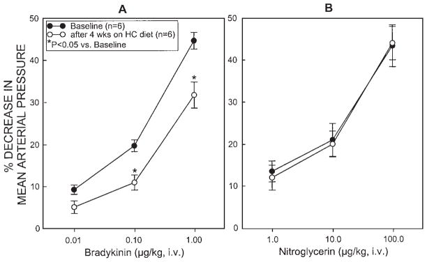
Change in mean arterial pressure in response to a bolus i.v. injection of (A) 0.01, 0.10 or 1.0 μg/kg of the endothelium-dependent vasodilator bradykinin and (B) 1, 10, or 100 μg/kg of the endothelium-independent vasodilator nitroglycerin in rabbits (n = 6) at baseline (before cholesterol diet) and after 4 weeks on a 1% cholesterol diet (just prior to sacrifice). Data are means ± SEM
Region at risk and infarct size
There were no significant differences among the 4 groups with respect to the weight of the region at risk (0.90 ± 0.18 g [18.2 ± 3.2% of LV weight], 0.87 ± 0.07 g [22.3 ± 2.5% of LV weight], 1.09 ±0.15 g [23.1 ± 3.2% of LV weight], and 1.10 ± 0.16 g [21.9 ± 3.3% of LV weight], respectively, in groups I, II, III, and IV). The average infarct size was 52% smaller in group II (NC-DETA/NO group) compared with group I (NC-control group) (29.7 ± 3.4% vs. 62.4 ± 4.0% of the region at risk, respectively; P < 0.05 [Fig. 2]), indicating that DETA/NO induced delayed protection against myocardial infarction in healthy rabbits, which confirms our previous observations [34, 44]. Infarct size in group III (HC-control group) was comparable to that in group I (NC-control group) (68.1 ± 4.5% vs. 62.4 ± 4.0% of the region at risk, respectively, P = NS [Fig. 2]). However, although group IV (HC-DETA/NO group) received the same dose of DETA/NO, infarct size was not smaller (61.0 ± 5.7% of the region at risk) than in group III (Fig. 2), suggesting that DETA/NO failed to induce a delayed protective effect against myocardial infarction in HC rabbits.
Fig. 2.

Myocardial infarct size in groups I (n = 5, NC control group), II (n = 7, NC DETA/NO group), III (n = 5, HC control group), and IV (n = 6, HC DETA/NO group). Infarct size is expressed as a percentage of the region at risk of infarction. Open circles represent individual rabbits, whereas solid circles represent means ± SEM. NC normocholesterolemia; HC hypercholesterolemia. *P < 0.05 vs. group I (NC control group)
In all groups, the size of the infarction was positively and linearly related to the size of the region at risk (r = 0.95 in group I [NC-control group], 0.80 in group II [NC-DETA/NO group], 0.93 in group III [HC-control group], and 0.90 in group IV [HC-DETA/NO group]) (Fig. 3). As expected [44], the regression line was shifted to the right in the NC-DETA/NO group as compared with the NC-control group (P < 0.05 by ANCOVA) (Fig. 3), indicating that for any given size of the region at risk, the resulting infarct size was reduced by pretreatment with DETA/NO in NC rabbits. In contrast, in the HC-DETA/NO group (group IV), the regression line did not differ significantly from that observed in the HC-control group (group III) (Fig. 3), demonstrating that, in HC rabbits, DETA/NO fails to induce the infarct size-limiting effect seen in healthy rabbits.
Fig. 3.

Relationship between size of the region at risk and size of myocardial infarction. The figure illustrates both individual values and the regression lines obtained by linear regression analysis for the NC control group (group I, n = 5), the NC DETA/NO group (group II, n = 7), the HC control group (group III, n = 5), and the HC DETA/NO group (group IV, n = 6). In all groups, infarct size was positively and linearly related to risk region size. The linear regression equations were as follows: group I, y = 0.60 χ + 0.02 (r= 0.95); group II, y = 0.36 χ − 0.06 (r= 0.80); group III, y = 0.60 χ + 0.15 (r = 0.93); and group IV, y = 0.71 χ − 0.10 (r = 0.90). ANCOVA demonstrated that the regression line for the NC DETA/NO group was significantly different from that for the NC control group (P < 0.05), indicating that for any given risk region size, infarct size was smaller in the DETA/NO-pretreated NC rabbits compared with the vehicle-pretreated NC rabbits; in contrast, the line for the HC DETA/NO group was similar to those for the vehicle-pretreated HC rabbits and the NC control groups
Discussion
Although previous studies [1, 19, 22, 34, 44, 45, 49] have demonstrated that administration of NO donors, in lieu of ischemia, elicits late PC in healthy animals, no study has been performed to examine NO donor-induced late PC in animals with concurrent systemic disorders such as HC. Since it is well documented that NO plays a key role in triggering the development of late PC [7, 8, 34, 35] and that NO bioactivity is impaired by HC [5, 10, 21], it is reasonable to postulate that our previous finding [48] that the infarct-sparing effect of ischemia-induced late PC is abrogated in hypercholesterolemic rabbits may be the result of the detrimental effect of HC on the formation of the NO that triggers ischemic PC. If this were the case, then exogenous NO should act as a trigger of late PC and elicit delayed cardioprotection in hypercholesterolemic animals. Instead, in the present study we found that administration of DETA/NO induced delayed cardio-protection against infarction in healthy rabbits but failed to do so in hypercholesterolemic rabbits. This finding suggests that, instead of or in addition to impairing the generation of the NO that triggers late PC, HC interferes with downstream biochemical pathways that underlie the late PC response.
Effect of HC on myocardial infarction in the nonpreconditioned state
The studies reported herein were performed in conscious animals in an effort to test the potential cardioprotective action of NO donors under physiological conditions. Since HC is one of the most prevalent risk factors for coronary artery disease, examining the PC phenomenon in the presence of HC is clinically relevant. Given that the focus of the present study was to examine the role of NO in triggering late PC, the use of a chronically instrumented conscious rabbit model was particularly important because the trauma and inflammatory reaction associated with a thoracotomy may elicit release of cytokines which, in turn, could modulate the activity of NOS and confound NO generation. Accordingly, we felt that studying late PC in a conscious hypercholesterolemic rabbit model would be both physiologically and clinically relevant. The duration of the cholesterol diet (4 weeks) was selected to avoid the development of atherosclerotic lesions while achieving high and steady plasma cholesterol levels so as to enable us to test the effect of HC in the absence of coexisting atherosclerosis. Indeed, this protocol induced high plasma cholesterol levels without histological evidence of atherosclerosis as demonstrated by postmortem oil red O staining of the aortas. As shown in Fig. 1, this hypercholesterolemic rabbit model exhibited impaired NO bioactivity, whereby the hypotensive response to the endothelium-dependent vasodilator bradykinin was blunted.
Despite the high cholesterol levels and the blunted pressure response to bradykinin, however, infarct size was not significantly different from that in normocholesterolemic rabbits (Fig. 2), suggesting that HC per se did not affect the susceptibility to ischemia/reperfusion injury. This finding is apparently in contrast with studies by Jung et al. [24] and Wang et al. [52] in anesthetized open-chest rabbit preparations, which concluded that hypercholesterolemic myocardium is more susceptible to ischemia/reperfusion injury. In the study by Jung et al. [24], rabbits were fed an atherogenic diet (0.25% cholesterol and 3% coconut oil) for 4 weeks, after which myocardial infarction was induced by a 30-min coronary occlusion followed by 2 h of reperfusion. Infarct size in their model was increased by 52% compared with normal diet-fed rabbits. Similar findings have been reported by Wang et al. [52] in rabbits fed 0.5% cholesterol and 10% coconut oil for 8 weeks followed by a 30-min coronary occlusion/4-h reperfusion sequence. The reason for the apparent discrepancy between these findings and our present results is unknown. The divergent results may be secondary to differences in experimental preparations (open-chest vs. conscious animals), diet components (cholesterol plus coconut oil vs. cholesterol only), or experimental protocols (2–4 h vs. 24 h reperfusion).
Effect of HC on PC
A number of reports [15, 17, 24–27, 37, 39, 48, 50] have examined the PC response in hypercholesterolemic animals. These few studies have dealt mainly with the early phase of PC [15, 17, 24–27, 39, 50] and have yielded conflicting results. Szilvassy et al. [39] first reported that HC abrogated the early protective effects induced by rapid cardiac pacing in a conscious rabbit model. Interestingly, normalization of serum cholesterol by re-feeding normal chow over 6 additional weeks reinstated the PC phenomenon even though intimal lesions were still present [39]. This finding was subsequently confirmed in hearts isolated from hypercholesterolemic rats, in which the early PC protection induced by rapid ventricular pacing was lost [17]. These studies [17, 39] indicate that HC per se, without atherosclerosis, inhibits the early PC response. Komamura et al. [26] and Ueda et al. [50] reported that the infarct-limiting effect of early PC was blunted by HC in rabbits fed a cholesterol diet. Kocic et al. [25] found that the protective effect of early PC on contractility and responsiveness to phenylephrine was prevented by experimental hyperlipidemia in isolated stunned rat papillary muscle. A recent study [29] in patients found that hyperlipidemia prevented the expected decrease in ST-segment elevation after repeated balloon inflations during coronary angioplasty, suggesting that HC blocks early PC in humans as well. In contrast, Kremastinos et al. [27] and Jung et al. [24] reported that the infarct size-limiting effect of early ischemic PC was not altered in hypercholesterolemic rabbits. Thus, data on the effect of HC on early PC are conflicting.
With regard to the effect of HC on late PC, only two reports [37, 38] are available in addition to our recent study [48]. In the first report, Szekeres et al. [37] found that the protection induced by rapid ventricular pacing against the hemodynamic and electrophysiological changes caused by subsequent rapid overpacing was still present in cholesterol-fed rabbits but a greater PC stimulus was required. These results suggest that HC elevates the threshold for the PC response. In another study, Szilvassy et al. [38] reported that the LPS-induced late PC effect on the hemodynamic and electrophysiological changes caused by ventricular overpacing was not abrogated in cholesterol-fed conscious rabbits. However, in both studies [37, 38], the experiments were performed in HC rabbits with atherosclerotic lesions, which may have confounded the effect of HC per se; furthermore, the endpoints were ST-segment elevation, left ventricular end-diastolic pressure, and shortening of the ventricular effective refractory period, not infarct size. Therefore, the findings are difficult to evaluate because the animal model was not one of pure hypercholesterolemia and because the significance of the end-points selected is unclear. Using a more straightforward experimental protocol and a HC rabbit model without significant atherosclerotic alterations, we have found that a sequence of six 4-min coronary occlusion/4-min reperfusion cycles, which induces delayed protection 24 h later against stunning [7, 9, 11, 12, 34, 41, 47, 53] and infarction [34, 35, 42] in healthy rabbits, failed to induce delayed protection in rabbits fed a 1% cholesterol-enriched diet for 6 weeks [48], indicating that HC abrogates the cardioprotective effect of the late phase of ischemic PC. The present study is the first to deal with the effect of HC on NO donor-induced late PC. Because the NO donor DETA/NO releases NO spontaneously and triggers the development of late PC in healthy rabbits [34, 44, 45], we expected that exogenous NO would reinstate the late PC effect in HC rabbits. Surprisingly, our present data show that administration of the NO donor DETA/NO that induces the late PC effect in NC rabbits failed to do so in HC rabbits, suggesting that HC blunts NO donor-induced late PC as well. However, because our present study did not examine another NO donor or a higher dose of DETA/NO, it is unknown whether other NO donors or higher doses of DETA/NO would reinstate the late PC effect in HC rabbits.
Possible mechanism whereby HC blunts NO donor-induced late preconditioning
Since it has been well documented that HC impairs NO bioactivity [5, 10, 21, 36] and that NO acts as a trigger of late PC [8], it is plausible that HC could impair the triggering role of NO in PC. If loss of late PC protection in hypercholesterolemic animals results from an impairment of the NO triggering mechanism, one would expect that NO donors would overcome this block and elicit the cardioprotection of late PC; that is, the loss of late PC should be rescued by exogenous NO. Our present results, however, show that administration of an NO donor fails to induce late PC, suggesting that the loss of PC protection in HC is not due, or at least is not due solely, to loss of the trigger. Since iNOS-derived NO plays an essential role in mediating the cardioprotection of late PC [8, 9, 20], one possibility is that HC may interfere with the role of NO as a mediator in lieu of or in addition to the role of NO as a trigger. That is, even though exogenous NO (via administration of an NO donor) initiates the late PC response, cardioprotection would not be manifest because HC also impairs the formation of the NO that mediates late PC. In this regard, it is well known that tetrahydrobiopterin (BH4) is an essential cofactor for iNOS to generate NO [18]. Since HC is known to be associated with increased production of superoxide anion [33], which may inhibit the biosynthesis or prevent recycling of BH4 [32], we speculate that HC may undermine the bioavailability of BH4, leading to impairment of the iNOS-derived NO formation that is required to mediate NO donor-induced late PC protection. In addition, we have previously found that administration of N-acetyl-serotonin, a BH4 synthesis inhibitor, to ischemically pre-conditioned rabbits abrogates late PC against both myocardial stunning [43] and infarction [48] and that ischemic PC elevates myocardial levels of BH4 in NC rabbits, but not in HC rabbits [48], indicating that BH4 plays an essential role in mediating ischemia-induced late PC and that HC prevents ischemic PC-induced upregulation of myocardial BH4. However, it remains to be determined whether and how, by undermining the bioavailability of BH4, HC affects the formation of NO that mediates NO donor-induced late PC.
Conclusions
In conclusion, the present study demonstrates that administration of the NO donor DETA/NO, which induces late PC protection against myocardial ischemia/reperfusion injury in healthy animals, fails to elicit cardioprotection in hypercholesterolemic rabbits. This finding suggests that the inhibitory effect of HC on the late PC response elicited by either ischemia or NO donors are due, at least in part, to a disruption of the signaling cascades and/or mediator proteins downstream of the NO that triggers these two adaptive responses.
Acknowledgments
We gratefully acknowledge Qiuli Bi for expert technical assistance. This study was supported in part by NIH R01 grants HL74351, HL-55757, HL-68088, HL-70897, and HL-76794, by AHA grants 0355391B, by the Medical Research Grant Program of the Jewish Hospital Foundation, Louisville, KY, and by the Commonwealth of Kentucky Research Challenge Trust Fund.
Contributor Information
Xian-Liang Tang, Division of Cardiology, University of Louisville, Louisville, Kentucky 40292, USA, Tel.: +1-502/852-1837, Fax: +1-502/852-6474.
Adam B. Stein, Division of Cardiology, University of Louisville, Louisville, Kentucky 40292, USA, Tel.: +1-502/852-1837, Fax: +1-502/852-6474
Gregg Shirk, Division of Cardiology, University of Louisville, Louisville, Kentucky 40292, USA, Tel.: +1-502/852-1837, Fax: +1-502/852-6474.
Roberto Bolli, Email: rbolli@louisville.edu, Division of Cardiology, University of Louisville, Louisville, Kentucky 40292, USA, Tel.: +1-502/852-1837, Fax: +1-502/852-6474.
References
- 1.Banerjee S, Tang XL, Qiu Y, Takano H, Manchikalapudi S, Dawn B, Shirk G, Bolli R. Nitroglycerin induces late preconditioning against myocardial stunning via a PKC-dependent pathway. Am J Physiol. 1999;277:H2488–H2494. doi: 10.1152/ajpheart.1999.277.6.H2488. [DOI] [PubMed] [Google Scholar]
- 2.Baxter GF, Goma FM, Yellon DM. Involvement of protein kinase C in the delayed cytoprotection following sublethal ischaemia in rabbit myocardium. Br J Pharmacol. 1995;115:222–224. doi: 10.1111/j.1476-5381.1995.tb15866.x. [DOI] [PMC free article] [PubMed] [Google Scholar]
- 3.Baxter GF, Goma FM, Yellon DM. Characterisation of the infarct-limiting effect of delayed preconditioning: time-course and dose-dependency studies in rabbit myocardium. Basic Res Cardiol. 1997;92:159–167. doi: 10.1007/BF00788633. [DOI] [PubMed] [Google Scholar]
- 4.Baxter GF, Marber MS, Patel VC, Yellon DM. Adenosine receptor involvement in a delayed phase of myocardial protection 24 hours after ischemic preconditioning. Circulation. 1994;90:2993–3000. doi: 10.1161/01.cir.90.6.2993. [DOI] [PubMed] [Google Scholar]
- 5.Best PJ, Lerman LO, Romero JC, Richardson D, Holmes DR, Jr, Lerman A. Coronary endothelial function is preserved with chronic endothelin receptor antagonism in experimental hypercholesterolemia in vitro. Arterioscler Thromb Vasc Biol. 1999;19:2769–2775. doi: 10.1161/01.atv.19.11.2769. [DOI] [PubMed] [Google Scholar]
- 6.Bolli R. The early and late phases of preconditioning against myocardial stunning and the essential role of oxyradicals in the late phase: an overview. Basic Res Cardiol. 1996;91:57–63. doi: 10.1007/BF00788866. [DOI] [PubMed] [Google Scholar]
- 7.Bolli R, Bhatti ZA, Tang XL, Qiu Y, Zhang Q, Guo Y, Jadoon AK. Evidence that late preconditioning against myocardial stunning in conscious rabbits is triggered by the generation of nitric oxide. Circ Res. 1997;81:42–52. doi: 10.1161/01.res.81.1.42. [DOI] [PubMed] [Google Scholar]
- 8.Bolli R, Dawn B, Tang XL, Qiu Y, Ping P, Xuan YT, Jones WK, Takano H, Guo Y, Zhang J. The nitric oxide hypothesis of late preconditioning. Basic Res Cardiol. 1998;93:325–338. doi: 10.1007/s003950050101. [DOI] [PMC free article] [PubMed] [Google Scholar]
- 9.Bolli R, Manchikalapudi S, Tang XL, Takano H, Qiu Y, Guo Y, Zhang Q, Jadoon AK. The protective effect of late preconditioning against myocardial stunning in conscious rabbits is mediated by nitric oxide synthase. Evidence that nitric oxide acts both as a trigger and as a mediator of the late phase of ischemic preconditioning. Circ Res. 1997;81:1094–1107. doi: 10.1161/01.res.81.6.1094. [DOI] [PubMed] [Google Scholar]
- 10.Britten MB, Zeiher AM, Schachinger V. Clinical importance of coronary endothelial vasodilator dysfunction and therapeutic options. J Intern Med. 1999;245:315–327. doi: 10.1046/j.1365-2796.1999.00449.x. [DOI] [PubMed] [Google Scholar]
- 11.Dawn B, Takano H, Tang XL, Kodani E, Banerjee S, Rezazadeh A, Qiu Y, Bolli R. Role of Src protein tyrosine kinases in late preconditioning against myocardial infarction. Am J Physiol Heart Circ Physiol. 2002;283:H549–H556. doi: 10.1152/ajpheart.00873.2001. [DOI] [PubMed] [Google Scholar]
- 12.Dawn B, Xuan YT, Qiu Y, Takano H, Tang XL, Ping P, Banerjee S, Hill M, Bolli R. Bifunctional role of protein tyrosine kinases in late preconditioning against myocardial stunning in conscious rabbits. Circ Res. 1999;85:1154–1163. doi: 10.1161/01.res.85.12.1154. [DOI] [PubMed] [Google Scholar]
- 13.Eto Y, Shimokawa H, Tanaka E, Morishige K, Fuchigami M, Ishiwata Y, Matsushima K, Takeshita A. Long-term treatment with propagermanium suppresses atherosclerosis in WHHL rabbits. J Cardiovasc Pharmacol. 2003;41:171–177. doi: 10.1097/00005344-200302000-00004. [DOI] [PubMed] [Google Scholar]
- 14.Ferdinandy P. Myocardial ischaemia/reperfusion injury and preconditioning: effects of hypercholesterolaemia/hyperlipidaemia. Br J Pharmacol. 2003;138:283–285. doi: 10.1038/sj.bjp.0705097. [DOI] [PMC free article] [PubMed] [Google Scholar]
- 15.Ferdinandy P, Csonka C, Csont T, Szilvassy Z, Dux L. Rapid pacing-induced preconditioning is recaptured by farnesol treatment in hearts of cholesterol-fed rats: role of polyprenyl derivatives and nitric oxide. Mol Cell Biochem. 1998;186:27–34. [PubMed] [Google Scholar]
- 16.Ferdinandy P, Szilvassy Z, Baxter GF. Adaptation to myocardial stress in disease states: is preconditioning a healthy heart phenomenon? Trends Pharmacol Sci. 1998;19:223–229. doi: 10.1016/s0165-6147(98)01212-7. [DOI] [PubMed] [Google Scholar]
- 17.Ferdinandy P, Szilvassy Z, Horvath LI, Csont T, Csonka C, Nagy E, Szentgyorgyi R, Nagy I, Koltai M, Dux L. Loss of pacing-induced preconditioning in rat hearts: role of nitric oxide and cholesterol-enriched diet. J Mol Cell Cardiol. 1997;29:3321–3333. doi: 10.1006/jmcc.1997.0557. [DOI] [PubMed] [Google Scholar]
- 18.Gross SS, Levi R. Tetrahydrobiopterin synthesis. An absolute requirement for cytokine-induced nitric oxide generation by vascular smooth muscle. J Biol Chem. 1992;267:25722–25729. [PubMed] [Google Scholar]
- 19.Guo Y, Bao W, Tang XL, Wu WJ, Bolli R. Nitric oxide donors induce late preconditioning against myocardial infarction in mice. J Mol Cell Cardiol. 1999;31:A–9 (abstr). [Google Scholar]
- 20.Guo Y, Jones WK, Xuan YT, Tang XL, Bao W, Wu WJ, Han H, Laubach VE, Ping P, Yang Z, Qiu Y, Bolli R. The late phase of ischemic preconditioning is abrogated by targeted disruption of the inducible NO synthase gene. Proc Natl Acad Sci U S A. 1999;96:11507–11512. doi: 10.1073/pnas.96.20.11507. [DOI] [PMC free article] [PubMed] [Google Scholar]
- 21.Harrison DG, Ohara Y. Physiologic consequences of increased vascular oxidant stresses in hypercholesterolemia and atherosclerosis: implications for impaired vasomotion. Am J Cardiol. 1995;75:75B–81B. doi: 10.1016/0002-9149(95)80018-n. [DOI] [PubMed] [Google Scholar]
- 22.Hill M, Takano H, Tang XL, Kodani E, Shirk G, Bolli R. Nitroglycerin induces late preconditioning against myocardial infarction in conscious rabbits despite development of nitrate tolerance. Circulation. 2001;104:694–649. doi: 10.1161/hc3201.092218. [DOI] [PubMed] [Google Scholar]
- 23.Imagawa J, Baxter GF, Yellon DM. Genistein, a tyrosine kinase inhibitor, blocks the “second window of protection” 48 h after ischemic preconditioning in the rabbit. J Mol Cell Cardiol. 1997;29:1885–1893. doi: 10.1006/jmcc.1997.0428. [DOI] [PubMed] [Google Scholar]
- 24.Jung O, Jung W, Malinski T, Wiemer G, Schoelkens BA, Linz W. Ischemic preconditioning and infarct mass: the effect of hypercholesterolemia and endothelial dysfunction. Clin Exp Hypertens. 2000;22:165–179. doi: 10.1081/ceh-100100070. [DOI] [PubMed] [Google Scholar]
- 25.Kocic I, Konstanski Z, Kaminski M, Dworakowska D, Dworakowski R. Experimental hyperlipidemia prevents the protective effect of ischemic preconditioning on the contractility and responsiveness to phenylephrine of rat-isolated stunned papillary muscle. Gen Pharmacol. 1999;33:213–219. doi: 10.1016/s0306-3623(99)00017-8. [DOI] [PubMed] [Google Scholar]
- 26.Komamura K, Funaya H, Hori M. Infarct size-limiting effect of ischemic preconditioning is attenuated in cholesterol-fed rabbit. Circulation. 1995;92:I-390 (abstr). [Google Scholar]
- 27.Kremastinos DT, Bofilis E, Karavolias GK, Papalois A, Kaklamanis L, Iliodromitis EK. Preconditioning limits myocardial infarct size in hypercholesterolemic rabbits. Atherosclerosis. 2000;150:81–89. doi: 10.1016/s0021-9150(99)00389-5. [DOI] [PubMed] [Google Scholar]
- 28.Kuzuya T, Hoshida S, Yamashita N, Fuji H, Oe H, Hori M, Kamada T, Tada M. Delayed effects of sublethal ischemia on the acquisition of tolerance to ischemia. Circ Res. 1993;72:1293–1299. doi: 10.1161/01.res.72.6.1293. [DOI] [PubMed] [Google Scholar]
- 29.Kyriakides ZS, Psychari S, Iliodromitis EK, Kolettis TM, Sbarouni E, Kremastinos DT. Hyperlipidemia prevents the expected reduction of myocardial ischemia on repeated balloon inflations during angioplasty. Chest. 2002;121:1211–1215. doi: 10.1378/chest.121.4.1211. [DOI] [PubMed] [Google Scholar]
- 30.Marber MS, Latchman DS, Walker JM, Yellon DM. Cardiac stress protein elevation 24 hours after brief ischemia or heat stress is associated with resistance to myocardial infarction. Circulation. 1993;88:1264–1272. doi: 10.1161/01.cir.88.3.1264. [DOI] [PubMed] [Google Scholar]
- 31.Marber MS, Yellon DM. Myocardial adaptation, stress proteins, and the second window of protection. Ann N Y Acad Sci. 1996;793:123–141. doi: 10.1111/j.1749-6632.1996.tb33510.x. [DOI] [PubMed] [Google Scholar]
- 32.Milstien S, Katusic Z. Oxidation of tetrahydrobiopterin by peroxynitrite: implications for vascular endothelial function. Biochem Biophys Res Commun. 1999;263:681–684. doi: 10.1006/bbrc.1999.1422. [DOI] [PubMed] [Google Scholar]
- 33.Ohara Y, Peterson TE, Harrison DG. Hypercholesterolemia increases endothelial superoxide anion production. J Clin Invest. 1993;91:2546–2551. doi: 10.1172/JCI116491. [DOI] [PMC free article] [PubMed] [Google Scholar]
- 34.Ping P, Takano H, Zhang J, Tang XL, Qiu Y, Li RC, Banerjee S, Dawn B, Balafonova Z, Bolli R. Isoform-selective activation of protein kinase C by nitric oxide in the heart of conscious rabbits: a signaling mechanism for both nitric oxide-induced and ischemia-induced preconditioning. Circ Res. 1999;84:587–604. doi: 10.1161/01.res.84.5.587. [DOI] [PubMed] [Google Scholar]
- 35.Qiu Y, Rizvi A, Tang XL, Manchikalapudi S, Takano H, Jadoon AK, Wu WJ, Bolli R. Nitric oxide triggers late preconditioning against myocardial infarction in conscious rabbits. Am J Physiol. 1997;273:H2931–H2936. doi: 10.1152/ajpheart.1997.273.6.H2931. [DOI] [PubMed] [Google Scholar]
- 36.Schwemmer M, Sommer O, Koeckerbauer R, Bassenge E. Cardiovascular dysfunction in hypercholesterolemia associated with enhanced formation of AT1-receptor and of eicosanoids. J Cardiovasc Pharmacol Ther. 2000;5:59–68. doi: 10.1177/107424840000500108. [DOI] [PubMed] [Google Scholar]
- 37.Szekeres L, Szilvassy Z, Ferdinandy P, Nagy I, Karcsu S, Csati S. Delayed cardiac protection against harmful consequences of stress can be induced in experimental atherosclerosis in rabbits. J Mol Cell Cardiol. 1997;29:1977–1983. doi: 10.1006/jmcc.1997.0418. [DOI] [PubMed] [Google Scholar]
- 38.Szilvassy Z, Ferdinandy P, Cluff CW, Elliott GT. Antiischemic effect of monophosphoryl lipid A in conscious rabbits with hypercholesterolemia and atherosclerosis. J Cardiovasc Pharmacol. 1998;32:206–212. doi: 10.1097/00005344-199808000-00006. [DOI] [PubMed] [Google Scholar]
- 39.Szilvassy Z, Ferdinandy P, Szilvassy J, Nagy I, Karcsu S, Lonovics J, Dux L, Koltai M. The loss of pacing-induced preconditioning in atherosclerotic rabbits: role of hypercholesterolaemia. J Mol Cell Cardiol. 1995;27:2559–2569. doi: 10.1006/jmcc.1995.0043. [DOI] [PubMed] [Google Scholar]
- 40.Takano H, Manchikalapudi S, Tang XL, Qiu Y, Rizvi A, Jadoon AK, Zhang Q, Bolli R. Nitric oxide synthase is the mediator of late preconditioning against myocardial infarction in conscious rabbits. Circulation. 1998;98:441–449. doi: 10.1161/01.cir.98.5.441. [DOI] [PubMed] [Google Scholar]
- 41.Takano H, Tang XL, Bolli R. Differential role of K(ATP) channels in late preconditioning against myocardial stunning and infarction in rabbits. Am J Physiol Heart Circ Physiol. 2000;279:H2350–2359. doi: 10.1152/ajpheart.2000.279.5.H2350. [DOI] [PubMed] [Google Scholar]
- 42.Takano H, Tang XL, Kodani E, Bolli R. Late preconditioning enhances recovery of myocardial function after infarction in conscious rabbits. Am J Physiol Heart Circ Physiol. 2000;279:H2372–2381. doi: 10.1152/ajpheart.2000.279.5.H2372. [DOI] [PubMed] [Google Scholar]
- 43.Takano H, Tang XL, Qiu Y, Banerjee S, Dawn B, Bolli R. The tetrahydrobiopterin synthesis inhibitor N-acetylsertonin abrogates late preconditioning against myocardial stunning in conscious rabbits. Circulation. 1998;98:I-586 (abstr). [Google Scholar]
- 44.Takano H, Tang XL, Qiu Y, Guo Y, French BA, Bolli R. Nitric oxide donors induce late preconditioning against myocardial stunning and infarction in conscious rabbits via an antioxidant-sensitive mechanism. Circ Res. 1998;83:73–84. doi: 10.1161/01.res.83.1.73. [DOI] [PMC free article] [PubMed] [Google Scholar]
- 45.Tang XL, Kodani E, Takano H, Hill M, Shinmura K, Vondriska TM, Ping P, Bolli R. Protein tyrosine kinase signaling is necessary for NO donor-induced late preconditioning against myocardial stunning. Am J Physiol Heart Circ Physiol. 2003;284:H1441–1448. doi: 10.1152/ajpheart.00789.2002. [DOI] [PubMed] [Google Scholar]
- 46.Tang XL, Qiu Y, Park SW, Sun JZ, Kalya A, Bolli R. Time course of late preconditioning against myocardial stunning in conscious pigs. Circ Res. 1996;79:424–434. doi: 10.1161/01.res.79.3.424. [DOI] [PubMed] [Google Scholar]
- 47.Tang XL, Takano H, Rizvi A, Turrens JF, Qiu Y, Wu WJ, Zhang Q, Bolli R. Oxidant species trigger late preconditioning against myocardial stunning in conscious rabbits. Am J Physiol Heart Circ Physiol. 2002;282:H281–H291. doi: 10.1152/ajpheart.2002.282.1.H281. [DOI] [PubMed] [Google Scholar]
- 48.Tang XL, Takano H, Xuan YT, Kodani E, Zhu Y, Wu WJ, Bolli R. Hypercholesterolemia abrogates late preconditioning via a tetrahydrobiopterin (BH4)-dependent mechanism in conscious rabbits. Circulation. 2002;106:II-313 (abstr). doi: 10.1161/CIRCULATIONAHA.105.566190. [DOI] [PMC free article] [PubMed] [Google Scholar]
- 49.Tissier R, Aouam K, Berdeaux A, Ghaleh B. Evidence for a ceiling of cardio-protection with a nitric oxide donor-induced delayed preconditioning in rabbits. J Pharmacol Exp Ther. 2003;306:528–531. doi: 10.1124/jpet.103.052563. [DOI] [PubMed] [Google Scholar]
- 50.Ueda Y, Kitakaze M, Komamura K, Minamino T, Asanuma H, Sato H, Kuzuya T, Takeda H, Hori M. Pravastatin restored the infarct size-limiting effect of ischemic preconditioning blunted by hypercholesterolemia in the rabbit model of myocardial infarction. J Am Coll Cardiol. 1999;34:2120–2125. doi: 10.1016/s0735-1097(99)00440-4. [DOI] [PubMed] [Google Scholar]
- 51.Vondriska TM, Zhang J, Song C, Tang XL, Cao X, Baines CP, Pass JM, Wang S, Bolli R, Ping P. Protein kinase C epsilon-Src modules direct signal transduction in nitric oxide-induced cardioprotection: complex formation as a means for cardioprotective signaling. Circ Res. 2002;88:306–1313. doi: 10.1161/hh1201.092994. [DOI] [PubMed] [Google Scholar]
- 52.Wang TD, Chen WJ, Mau TJ, Lin JW, Lin WW, Lee YT. Attenuation of increased myocardial ischaemia-reperfusion injury conferred by hypercholesterolaemia through pharmacological inhibition of the caspase-1 cascade. Br J Pharmacol. 2003;138:291–300. doi: 10.1038/sj.bjp.0705098. [DOI] [PMC free article] [PubMed] [Google Scholar]
- 53.Xuan YT, Tang XL, Banerjee S, Takano H, Li RC, Han H, Qiu Y, Li JJ, Bolli R. Nuclear factor-kappaB plays an essential role in the late phase of ischemic preconditioning in conscious rabbits. Circ Res. 1999;84:1095–1109. doi: 10.1161/01.res.84.9.1095. [DOI] [PubMed] [Google Scholar]
- 54.Yang XM, Baxter GF, Heads RJ, Yellon DM, Downey JM, Cohen MV. Infarct limitation of the second window of protection in a conscious rabbit model. Cardiovasc Res. 1996;31:777–783. doi: 10.1016/0008-6363(96)00026-0. [DOI] [PubMed] [Google Scholar]