


Figure 1. CU and SA pilus and fiber assembly components.
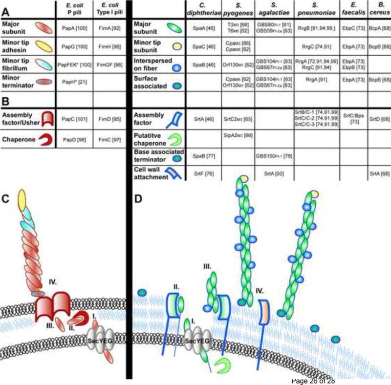
(a)–(b) E. coli prototype CU assembled pili. (a) Pilus subunit location within wild-type pilus fibers are depicted, as determined by immunoelectron microscopy. “*” indicates subunits whose location was deduced from biochemical and/or structural studies and not by immunoelectron microscopy. Empty boxes indicate no available data for that pilus biogenesis component. (b) Schematic of pilus biogenesis machinery for CU assembled pili. E. coli CU pilin subunits are translocated via the Sec machinery through the inner membrane (i), after which they associate with dedicated chaperone proteins in the periplasm (ii) which prevent subunit misfolding and facilitate delivery of the subunits to the outer membrane usher protein (iii) through which subunits are secreted and which serves as a platform for ordered pilus assembly (iv). (c)–(d) Gram-positive SA pili. (c) Table of pilus subunits for Gram-positive SA pili. Subscript for S. pyogenes indicates M1, M3 or M6 serotype and for S. agalactiae indicates the PI-1 or PI-2a pilus island. Slash marks (/) indicate differing names for the same protein in the literature. (d) Schematic of Gram-positive SA pili. Gram-positive pilin subunits are targeted for translocation across the cell membrane by the general secretion machinery via an N-terminal signal sequence (i). Presumably, subunits are then retained in the cell membrane by a transmembrane domain (ii) prior to LPXTG recognition and cleavage by sortase enzymes leading to ordered pilus fiber assembly (iii) and attachment to the cell wall (iv).