


Fig. 4. Immunodepletion of IKKα from cellular lysates decreases IκBα phosphorylation in vitro.
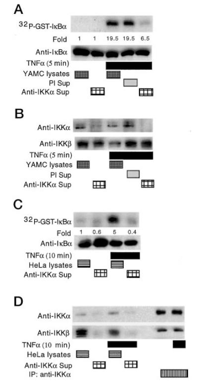
Kinase assays were performed in vitro by incubating lysates from untreated or TNFα-treated (100 ng/ml) YAMC cells, or the supernatants (Sup) remaining after immunoprecipitation with either anti-IKKα or preimmune (PI) serum. GST-IκBα was added to the kinase reaction mixture with 5 μCi of [γ-32P]ATP as before for detection of phosphorylation by autoradiography or IκBα by Western blot analysis (A). Cellular lysates or supernatants from the indicated immunoprecipitations were separated by SDS-PAGE for Western blot analysis with antibodies to either IKKα or IKKβ, as indicated (B). Kinase assays were performed using lysates or supernatants of immunoprecipitates from HeLa cells as above (C). HeLa cells were treated with TNFα and lysates, supernatants, or immunoprecipitates of IKKα studied for IKKα and IKKβ by Western blot analysis (D). Lane 5 was loaded with sample buffer only, to prevent any cross-contamination between immunoprecipitates and cellular lysates.