


Figure 6.
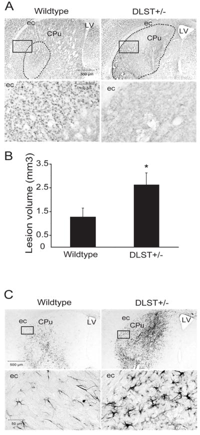
DLST-deficiency enhances malonate toxicity. A. NeuN-stained sections through the caudate putamen lesioned with malonate show an exacerbation of the lesion in DLST+/- mice. Broken lines show demarcation of the areas of NeuN-stained neuron loss. Boxed areas in the photomicrographs are magnified in the lower panel showing the severity of loss of NeuN-labeled neurons. B. Lesion volume analysis using stereological cavalieri method shows an increase of lesion volumes in DLST+/- mice. Values are means ± SEM. * p < 0.05. n = 10-11. C. CD40L immunoreactivity in the caudate putamen lesioned with malonate. There is marked proliferation and hypertrophy of CD40L-labeled astrocytes in DLST+/- mice. Boxed areas in the photomicrographs are magnified in the lower panel. ec= external capsule, LV= lateral ventricle, CPu= caudate putamen.