


Fig. 2. Stable formation of the force generating system of myocytes.
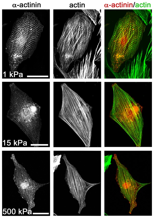
Cardiomyocytes were grown on ∼1 kPa, 15 kPa and 500 kPa stiff elastomeric substrates for 3 days and analyzed after fixation for myofibril formation using α-actinin (left) and actin (middle) as marker proteins. Overlay images are given (right). Scale bars: 50 µm.