


Figure 1.
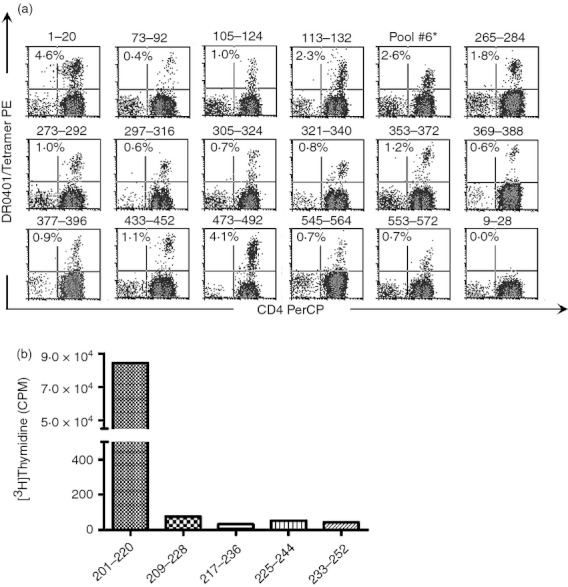
Identification of HLA‐DR0401 restricted epitopes by tetramer guided epitope mapping. (a) CD25‐depleted T cells from HLA‐DR0401 donors were stimulated with GAD65 peptides followed by tetramer staining. Each panel shows a representative result (combined from multiple experiments using different subjects) for single antigenic peptides that were identified as epitopes: GAD1–20 (pool 1), GAD73–92 (pool 2), GAD105–124 (pool 3), GAD113–132 (pool 3), Pool 6 (further characterized in b), GAD265–284 (pool 7), GAD273–292 (pool 7), GAD273–292 (pool 7), GAD297–316 (pool 8), GAD305–324 (pool 8), GAD321–340 (pool 9), GAD353–372 (pool 9), GAD369–388 (pool 10), GAD377–396 (pool 10), GAD433–452 (pool 11), GAD473–492 (pool 12), GAD545–564 (pool 14), and GAD553–572 (pool 14). DR0401/GAD9–28 is included as a representative negative result. Each percentage indicates the level of tetramer staining above background. (b) To identify the antigenic peptide within pool #6, the proliferation of a T‐cell line was assessed following stimulation with each individual peptide within the peptide pool. Only GAD201–220 elicited proliferation above background.